
記住我


A previously healthy 11-year-old boy had visited his pediatrician with fever, headache, abdominal pain, diarrhea, pollakiuria, painful micturition and gross hematuria 2 days before admission to our hospital. He had no notable medical history or allergies. He had eaten a soft-boiled egg 2 days before the onset of symptoms. He had had no known close contact with gastroenteritis.
His heart rate at admission was 136 beats/min, respiratory rate of 26 breaths/min, blood pressure of 92/52 mm Hg, body temperature of 40.3°C and oxygen saturation of 97% on room air. His body weight on admission was 33 kg, which was 5% below average. On physical examination, there was widespread tenderness in the lower abdomen and at the left costovertebral angle.
Initial laboratory investigations revealed a white blood cell count of 40.3 × 10⁹/L (normal: 4.0–11.9) with a predominance of granulocytes (92.5%), platelet count of 216 × 109/L (normal: 180–460), C-reactive protein level of 164.8 mg/L (normal: ≤3), blood urea nitrogen value of 24.9 mg/dL (normal: 6.8–19.3) and serum creatinine level of 1.42 mg/dL (normal: 0.35–0.58). Urinalysis revealed microscopic hematuria, proteinuria, pyuria and ketonuria. The fractional excretion of sodium was 0.12%. Abdominal ultrasonography revealed grade I hydronephrosis in the left kidney and a wedge-shaped hyperechoic region in the upper pole.
On the first day of admission, he was started on ceftriaxone (120 mg/kg/day) intravenously and fluid replacement. Furosemide was started on day 3 when a decrease in urine volume and an increase in the serum creatinine level to 1.78 mg/dL were noted.
Culture results and further imaging studies revealed the diagnosis.
For Denouement see P. 939.
DENOUEMENT(Pediatr Infect Dis J 2022;41:939–940)Continued from P. 938.
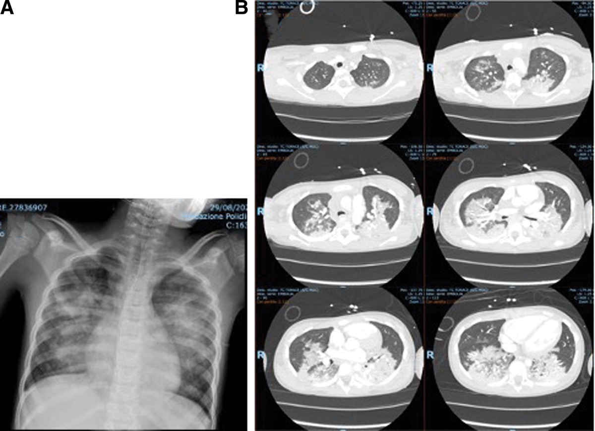
Salmonella enteritidis was isolated from urine, stool and blood cultures. The isolate was sensitive to ceftriaxone and ampicillin. His left back pain increased even after the start of antibiotic treatment. From left hydronephrosis found by abdominal ultrasonography, we considered complications, such as urinary stones or an intra-abdominal tumor. On day 3rd, we decided to perform a nonenhanced computed tomography (CT) scan of the abdomen. We did not perform contrast because of decreased renal function. This showed swelling of the left kidney and gas localized in the renal pelvis, suggesting emphysematous pyelonephritis (EPN; Fig. 1). By day 6, his left back pain had disappeared, and urine output had increased. Given the improvement in symptoms, surgical intervention was deferred. On day 18, antimicrobial treatment was changed to ampicillin after he developed acute cholecystitis, which was considered an adverse reaction to ceftriaxone. Abdominal magnetic resonance imaging on day 21 showed atrophy of the left kidney and scar formation on both poles but no morphological abnormality of the urethra or hydronephrosis. Imaging showed no abscess around the kidney, so the antibiotic was discontinued after 3 weeks, with no subsequent relapse of symptoms. On follow-up 2 months after discharge, the serum creatinine level had improved to 0.61 mg/dL.
 FIGURE 1.:
 Computed tomography scan of the abdomen on the third day of hospitalization showing swelling of the left kidney (major axis, 103 mm) and gas localized in the renal pelvis.
EPN is a rare, severe gas-producing necrotizing infection of the renal parenchyma and surrounding areas. Most reported cases have been in diabetic patients in their 40s and 50s, and pediatric cases are rare.1 Although the outcome for patients with EPN has improved due to advances in treatment, the prognosis is still poor with a reported mortality rate of 18%.1 In the present case, there was transient hydronephrosis in the absence of any congenital malformation of the urinary tract.
Although an enterobacterium is usually responsible for EPN, S. enteritidis was detected in stool, urine and blood samples in this case. Urinary tract infection caused by nontyphoidal Salmonella (NTS) is very rare, with a reported frequency in the range of 0.015%–0.118%.2 To our knowledge, there have been no reports of pediatric EPN caused by Salmonella. The patient had eaten a soft-boiled egg 2 days before the onset of the illness, which might have caused S. enteritidis infection given the incubation period.
The route of entry of S. enteritidis into the kidney, in this case, could have been hematogenous or retrograde via the urethra. Urethral infections caused by NTS are more likely to occur in the presence of a urethral disorder,3 which may reflect the fact that the route of entry is often via the urethra. In this case, bacteremia was thought to have occurred secondary to urinary tract infection and the development of EPN.
The diagnostic accuracy of renal ultrasound for EPN has been reported to be 50%–86%.4 Huang et al1 found that abdominal CT was important for the diagnosis of EPN and assessment of its severity. In our case, we could not make the diagnosis by renal ultrasound but could do so by abdominal CT, which showed gas in the renal pelvis.
Huang and Tseng proposed a therapeutic strategy for EPN based on a combination of clinicoradiologic classification and risk factors. EPN is classified according to the location of gas seen on CT as follows: confined to the collecting system (class 1); confined to the renal parenchyma (class 2); extending outside the kidney (class 3) and bilateral kidney involvement (class 4).1 Risk factors for aggravation of EPN are thrombocytopenia, shock, impaired consciousness and acute renal failure.1 Antibiotic therapy combined with percutaneous drainage (PCD) is recommended for patients with class 1 or 2 EPN and in those with class 3 or 4 with less than 2 risk factors, whereas nephrectomy is recommended for patients with class 3 or 4 EPN and 2 or more risk factors. Our patient had class 1 EPN because the emphysema was confined to the collecting system as well as 1 risk factor (acute renal failure). Therefore, a combination of antibiotic therapy and PCD would be recommended. However, in this case, the emphysematous pyelonephritis was too small for surgical drainage, so we treated with antimicrobial therapy alone, which fortunately controlled the infection. The blood creatinine level normalized, but the atrophy of the affected kidney remained, reflecting the severity of the EPN.
Seven cases of EPN in children from 2010 to 2021 can be searched on PubMed.4–9 Four patients had urinary disorders,5–8 1 had immunodeficiency4 and the 2 did not have an underlying illness.4,9 No complications of diabetes including this disease were observed. This is a major difference from adult cases with a high complication rate of diabetes.1
There were 3 cases that recovered only by antimicrobial therapy.5–7 There was one class 1 and one class 2, and the remaining one had only ultrasonography. From the accumulation of findings, it has been reported that class 1 without abscess or urinary tract obstruction can be treated with antimicrobial therapy alone.10 However, pediatric deaths have also been reported, and careful management is still important.4,9
S. enteritidis is rarely associated with urinary tract infection, even in healthy children. Urinary tract infection caused by S. enteritidis can lead to EPN, and abdominal CT is important for diagnosis and to inform management.
REFERENCES 1. Huang JJ, Tseng CC. Emphysematous pyelonephritis: clinicoradiological classification, management, prognosis, and pathogenesis. Arch Intern Med. 2000;160:797–805. 2. Tena D, González-Praetorius A, Bisquert J. Urinary tract infection due to non-typhoidal Salmonella: report of 19 cases. J Infect. 2007;54:245–249. 3. Gorelik Y, Paul M, Geffen Y, et al. Urinary tract infections due to nontyphoidal Salmonella. Am J Med Sci. 2017;353:529–532. 4. Ambaram PR, Kala UK, Petersen KL. Emphysematous pyelonephritis in children. Pediatr Infect Dis J. 2016;35:1159–1161. 5. Girgenti V, Pelizzo G, Amoroso S, et al. Emphysematous pyelonephritis following ureterovesical reimplantation for congenital obstructive megaureter. Pediatric case report and review of the literature. Front Pediatr. 2019;7:2. 6. Gross IT, Ford R. Emphysematous pyelonephritis in a child with nephrolithiasis. J Pediatr. 2016;168:250–250.e1. 7. Siddique K, Seikaly MG. Emphysematous pyelonephritis in an infant. Pediatr Infect Dis J. 2013;32:1157–1158. 8. Kitano H, Hieda K, Kitagawa H, et al. Case Report: Emphysematous Pyelonephritis With a Congenital Giant Ureterocele. Front Pediatr. 2021;9:775468. 9. Jiya FB, Ibitoye PK, Jiya NM, et al. Emphysematous pyelonephritis in an infant from Sokoto, north-western Nigeria. Afr J Lab Med. 2021;10:1181. 10. Demet A, Özgür ÖŞ, et al. Unexpected cause and successful management of typical urinary tract infection symptoms: Answers. Pediatr Nephrol. 2021;36:3647–3651.
留言 (0)