This study compared oropharynx and hyoid bone changes between female extraction patients and female non-extraction patients; it also explored differences in oropharynx and hyoid bone changes among four skeletal patterns. Female extraction patients exhibited increases in oropharynx volume and posterior movement of the hyoid bone compared with female non-extraction patients; the increase in oropharynx volume was greater in skeletal class I patients and moderate retraction patients.
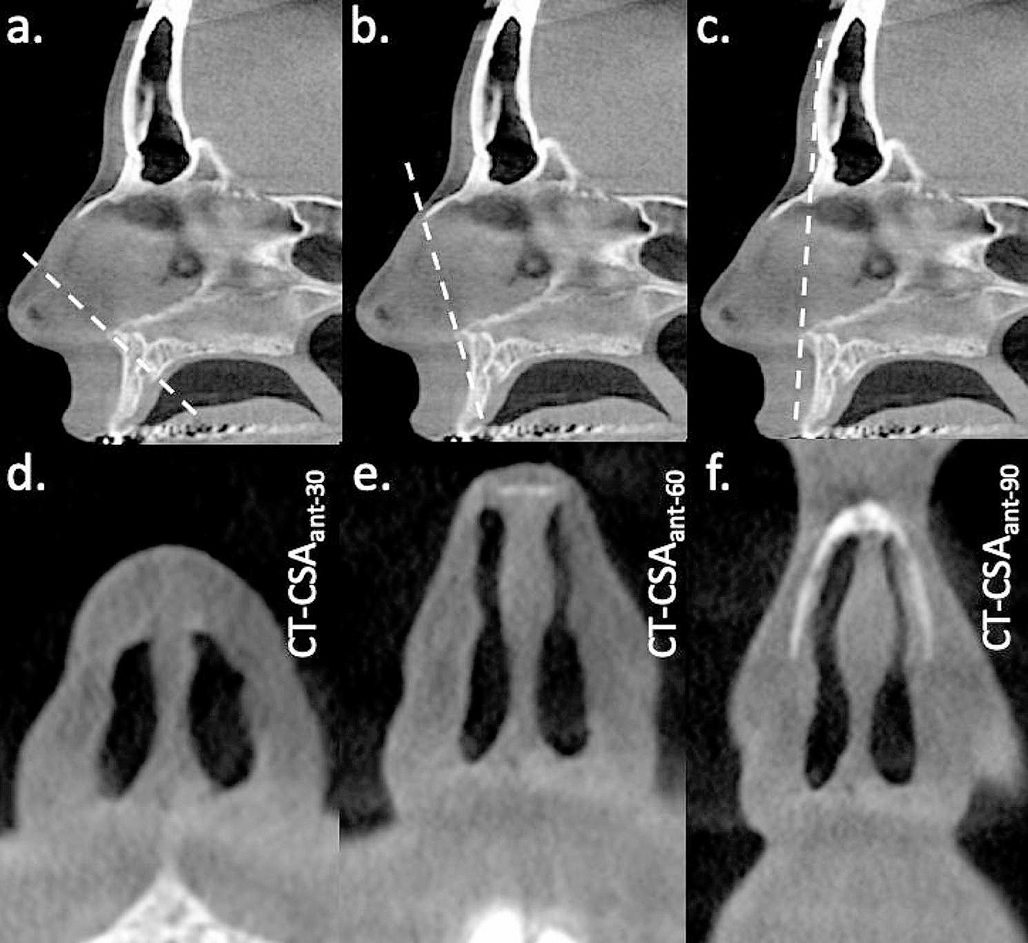
Extraction treatment can alleviate crowding and reduce facial convexity. However, an important concern during dental extraction is respiratory function, particularly in the upper airway (i.e. nasopharynx, oropharynx, hypopharynx). The nasopharynx and hypopharynx are supported by bone and cartilage, and are located far from the oral cavity; they are not easily influenced by extraction treatment. In contrast, the oropharynx comprises soft tissue and tongue; it is directly connected to the oral cavity. Besides, Jason et al. assessed oropharyngeal airway volume and demonstrated excellent intra-examiner and inter-examiner reliabilities, while the reliabilities of nasopharynx and hypopharynx assessments via CBCT have been low [18]. Hence, the oropharynx was the main focus of measurements in the present study.
Several previous studies have used CBCT to analyse the effect of extraction on upper airway size. However, their results have been inconsistent [5, 9, 10, 17, 19]. Sun et al. and Zheng et al. both reported significant decreases in oropharynx volume after maximum incisor retraction in class I bimaxillary protrusion patients [20, 21]. They presumed that dental extraction reduced arch length and oral cavity, which led to posterior movement of the tongue, followed by oropharyngeal narrowing. In contrast, Joy et al. and Pliska et al. [17, 19]. reported no clinically significant changes in oropharynx volume. Heterogeneity in patient characteristics (e.g. age, sex, skeletal pattern, extraction indication) among the studies might have contributed to these disparate findings. At present, no strong evidence is available regarding the effects of extraction on upper airway size. In this retrospective study with a large sample size, we found that oropharyngeal changes did not differ significantly between extraction and non-extraction groups, consistent with reports by Joy et al. and Pliska et al. [17, 19]. However, we also found that the oropharynx volume in the extraction group significantly increased from T0 to T1. Notably, the transverse diameter of the oropharynx (PNS-lateral and E-lateral) exhibited greater changes in the extraction group, indicating that the lateral wall of the oropharynx is easily changed. Da Costa et al. suggested that the lateral wall of oropharynx was the main area affected during orthognathic surgery and orthodontic treatment [22].
An enlarged upper airway after extraction has been reported previously in adolescent patients because of ongoing craniofacial growth; this change has rarely been reported in adults. Several factors might contribute to an increased oropharynx size in female adults. First, class II elastics were commonly used in the extraction group to reinforce the maxillary anchorage. Class II elastics could move the mandible forward, while increasing the oropharynx size. Second, patients with bimaxillary protrusion usually present with a narrow dental arch [23]. The constricted dental arch could lead to tongue crowding, which might be associated with a narrow oropharynx. After extraction treatment, the expanded dental arch leads to a larger oropharynx. Increased upper airway dimensions have been reported after maxillary expansion [24, 25]. Finally, the mesial movement of posterior teeth, which provides a posterior space for the tongue, could lead to greater airway volume. In the present study, we found that increases in the oropharynx volume and MCA were greater in the moderate retraction group than in the maximum retraction group, which confirmed this hypothesis. The mechanism of oropharyngeal changes should be investigated in future studies.
Some studies have theorized that dental extraction can reduce upper airway size and increases the risk of obstructive sleep apnoea (OSA), but there are inherent disadvantages as these were performed with lateral cephalogram [6,7,8, 14, 15]. The sagittal information provided by lateral cephalogram may be misleading. Furthermore, the upper airway varies among patients, and previous studies have included small sample sizes. In contrast, we included a larger sample size and found larger oropharynx size upon CBCT analysis. It is also important to note that there is no direct link between a narrow airway and OSA. The American Association of Orthodontists has indicated that a narrow airway does not result in OSA [26]. CBCT cannot provide information regarding airway function; OSA should be diagnosed via polysomnography or an out-of-centre sleeping test. Extraction safety in OSA patients should be investigated in future studies.
The hyoid bone is connected with the pharynx, cranial base, and mandibular symphysis via muscles and ligaments; this complex can maintain airway stability. In the present study, we found significant posterior movement of the hyoid bone in the extraction group and slight anterior movement of the hyoid bone in the non-extraction group, especially in class I-hyper patients; posterior movement of the hyoid bone was greater in the maximum retraction group than in the moderate retraction group. The vertical position of the hyoid bone was stable in all groups. Our results are consistent with those of Bhatia et al. [6], who reported that the hyoid bone moved posteriorly during anterior teeth retraction. In contrast, Maaitah et al. and Germec-Cakan et al. [7, 8]. reported no significant changes in hyoid bone position. Although many studies have reported associations between the hyoid bone and oropharynx, the mechanism of hyoid bone compensation during oropharyngeal changes remains unclear. Wang et al. suggested that the hyoid bone moves both posteriorly and inferiorly after extraction treatment, which could prevent encroachment of the tongue into the oropharynx [27].
The differences in oropharynx among patients with distinct skeletal patterns have been reported previously [11,12,13]. With regard to the vertical skeletal patterns, the hyperdivergent patients had smaller upper airway volumes compared with normodivergent and hypodivergent patients [11]. With regard to sagittal skeletal patterns, the skeletal class II patients had a narrow upper airway [13, 28]. Our results confirmed that skeletal class II and hyperdivergent patients had a narrow oropharynx volume compared with other skeletal pattern patients. To our knowledge, dental extraction involving the oropharynx has not been investigated among patients with distinct skeletal patterns. We observed significant differences in premolar extraction effects on oropharynx volume (between class I-norm and class II-norm patients) and MCA of the oropharynx (between class I-hyper and class II-hyper patients). The oropharynx volumes from T0 to T1 increased significantly in the class I group, but not in the class II group. These results suggest that, compared with vertical skeletal patterns, the effects of sagittal skeletal patterns might be more important in the context of oropharyngeal changes in female patients.
Class II and hyperdivergent patients reportedly have a narrow upper airway and a high risk of OSA [29, 30]. In our study, class II and hyperdivergent patients with a narrow oropharynx and posterior hyoid bone position did not exhibit oropharynx collapse, but they tended to exhibit increases in oropharynx volume and MCA after extraction treatment. Zhang et al. also analysed the effects of extraction on upper airway changes in skeletal class II and hyperdivergent patients; consistent with our results, they found that the volume and MCA did not change significantly [10]. Hence, changes in oropharynx variables might not be associated with baseline condition; skeletal class II and hyperdivergent patients without OSA did not exhibit contraindications for premolar extraction.
In addition to skeletal pattern, changes in the upper airway after extraction treatment may have been inconsistent among previous studies because of variations in anterior teeth retraction. The extraction space could be closed by anterior teeth retraction and posterior teeth mesial movement. We divided extraction patients into maximum and moderate retraction groups based on changes in U1-X. Changes in oropharynx variables did not differ significantly between groups, consistent with a study by Valiathan et al., who found that changes in incisor angulation and position did not cause significant differences in oropharynx volumes [31]. In the present study, the volume and MCA of the oropharynx from T0 to T1 increased significantly in the moderate retraction group, but not in the maximum retraction group. This finding was consistent with our expectations because the maxillary mini-screw was commonly used in the maximum retraction group to achieve considerable retraction of anterior teeth, while class II elastic was commonly used in the moderate retraction group. The mesial movement of posterior teeth and the use of class II elastic might have contributed to a significant increase in oropharynx size in the moderate retraction group.
There are some limitations in this study. Firstly, the number of non-extraction patients was limited in this study due to ethical considerations. We used CBCT to confirm the amount of anterior tooth retraction in extraction patients according to pre-treatment alveolar bone morphology; teeth were kept in alveolar bone after orthodontic treatment. Most non-extraction patients in our study used CBCT only because of factors related to implant restoration and impacted teeth, which caused a small sample size of control group. Secondly, the airway could be easily influenced by factors such as age, sex, weight, head position, breathing mode, and tongue position [32, 33]. The upper airway size increases until 20 years of age and decreases thereafter [34, 35]. Our study population was limited to fully grown females in an attempt to avoid these confounding factors. While, the OSA is more prevalent in male patients than in female patients [36]. In this study, we only included female patients. The effects of first premolar extraction on the oropharynx and hyoid bone positions in male patients should be further investigated. Besides, body mass index was not recorded after orthodontic treatment, so the effects of weight changes during orthodontic treatment could not be analysed. Thirdly, due to the relatively low prevalence of four premolar extraction in skeletal class III patients and hypodivergent patients in Asian, only skeletal class I and class II patients with hyperdivergent and normodivergent patterns were included. Importantly, the ability for upper airway adaptation should be considered when interpreting our findings. Our results only reflect changes in the size and morphology of the oropharynx and the hyoid bone position in female extraction patients. Respiratory function and the long-term stabilities of the oropharynx and hyoid bone should be investigated in future studies.


留言 (0)