
記住我


Perioperative hemodynamic management is critical to patient and graft outcomes in transplantation medicine.1 Maintaining hemodynamic stability during and after renal transplantation is particularly important in deceased-donor kidney transplants. Deceased-donor kidneys have reduced long-term survival2 and the cold ischemic time is associated with significantly impaired graft function compared with living-donor kidney transplants.3,4
Examining the effects of intraoperative fluid management on outcomes after kidney transplantation is challenging because fluid therapy and hemodynamics are inseparably linked and both are critical for renal perfusion. Although fluid administration remains the dominant therapeutic approach to maintain or restore circulatory function, an evidence-based perioperative strategy for fluid management has been elusive.5 We developed hemodynamic monitoring software that was utilized in a high-risk kidney transplant recipient. We describe how continuous visualization of trends in stroke volume (SV) and systemic elastance allowed differentiation of the roles of the heart and vasculature and provided a means for choosing and titrating fluid, inotropes, or vasopressors. Written informed patient consent was obtained for the publication of this report. This article adheres to the applicable EQUATOR (Enhancing the QUAlity and Transparency Of health Research) guidelines.
CASE REPORTA 68-year-old man with diabetic nephropathy on hemodialysis was referred for pretransplantation assessment. His history included chronic atrial fibrillation and expressive dysphasia from a stroke 8 years prior. He was receiving long-term warfarin. He had multiple concerning preoperative findings, including elevated pulmonary pressures on cardiac catheterization (right ventricular pressure 80/8 mm Hg, pulmonary artery pressure 77/27 mm Hg) and right ventricular dysfunction on echocardiography (tricuspid annular plane systolic excursion 12 mm).6
A cadaveric donor kidney became available within the same metropolitan area as the patient. The allograft was in the “worst 31%” of donors,7 and the recipient’s mortality risk was in the worst 4%.8 The patient was dialyzed and prothrombin complex concentrate was administered to normalize the international normalized ratio. The Table shows the preoperative (postdialysis) laboratory results.
Table 1. - Preoperative and postoperative values for hemoglobin, urea, creatinine and phosphate Preoperative Postoperative Day 1 Day 2 am Day 2 pm Day 3 am Day 3 pm Day 4 Day 5 Day 6 Hemoglobin (g/L) 116 122 109 105 98 – 89 – 99 PO4 (mmol/L) 1.4 – 2.0 1.8 1.4 – 0.7 0.6 0.6 Urea (mmol/L) 9.8 – 9.7 11.3 13.8 15 12.4 10.6 10.6 Creatinine (μmol/L) 509 – 370 256 205 180 140 109 115Reference ranges: PO4, 0.97–1.45 mmol/L; urea, 2–7 mmol/L; creatinine, 60–115 µmol/L. Excellent graft function was shown by near normalization of values without further dialysis. Renal phosphate wasting occurred early after kidney transplantation.
In the operating room, peripheral venous and radial arterial catheters were placed in the patient’s nonfistula arm. A 3-lumen subclavian catheter was inserted, and the distal lumen was connected via a TruWave transducer to the EV1000 monitoring system (Edwards Lifesciences, Irvine, CA). A FloTrac sensor (FloTrac System 4.0, Edwards Lifesciences) was connected to the arterial cannula and the EV1000 hemodynamic monitor. The arterial and central line pressure bags were maintained at 300 mm Hg, with both transducer sensor stopcocks kept level to the phlebostatic axis, located at the fourth intercostal space (corresponding to the right atrium). A serial-to-USB cable transmitted hemodynamic data every 20 seconds from the EV1000 monitor to a computer with software to calculate and display the patient’s “pressure field” in synchrony with the Flotrac transducer.
The pressure field is an investigational tool for continuously resolving mean arteriovenous pressure into its cardiac and vascular components and displaying this visually to enable a clinical decision. The pressure field equation relates mean arterial pressure (MAP) and central venous pressure (CVP) to SV and systemic elastance (Es) as follows:
[MAP−CVP]=SV×Es
This equation differs from the approach of Starling and Guyton, where the heart’s contribution is understood as cardiac output, and ventricular afterload is represented by systemic vascular resistance.
The patient was monitored to determine the resting pressure field before induction of anesthesia (Figure) to guide intraoperative management. Anesthesia was then induced with midazolam 2.5 mg, fentanyl 100 μg, propofol 100 mg, and rocuronium 100 mg. On induction, a norepinephrine infusion was started at 4 μg/min. Anesthesia was maintained with a propofol target-controlled infusion (Marsh protocol, target concentration 3 μg/mL) and a remifentanil target-controlled infusion (Minto model, target concentration 4 ng/mL). Methylprednisolone (1000 mg in 100 mL) was given postinduction. Furosemide (40 mg), mannitol (20 g in 100 mL), and acetaminophen (1000 mg in 100 mL) were administered intraoperatively.
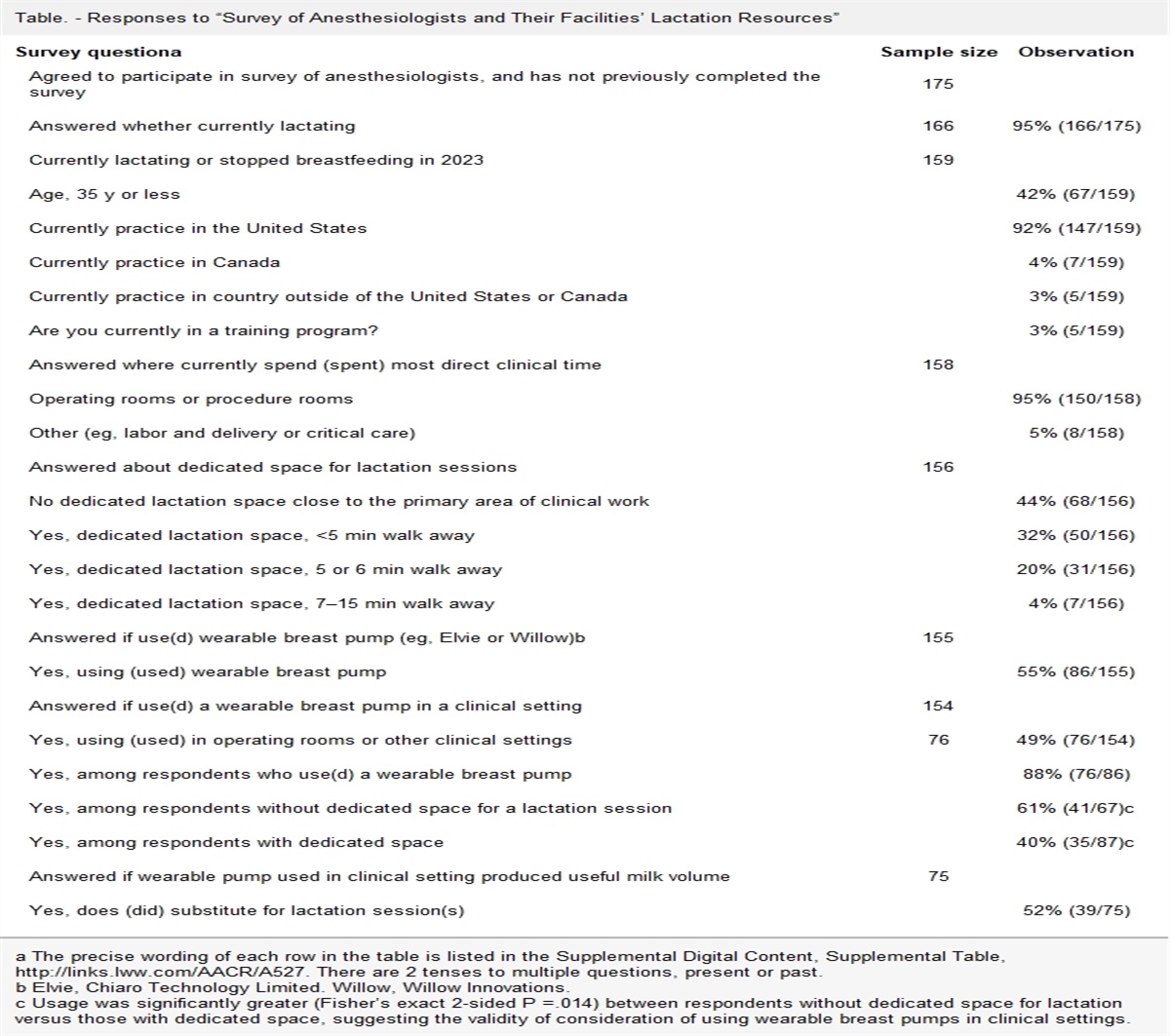
 Figure.:
 Pressure field before and during surgery and after reperfusion. Systemic elastance (x-axis) is a measure of vasomotor tone; stroke volume (y-axis) varies with preload and contractility. The curved lines represent mean arteriovenous pressure at 15 mm Hg intervals.
Surgery lasted 3 hours and 20 minutes. Management was based on plotting the pressure field and varying norepinephrine and fluid to mimic the preoperative pattern (Supplemental Digital Content, Video, https://links.lww.com/AACR/A493). The norepinephrine infusion was titrated to regulate movement of the pressure field, that is to minimize changes in SV (y-axis) and Es (x-axis; Figure). A transient decrease in SV occurred at induction and was assumed to represent a fall in preload due to redistribution; it was not treated with fluid and spontaneously recovered over a period of minutes. Blood loss was 300 mL, and no intravenous fluid was administered, apart from medications, during surgery. Urine output was 200 mL between reperfusion of the transplanted kidney and surgery completion. The total fluid volume administered was estimated at 500 mL and was similar to the blood loss and urine volumes. Norepinephrine was infusing at 4.5 μg/min on completion of surgery and was ceased in the recovery unit. No additional fluid was given. The postoperative hemoglobin was 122 g/L and the net fluids were zero.
The patient was monitored overnight in the intensive care unit and discharged to the ward the following day. Between surgery and discharge on day 6, the hemoglobin fell to 99 g/L. No dialysis was required. The hemoglobin, phosphate, urea, and creatinine over the first 5 postoperative days indicated excellent donor kidney function (Table). At 6 months, the donor kidney was functioning normally.
DISCUSSIONThe current approach for managing the circulation relies heavily on arterial pressure as a surrogate for tissue perfusion, with fluid loading the default response to low arterial pressures. Most authors advocate fluid loading to optimize the donor kidney before it is explanted and to increase renal blood flow in the recipient after reperfusion while also avoiding hypotension.9,10 A recent retrospective study found an average fluid load of 3 L,11 while another author reported an average fluid load of 40 mL/kg.12 Although there is controversy around the merits of potassium-free or balanced crystalloid solutions and colloid in this population,13,14 there is a unanimous view favoring the liberal use of volume expanders over pressors. A recent consensus statement from the Committee on Transplant Anesthesia of the American Society of Anesthesiologists on fluid management during kidney transplantation reported no difference in outcomes between crystalloids and colloids but affirmed the role of liberal intravenous fluids.14 Despite a net zero fluid balance, our patient’s postoperative recovery was complication free.
The pressure field addresses the need to identify if a fall in pressure is due to a change in preload, contractility, or vasomotor tone. Every pressure value is deconstructed into elastance as the X-axis value and SV on the Y-axis, where SV reflects both preload and contractility. Changes in preload may reflect volume loss from the circulation or changes in blood volume distribution. Observing the response to a fluid bolus of 1 mL/kg is typically sufficient to enable identification of an issue of volume loss, administration of an inotrope enables identification of a contractility issue, and administration of a vasopressor will reverse volume redistribution within the circulation. Management remains an empirical process, but with more precise control and smaller and earlier interventions. Initiating monitoring before induction enables a patient-specific template to guide management. This identifies specific patterns of change in the regulation of pressure and flow and allows clinicians to prescribe highly individualized solutions. Management is always dictated by the patient’s physiology.
The pressure field method in this patient primarily involved the titration of norepinephrine. Although fluid administration is sometimes critical to managing circulation, the early decrease in SV was transient and there was no subsequent evidence to indicate hypovolemia.
The pressure field extends the study of Sunagawa et al.15 Sunagawa et al15 proposed that effective arterial elastance is a better measure than SVR of ventricular afterload. Where mean arteriovenous pressure is the difference between mean ventricular outflow pressure and mean right atrial inflow pressure, Starling’s pressure equation can be simplified to the relationship between SV and Es. When combined with high-frequency measurement, this alternative interpretation provides a different methodology for optimizing circulation.
In conclusion, the pressure field allows differentiation of the roles of the heart and vasculature in generating the arteriovenous pressure gradient. It measures real-time ventricular-vascular interaction and provides a means for titrating fluid, inotropes, or vasopressors. Combining the method with large data sets may provide a basis for improved diagnosis of cardiovascular disease and treatment recommendations. Although the pressure field method was helpful in this patient, it needs to be formally evaluated as a method for optimizing perfusion during renal transplantation.
DISCLOSURESName: Stephen F. Woodford, MBBS.
Contribution: This author helped write the first draft manuscript and create the supplemental video.
Conflicts of Interest: S. F. Woodford received honoraria from Edwards Lifesciences. This author holds patents related to the pressure field.
Name: Lachlan F. Miles, MBBS, PhD.
Contribution: This author helped edit the manuscript.
Conflicts of Interest: L. F. Miles received honoraria from Edwards Lifesciences.
Name: Dong-Kyu Lee, MD.
Contribution: This author helped edit the manuscript and the Figure.
Conflicts of Interest: D.-K. Lee received honoraria from Edwards Lifesciences.
Name: Laurence Weinberg, MD, PhD.
Contribution: This author helped edit the manuscript, create the Table, and edit the Figure.
Conflicts of Interest: L. Weinberg received honoraria from Edwards Lifesciences.
This manuscript was handled by: BobbieJean Sweitzer, MD, FACP.
REFERENCES 1. Michelet D, Brasher C, Marsac L, et al. Intraoperative hemodynamic factors predicting early postoperative renal function in pediatric kidney transplantation. Paediatr Anaesth. 2017;27:927–934. 2. Bellamy MC, Scott A. Therapeutic issues in transplant patients. Review. Anaesth Intensive Care Med. 2015;16:344–348. 3. Troppmann C, Gillingham KJ, Benedetti E, et al. Delayed graft function, acute rejection, and outcome after cadaver renal transplantation. The multivariate analysis. Transplantation. 1995;59:962–968. 4. Swanson EA, Patel MS, Groat T, et al. Vasopressor selection during critical care management of brain dead organ donors and the effects on kidney graft function. J Trauma Acute Care Surg. 2020;88:783–788. 5. Weinberg L, Collins MG, Peyton P. Urine the right direction: the consensus statement from the committee on transplant anesthesia of the American Society of Anesthesiologists on fluid management during kidney transplantation. Transplantation. 2021;105:1655–1657. 6. Mendes LA, Dec GW, Picard MH, Palacios IF, Newell J, Davidoff R. Right ventricular dysfunction: an independent predictor of adverse outcome in patients with myocarditis. Am Heart J. 1994;128:301–307. 7. Senanayake S, Graves N, Healy H,, et al. Donor kidney quality and transplant outcome: an economic evaluation of contemporary practice. Value Health. 2020;23:1561–1569. 8. Clayton PA, McDonald SP, Snyder JJ, Salkowski N, Chadban SJ. External validation of the estimated posttransplant survival score for allocation of deceased donor kidneys in the United States. Am J Transplant. 2014;14:1922–1926. 9. Forbes RC, Concepcion BP, King AB. Intraoperative management of the kidney transplant recipient. Current Transplantation Reports. 2017;4:75–81. 10. Chapin JW, Bruda N, Snider S. Anesthesia for renal transplantation. Review. Semin Cardiothoracic Vascular Anesth. 1998;2:106–113. 11. Dost BB M, Kaya C, Ustun YB, Bilgin S, Koksal E, Bostanci Y. Anesthetic management of patients undergoing renal transplantation: a review of a two-year experience. Signa Vitae. 2021;17:95–100. 12. Robertson E, Logan N, Pace N. Anaesthesia for renal transplantation. Review. Anaesth Intensive Care Med. 2018;19:552–556. 13. Meredith S, Basavaraju A, Logan N. Anaesthesia for renal transplantation. Review. Anaesth Intensive Care Med. 2021;22:500–504. 14. Wagener G, Bezinover D, Wang C, et al. Fluid management during kidney transplantation: a consensus statement of the committee on transplant anesthesia of the American Society of Anesthesiologists. Transplantation. 2021;105:1677–1684. 15. Sunagawa K, Sagawa K, Maughan WL. Ventricular interaction with the loading system. Ann Biomed Eng. 1984;12:163–189.
留言 (0)