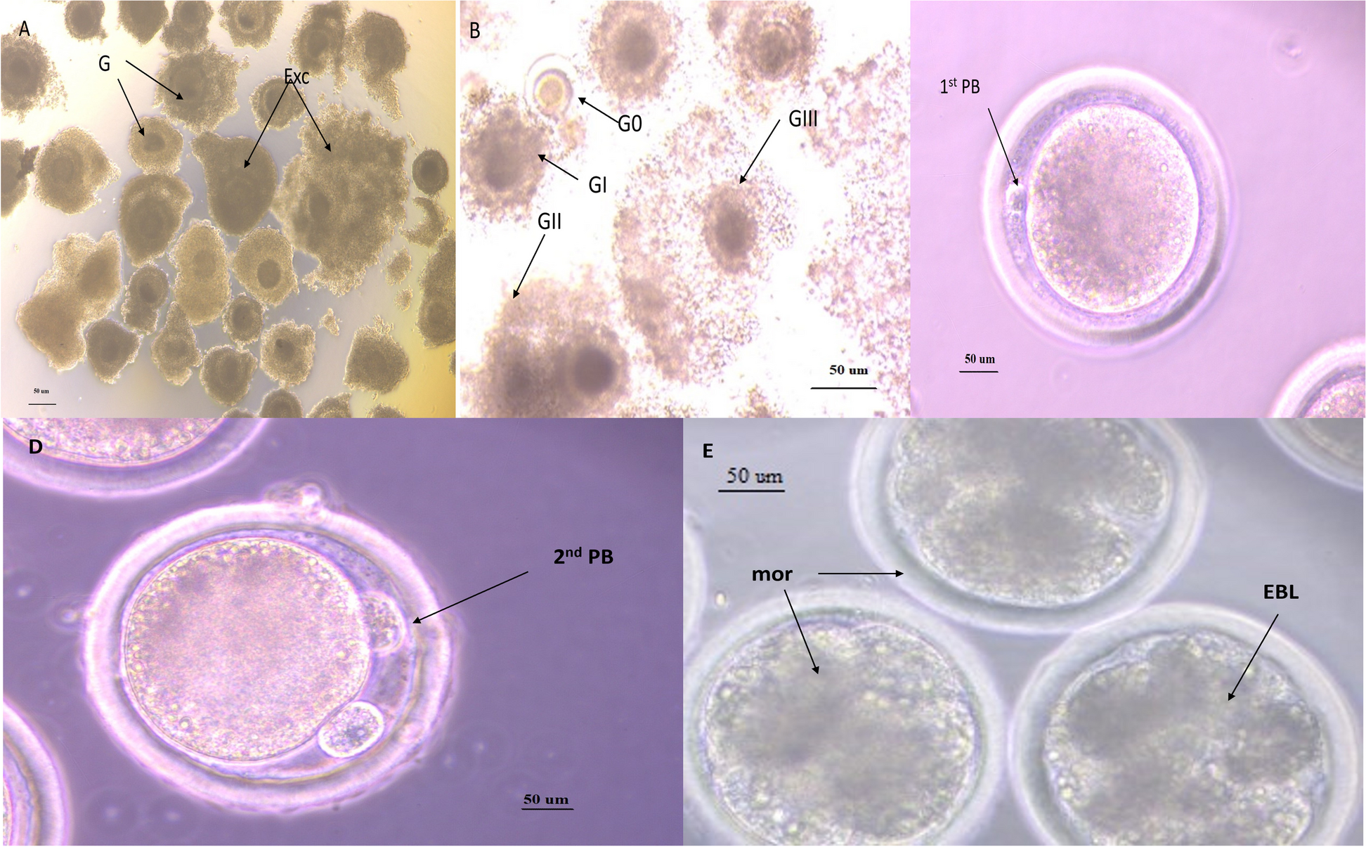
In this case report, we highlight the practical dilemma (i.e. to perform OTC surgery in a 45, X TS patient or not) by reporting on the presence of follicles in a 13-year-old female diagnosed with 45, X monosomy and an unmeasurable AMH. We compare our results with previous research, highlight the challenges we faced in this case and provide recommendations for daily practice. Hereby, we demonstrate that excluding certain subgroups of TS patients (e.g. monosomy patients, and/or girls with an AMH below 2.0 ug/l) may be premature, especially based on the current state of published research data.
From previous research [3, 20, 21], we know that the presence of a 46, XX cell line and a measurable AMH level have been associated with spontaneous pubertal development, the presence of follicles and spontaneous pregnancies in TS patients. However, optimal discriminative markers for the presence or absence of follicles in females with TS are currently still lacking. AMH serum levels can vary considerably due to biological variability and intertest variability of AMH assays, and there are limited prospective longitudinal data assessing AMH concentrations in prepubertal girls. Therefore, adequate interpretation of AMH levels, especially in prepubertal TS patients, is difficult. Furthermore, most research articles on TS, including key papers that are describing the presence or absence of follicles in females with TS, still lack a description of the methods used for genotyping [3, 22,23,24,25]. This is, however, essential information especially when linked to fertility or fertility preservation. Missing X chromosomal content in ovarian cells is likely to result in germ cell apoptosis and impaired folliculogenesis during foetal life [26, 27]. One could assume that the described cases where follicles were found or pregnancies have occurred in females with 45, X monosomy were actually examples of undetected germ cell mosaicism [28, 29].
In routine care, karyotyping of lymphocytes is the standard technique used to determine TS. most girls have been diagnosed with TS based on the examination of only one cell type. In most centres, the diagnosis is based on 20–30 lymphocytes only, although it has been recommended to determine the karyotype of at least 50 metaphases to exclude < 10% of mosaicism (CI 0.99) [30]. Interestingly, a recent cohort study in 142 adult patients with TS, revealed that the percentage of 45, X cells in lymphocytes and buccal cells was identical in less than one third of cases [31]. For this reason, some experts [15, 31, 32] recommend adding buccal cell FISH analysis for a better diagnosis-prognosis-treatment and guidance for some subgroups of TS patients.
Our patient had been diagnosed with 45, X monosomy but presented with spontaneous pubertal development and monthly periods of vaginal bleeding (5–7 days). Additional FISH examination of buccal smear revealed mosaicism. At the time she was referred to us, she was reporting irregular periods of vaginal bleeding since a few months and the endocrinological assessment repeatedly showed an AMH level under the reference value. Since the ovarian function in TS patients only declines with age, it seemed likely that this girl had diminished ovarian reserve. The counselling of the girl and her parents therefore included a difficult dilemma: should we perform OTC surgery or not?
During the counselling process by a dedicated team, different considerations were discussed and addressed.
First of all, we must be careful not to raise false hopes even if OTC might be the only possibility to preserve the ovarian follicles that are still left. In this patient, it was questionable whether the ovarian reserve in the normal-looking ovary would still be sufficient for fertility preservation purposes. In other words: if there is already an imminent premature ovarian insufficiency (POI), does it make sense to later transfer the frozen and thawed pieces back? Especially since the freezing and thawing of ovarian tissue will also have a negative effect on the number of viable oocytes and thus the chance of pregnancy and live birth. In addition, patients with TS are known to have a higher risk of having a miscarriage [20]) and a slightly higher risk of having a child with a congenital disorder [20, 33]. Therefore, not only the quantity but also the functionality of the oocytes and supporting ovarian cells must be considered. In vitro activation (IVA) [34] or in vitro maturation (IVM), eventually combined with preimplantation genetic diagnosis (PGD) [35] could possibly improve the chances of pregnancy and live birth in the future. However, both techniques (e.g. IVA and IVM) are at the moment still experimental. Furthermore, we should also be aware of the greater frequency of uterine morphological anomalies in patients with TS and the potential for compromised endometrial receptivity [20].
The second consideration in this case was whether we are not reducing her chances of a (complete) spontaneous pubertal development by performing an ovariectomy. Unfortunately, there are no follow-up studies that report on the effect of ovarian surgery on the chances of spontaneous pubertal development, pregnancy and risk of (earlier) menopause in patients with TS. In non-TS patients, an ovariectomy does not affect the chances of pregnancy [36] but may lead to 1–3 years earlier onset of menopause [37]. However, these data are obviously from girls and women with normal ovarian reserve, and the remaining ovary might ‘compensate’ for the removed ovary. The impact of this procedure on our patient is unknown, but most likely, her ovarian reserve would not be sufficient to become pregnant later, but possibly just enough to complete spontaneous pubertal development. We discussed that in the worst case, this process could be stagnated by the removal of the ‘normal’ looking ovary. This would mean that she, like many TS patients, would become dependent on hormone replacement therapy, but at an earlier time. Fortunately, this patient experienced complete pubertal development without hormone replacement therapy. In addition, she continued to have periods of (irregular) vaginal bleeding two years after surgery. However, these might have been anovulatory cycles with persistent follicles. Nevertheless, a repeatedly measured low oestrogen value based on a confirmed hypergonadotropic hypogonadism status was an indication to start hormone replacement therapy 3 years after surgery.
Lastly, the increased risk of foetal and maternal complications during future pregnancy and labour in females with TS should be considered. In comparison with healthy females, females with TS are at increased risk of intra-uterine growth restriction, preterm labour, thyroid dysfunction, diabetes, obesity, hypertension and pre-eclampsia [38]. In the past, women with TS were even discouraged to become pregnant due to the risk of aortic dissection. Recent studies have shown however, that this risk has decreased to 0.5% due to increased awareness of cardiovascular complications, improved preconception screening, and strict cardiovascular monitoring during pregnancy [39, 40]. Our patient had no history of aortic dissection, neither indications of an increased risk for aortic dissection, e.g. an increased aortic size index, bicuspid aortic valve, elongation of the transverse aorta, or coarctation of the aorta in combination with hypertension.
In conclusion, it seems that in this case, performing OTC was an appropriate decision. The ovariectomy did not affect her spontaneous pubertal development, and possibly we were just in time to preserve her fertility. Considering the low ovarian reserve, we found in the preserved ovarian fragments, it would have been very unlikely that she would have been able to conceive spontaneously in adulthood. Hereby, we demonstrate that excluding certain subgroups of TS patients from OTC (e.g. monosomy patients, and/or girls with an AMH below 2.0 ug/l) may be premature, especially based on the current state of published research data.
Our case is not the first case of a girl with TS who presented with a perioperative left–right difference in ovarian morphology during laparoscopic surgery [19]. However, this is the first case reporting on the presence of both a (morphological) streak ovary and normal ovary in a young female with TS. We recommend other clinicians to also discuss this rare outcome during fertility preservation counselling, and how to deal with it during laparoscopic surgery, even if we do not know yet what the clinical consequences are. In this patient, the left ovary could be visualized during transabdominal ultrasound (TUS). However, distinguishing precisely between anatomically normal ovaries and streak ovaries is not easy, and there might be a high chance that the ovaries cannot be visualized at all [41]. A Danish study showed that ovaries could be detected in 37% of young females with TS by TUS and in 55% by MRI (P = 0·1) [42], but they did not confirm their findings with laparoscopic surgery, which is considered to be the gold standard. We recommend further investigation of these imaging methods before applying these methods in daily practice.
As this is our 2nd case where we report on a girl who had been diagnosed with classical monosomy TS (45, X), but presented with a hidden X mosaicism in her ovarian cells [19], we want to highlight again the importance of buccal cell analysis for some subgroups of TS patients. In particular, in patients who have been diagnosed with 45, X monosomy to better differentiate between monosomy and mosaicism.
Moreover, we hope that future research reports will be more specific about the methods used for genotyping when reporting on TS and fertility or fertility preservation. We recommend including at least the type and number of cells on which the diagnosis is based. Lastly, by raising awareness about the possibilities of fertility preservation for girls with TS, we hope that more girls will be referred to a fertility specialist for fertility counselling. Since the current referral rates are rather low [14], we hope that by providing them with more information about their fertility and the full range of options available now and in the future to fulfil their desire to have children, will lead to better informed patients and less psychological burden.

留言 (0)