Tiwari P, Bhovi TV, Jaju PP, Gupta M, Shrivastava K. Frontal sinus-a useful personal identification tool. Oral Surg Oral Med Oral Pathol Oral Radiol. 2016;2(1):11. https://doi.org/10.5958/2395-6194.2016.00009.6.
Ahmed AG, Gataa IS, Fateh SM, Mohammed GN. CT scan images analysis of maxillary sinus dimensions as a forensic tool for sexual and racial detection in a sample of Kurdish population. Eur Sci J. 2015;11(18).
Sharma SK, Jehan M, Kumar A. Measurements of maxillary sinus volume and dimensions by computed tomography scan for gender determination. J Anat Soc India 2014;63(1):36–42. https://doi.org/10.1016/j.jasi.2014.04.007.
Hamed SS, El-Badrawy AM, Fattah SA. Gender identification from frontal sinus using multi-detector computed tomography. J. Forensic Radiol. Imaging. 2014;2(3):117–20. https://doi.org/10.1016/j.jofri.2014.03.006.
Ramaswamy P, Khaitan T. Frontal sinus index–a new tool for sex determination. J Forensic Radiol Imaging. 2014;2(2):77–9. https://doi.org/10.1016/j.jofri.2014.02.002.
Article
Google Scholar
Goyal M, Acharya AB, Sattur AP, Naikmasur VG. Are frontal sinuses useful indicators of sex? J Forensic Leg Med. 2013;20(2):91–4. https://doi.org/10.1016/j.jflm.2012.04.028.
Article
PubMed
Google Scholar
Sidhu R, Chandra S, Devi P, Taneja N, Sah K, Kaur N. Forensic importance of maxillary sinus in gender determination: a morphometric analysis from Western Uttar Pradesh, India. European J Gen Dent. 2014;3(1):53. https://doi.org/10.4103/2278-9626.126213.
Belaldavar C, Kotrashetti VS, Hallikerimath SR, Kale AD. Assessment of frontal sinus dimensions to determine sexual dimorphism among Indian adults. J Forensic Dent Sci. 2014;6(1):25. https://doi.org/10.4103/0975-1475.127766.
Article
PubMed
PubMed Central
Google Scholar
Uzabakiriho A. The role of forensic science in criminal investigation in Rwanda. Res J Forensic Sci. 2015;3:1–4.
Google Scholar
Nambiar P, Naidu MD, Subramaniam K. Anatomical variability of the frontal sinuses and their application in forensic identification. Clin Anat. 1999;12(1):16–9. https://doi.org/10.1002/(SICI)1098-2353(1999)12:1%3C16::AID-CA3%3E3.0.CO;2-D
Quatrehomme G, Fronty P, Sapanet M, Grévin G, Bailet P, Ollier A. Identification by frontal sinus pattern in forensic anthropology. Forensic Sci Int. 19962;83(2):147–53. https://doi.org/10.1016/s0379-0738(96)02033-6.
Akhlaghi M, Bakhtavar K, Moarefdoost J, Kamali A, Rafeifar S. Frontal sinus parameters in computed tomography and sex determination. Leg Med (Tokyo). 2016;1(19):22–7. https://doi.org/10.1016/j.legalmed.2016.01.008.
Article
Google Scholar
Zuckerkendl E. Anatomy of the frontal sinuses. In: Pathological Normal Anatomy of the Nasal Fans and their Annexes Pneumatics. Paris: G. Masson; 1895. p. 349–6.
Schuller A. A Note on the Identification of Skulls by X-ray Pictures of the Frontal Sinus. Med J Aust. 1943;1(1):554–6.
Article
Google Scholar
Jun BC, Song SW, Park CS, Lee DH, Cho KJ, Cho JH. The analysis of maxillary sinus aeration according to aging process; volume assessment by 3-dimensional reconstruction by high-resolutional CT scanning. Otolaryngol Head Neck Surg (1979). 2005;132(3):429–34. https://doi.org/10.1016/j.otohns.2004.11.012.
Amin MF, Hassan EI. Sex identification in Egyptian population using Multidetector Computed Tomography of the maxillary sinus. J Forensic Leg Med. 20121;19(2):65–9. https://doi.org/10.1016/j.jflm.2011.10.005.
Kanthem RK, Guttikonda VR, Yeluri S, Kumari G. Sex determination using maxillary sinus. J Forensic Dent Sci. 2015;7(2):163. https://doi.org/10.4103/0975-1475.154595.
Daraze A, Hoteit M, Youness H. Maxillary sinus size in different gender and sagittal skeletal classes: orthodontics and forensic interests. International Journal of Oral and Dental Sciences. 2016;2(1):27–34.
Google Scholar
Tambawala SS, Karjodkar FR, Sansare K, Prakash N. Sexual dimorphism of maxillary sinus using cone beam computed tomography. Egypt J Forensic Sci 20161;6(2):120–5. https://doi.org/10.1016/j.ejfs.2015.08.002.
Teke HY, Duran S, Canturk N, Canturk G. Determination of gender by measuring the size of the maxillary sinuses in computerized tomography scans. Surg Radiol Anat. 20071;29(1):9–13. https://doi.org/10.1007/s00276-006-0157-1.
Sidhu R, Chandra S, Devi P, Taneja N, Sah K, Kaur N. Forensic importance of maxillary sinus in gender determination: a morphometric analysis from Western Uttar Pradesh, India. European J Gen Dent. 2014;3(1):53. https://doi.org/10.4103/2278-9626.126213.
Article
Google Scholar
Michel J, Paganelli A, Varoquaux A, Piercecchi-Marti MD, Adalian P, Leonetti G, Dessi P. Determination of sex: interest of frontal sinus 3 D reconstructions. J Forensic Dent Sci. 2015;60(2):269–73. https://doi.org/10.1111/1556-4029.12630.
Article
Google Scholar
Mathur H, Mathur A, Ahmed J, Khorate M, Tripathi P. Conventional frontal sinus imaging in identification of sex: original study in sample of Udaipur City. India J med sci clin res. 2013;1(1):33–7.
Google Scholar
Culbert WL, LAW FM. Identification by comparison of roentgenograms: of nasal accessory sinuses and mastoid processes. JAMA. 1927;88(21):1634–6. https://doi.org/10.1001/jama.1927.02680470020009.
Gruber J, Kameyama MM. The role of legal dental radiology. Pesqui Odontol Bras. 2001;15:263–8.
CAS
Article
Google Scholar
Carvalho SP, Silva RH, Lopes-Júnior C, Peres AS. Use of images for human identification in forensic dentistry. Radiol Bras. 2009;42(2):125–30. https://doi.org/10.1001/jama.1927.02680470020009.
Article
Google Scholar
Cox M, Malcolm M, Fairgrieve SI. A new digital method for the objective comparison of frontal sinuses for identification. J Forensic Dent Sci. 2009;54(4):761–72. https://doi.org/10.1111/j.1556-4029.2009.01075.x.
Article
Google Scholar
Uthman AT, Al-Rawi NH, Al-Naaimi AS, Al-Timimi JF. Evaluation of maxillary sinus dimensions in gender determination using helical CT scanning. J Forensic Dent Sci. 2011;56(2):403–8. https://doi.org/10.1111/j.1556-4029.2010.01642.x.
Article
Google Scholar
Hounsfield GN. Computerized transverse axial scanning (tomography): part 1. Description of system. BJR Open 1973;46(552):1016–22. https://doi.org/10.1259/0007-1285-46-552-1016.
Fourie Z, Damstra J, Gerrits PO, Ren Y. Evaluation of anthropometric accuracy and reliability using different three-dimensional scanning systems. Forensic Sci Int. 2011;207(1):127–34. https://doi.org/10.1016/j.forsciint.2010.09.018.
Article
PubMed
Google Scholar
Guijarro-Martinez R, Swennen GRJ. Three-dimensional cone beam computed tomography definition of the anatomical sub regions of the upper airway: a validation study. Int J Oral Maxillofac Surg. 2013;42(1):1140–9. https://doi.org/10.1016/j.ijom.2013.03.007.
CAS
Article
PubMed
Google Scholar
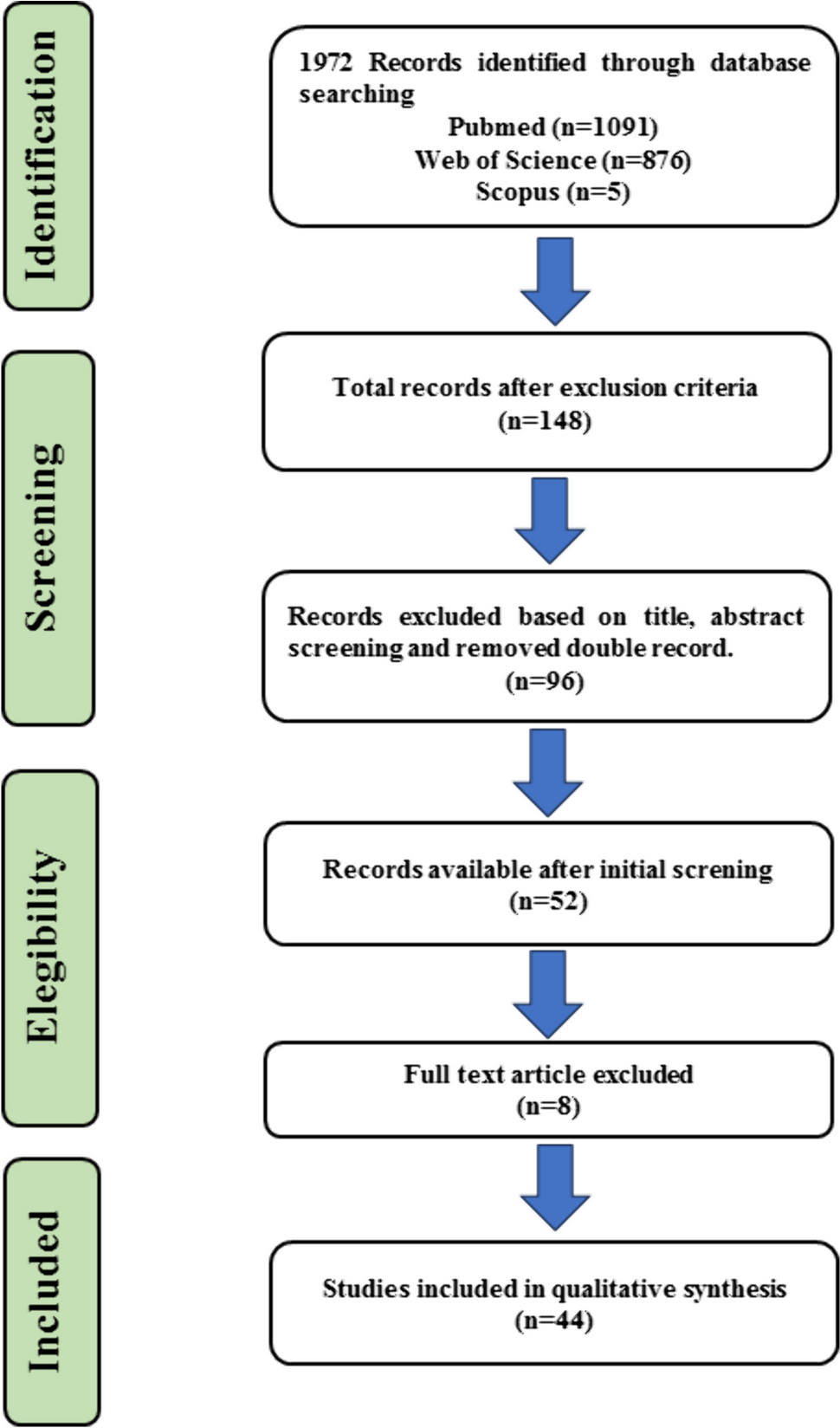
Page MJ, McKenzie J, Bossuyt P, Boutron I, Hoffmann T, mulrow cindy d, et al. Updating guidance for reporting systematic reviews: development of the PRISMA 2020 statement. Center for Open Science; 2020 Sep 14; https://doi.org/10.31222/osf.io/jb4dx.
Henry BM, Tomaszewski KA, Ramakrishnan PK, Roy J, Vikse J, Loukas M, et al. Development of the anatomical quality assessment (AQUA) tool for the quality assessment of anatomical studies included in meta-analyses and systematic reviews. Clin Anat. 2016;30(1):6–13. https://doi.org/10.1002/ca.22799.
Article
PubMed
Google Scholar
Zappalá M, Lightbourne S, Heneghan NR. The relationship between thoracic kyphosis and age, and normative values across age groups: a systematic review of healthy adults. J Orthop Surg Res. 2021 Jul 9;16(1). https://doi.org/10.1186/s13018-021-02592-2.
Henry BM, Tomaszewski KA, Walocha JA. Methods of evidence-based anatomy: a guide to conducting systematic reviews and meta-analysis of anatomical studies. Ann Anat. 2016;205:16–21. https://doi.org/10.1016/j.aanat.2015.12.002.
Article
PubMed
Google Scholar
Memarian A, Aghabiklooei A, Asl N dadashzadeh, Zarei E, Kordrostami R, Zavareh FN. Computed tomographic study of frontal sinuses in iraninan population for gender indentification. J Punjab Acad Forensic Med Toxicol. 2020;20(1):109. https://doi.org/10.5958/0974-083x.2020.00065.5.
Čechová M, Dupej J, Brůžek J, Bejdová Š, Horák M, Velemínská J. Sex estimation using external morphology of the frontal bone and frontal sinuses in a contemporary Czech population. Int J Legal Med. 2019;133(4):1285–94. https://doi.org/10.1007/s00414-019-02063-8.
Article
PubMed
Google Scholar
Chowdhuri S, Das S, GhoshZ R, Patra SS, Thassu I. Study of multidetector computed tomography images of the frontal sinuses for human identification: a study in regional Indian population. Int J Forensic Dent. 2019;4(2):73. https://doi.org/10.5958/0973-9130.2019.00180.4.
Article
Google Scholar
Chalkoo AH, Sharma P, Nazir N, Tariq S. Evaluation of the frontal sinuses dimensions in sex estimation among a sample of adult Kashmiri population using multidetector computed tomography. IP Int J Maxillofac Imaging. 2020;4(4):122–5. https://doi.org/10.18231/2581-3838.2018.0031.
Denny C, Jacob AS, Ahmed J, Natarajan S, Binnal A. Frontal sinus as an aid in gender identification in forensic dentistry: a retrospective study using cone beam computed tomography. World J Dent. 2018;9(1):34–7. https://doi.org/10.5005/jp-journals-10015-1503.
Article
Google Scholar
Tatlisumak E, Asirdizer M, Bora A, Hekimoglu Y, Etli Y, Gumus O, Keskin S. The effects of gender and age on forensic personal identification from frontal sinus in a Turkish population. Saudi Med J. 2017;38(1):41. https://doi.org/10.15537/smj.2017.1.16218.
Motawei SM, Wahba BA, Aboelmaaty WM, Tolba EM. Assessment of frontal sinus dimensions using CBCT to determine sexual dimorphism amongst Egyptian population. J Forensic Radiol Imaging. 2016;1(6):8–13. https://doi.org/10.1016/j.jofri.2016.07.001.
Article
Google Scholar
Benghiac AG, Thiel BA, Haba D. Reliability of the frontal sinus index for sex determination using CBCT. Rom J Leg Med. 2015;23(4):275–8. https://doi.org/10.4323/rjlm.2015.275.
Article
Google Scholar
Kim DI, Lee UY, Park SO, Kwak DS, Han SH. Identification using frontal sinus by three‐dimensional reconstruction from computed tomography. J Forensic Dent Sci. 2013;58(1):5–12. https://doi.org/10.1111/j.1556-4029.2012.02185.x.
Tatlisumak E, Ovali GY, Aslan A, Asirdizer M, Zeyfeoglu Y, Tarhan S. Identification of unknown bodies by using CT images of frontal sinus. Forensic Sci Int. 2007;166(1):42–8. https://doi.org/10.1016/j.forsciint.2006.03.023.
Article
PubMed
Google Scholar
ELbaz D, El-Shall O, El Kolaly H. Sexual dimorphism by analysis of maxillary sinus dimensions in a sample of Egyptian population using cone beam computed tomography. Al-Azhar Dent J Girls. 2019;6(4):385–90. https://doi.org/10.21608/adjg.2019.7177.1070.
Teixeira LC, Walewski LÂ, de Souza TE, Iwaki LC, Silva MC. Three-dimensional analysis of the maxillary sinus for determining sex and age in human identification. Forensic Imaging. 2020;1(22): 200395. https://doi.org/10.1016/j.fri.2020.200395.
Article
Google Scholar
Soares CB, Miranda-Viana M, Pontual AA, Ramos-Perez FM, Perez DE, Figueiroa JN, Pontual ML. Morphological and dimensional assessment of the maxillary sinus for human identification and sexual dimorphism: a study using CBCT. Forensic Imaging. 2020;1(23): 200409. https://doi.org/10.1016/j.fri.2020.200409.
Article
Google Scholar
Sathawane SR, Sukhadeve AV, Chandak MR, Lanjekar AB, Moon GV. Sex determination by maxillary sinus dimensions using cone-beam computed tomography and discriminant function: an analytical study. Int J Forensic Dent. 2020;5(1):19.
Google Scholar
Waluyo RF, Priaminiarti M, Yuniastuti M, Soedarsono N, Susilo BT. Measurements of sex-related differences in maxillary sinus and mandibular canal characteristic using cone beam computed tomography. Forensic Imaging. 2020;1(21): 200371. https://doi.org/10.1016/j.fri.2020.200371.
Article
Google Scholar
Najem SS, Safwat WM, ELAziz RA, Gaweesh YS. Maxillary sinus assessment for gender and age determination using cone beam computed tomography in an Egyptian sample. Alex Dent J. 2020 May 11. https://doi.org/10.21608/adjalexu.2020.88457.
Shah D, Patil PS, Jadhav PL, Agnihotri R, Patil N. Assessment of gender determination: by morphometric dimensions of the maxillary sinuses and computed tomography. Asian J Med Radiol Res. 2019;7(2):56. https://doi.org/10.21276/ajmrr.2019.7.2.13.
Khanal K, Menezes RG, Paudel RC, Singh PK. Maxillary sinus-a tool for sex determination in the Nepalese population. J Kathmandu Med Coll. 2019;8(2):87–91. https://doi.org/10.3126/jkmc.v8i2.28170.
Article
Google Scholar
Gomes AF, de Oliveira GT, Yamasaki MC, Groppo FC, Neto FH, de Fátima PR. Development and validation of a formula based on maxillary sinus measurements as a tool for sex estimation: a cone beam computed tomography study. Int J Legal Med. 2019;133(4):1241–9. https://doi.org/10.1007/s00414-018-1869-6.
Article
Google Scholar
Dhanak K, Ingale S, Kochar SR, Pathak A. CT scan of maxillary sinus: a useful tool for forensic identification. Indian J Forensic Med Toxicol. 2019;13(2). https://doi.org/10.5958/0973-9130.2019.00077.x.
Dangore-Khasbage S, Bhowate R. Utility of the morphometry of the maxillary sinuses for gender determination by using computed tomography. Dent Med Probl. 2018;55(4):411–7. https://doi.org/10.17219/dmp/99622.
Radulesco T, Michel J, Mancini J, Dessi P, Adalian P. Sex estimation from human cranium: forensic and anthropological interest of maxillary sinus volumes. J Forensic Dent Sci. 2018;63(3):805–8. https://doi.org/10.1111/1556-4029.13629.
Article
Google Scholar
Bangi BB, Ginjupally U, Nadendla LK, Vadla B. 3D evaluation of maxillary sinus using computed tomography: a sexual dimorphic study. Int J Dent. 2017;4:2017. https://doi.org/10.1155/2017/9017078.
Article
Google Scholar
Paknahad M, Shahidi S, Zarei Z. Sexual dimorphism of maxillary sinus dimensions using cone-beam computed tomography. J Forensic Dent Sci. 2017;62(2):395–8. https://doi.org/10.1111/1556-4029.13272.
Article
Google Scholar
de Oliveira GT, Yamasaki MC, Groppo FC, da Silveira HL, de Almeida Boscolo SM, Sanderink GC, Berkhout WE. Validation study of a new method for sexual prediction based on CBCT analysis of maxillary sinus and mandibular canal. Arch Oral Biol. 2017;1(83):118–23. https://doi.org/10.1016/j.archoralbio.2017.07.010.
Article
Google Scholar
Urooge A, Patil BA. Sexual dimorphism of maxillary sinus: a morphometric analysis using cone beam computed tomography. J Clin Diagn Res. 2017;11(3):ZC67. https://doi.org/10.7860/jcdr/2017/25159.9584.
Akhlaghi M, Bakhtavar K, Kamali A, Maarefdoost J, Sheikhazadi A, Mousavi F, Anary SH, Sheikhazadi E. The diagnostic value of anthropometric indices of maxillary sinuses for sex determination using CT-scan images in Iranian adults: a cross-sectional study. J Forensic Leg Med. 2017;1(49):94–100. https://doi.org/10.1016/j.jflm.2017.05.017.
Article
Google Scholar
Chaurasia A, Katheriya G. Morphometric evaluation of Bizygomatic distance and maxillary sinus width as dimorphic tool-a CBCT study. Int J Maxillofac Imaging. 2016;2(4):123–8. https://doi.org/10.18231/2455-6750.2016.0004.
Ahmed AG, Gataa IS, Fateh SM, Mohammed GN. CT scan images analysis of maxillary sinus dimensions as a forensic tool for sexual and racial detection in a sample of Kurdish population. Eur Sci J. 2015;1;11(18). https://doi.org/10.17656/sdj.10010.
Ekizoglu O, Inci E, Hocaoglu E, Sayin I, Kayhan FT, Can IO. The use of maxillary sinus dimensions in gender determination: a thin-slice multidetector computed tomography assisted morphometric study. J Craniofac Surg. 2014;25(3):957–60. https://doi.org/10.1097/scs.0000000000000734.
Article
PubMed


留言 (0)