
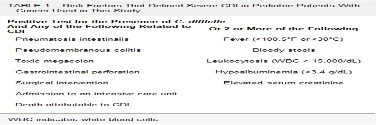
記住我

A 9-year-old male patient from Belo Horizonte, Brazil, presented to the Emergency Department with diarrhea, abdominal pain, coughing and vomiting for 7 days. The patient reported having swum in an artesian well 30 days before and developed a maculopapular rash on the limbs 15 days later. On physical examination, he was tachypneic and hypoxemic (peripheral O2 saturation: 92%) with expiratory wheezing. He received salbutamol and ipratropium, prednisone and O2 and was admitted to a referral hospital (Hospital Infantil João Paulo II, FHEMIG, Belo Horizonte, Brazil). Upon admission, a complete blood count showed a white blood cell count of 38.1 × 10⁹/L with eosinophilia of 23.6 × 10⁹/L eosinophils, 8.4 × 10⁹/L neutrophils, 3.5 × 10⁹/L lymphocytes, 1.9 × 10⁹/L monocytes, and 0.7 × 10⁹/L basophils, hemoglobin of 12.2 g/dL, and platelet count of 143 × 109/L. On the second day of hospitalization, he developed progressive irritability, enuresis, mental confusion and lower limb paresis and was transferred to the intensive care unit.
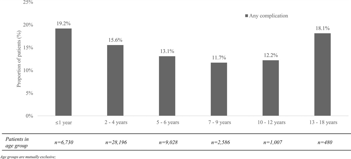
The cerebrospinal fluid (CSF) showed 1 white blood cell per mm3, glucose of 63 mg/100 mL and protein <10 mg/100 mL. Magnetic resonance imaging of the brain showed multiple areas of signal alteration involving white and gray matter, located supra and infratentorially, in the semioval centers, in both periventricular and subcortical areas, as well as in the thalamus, basal ganglia, and cerebellum. Such areas were characterized by hyperintensity on T2/FLAIR, discrete hyperintense signal on T1 before contrast, and diffusion restriction and contrast enhancement, without mass effect (Fig. 1). Although diffuse, the distribution of the lesions correlated with the areas of vascular borders, suggesting the possibility of small areas of infarction (“watershed infarction”).
 FIGURE 1.:
 Brain MRI—In the first line (A1–4), axial diffusion weighted sequence showing multiple areas of restriction to diffusion in the semioval centers, in both periventricular and subcortical areas, as well as in the right cerebellar hemisphere. In the second line (B1–4), axial T2-weighted sequence in the same levels of images A1–4, the areas disclose hyperintense signal. In the third line (C1–4), axial gadolinium-enhanced T1-weighted sequence in the same levels of the images above, some areas demonstrate contrast enhancement. MRI indicates magnetic resonance imaging.
Due to the epidemiologic history, clinical presentation and laboratory results, an additional study was performed which revealed the diagnosis.
For Denouement see P. 863.
DENOUEMENT(Pediatr Infect Dis J 2022;41:863)Continued from P. 862.
Given the epidemiologic history of contact with natural waters and the finding of extreme peripheral eosinophilia, it was important to consider the possibility of parasitic diseases, especially schistosomiasis. Indirect immunofluorescence for schistosomiasis was positive (1:64). Stool parasitologic examination did not reveal any parasites, and testing for circulating cathodic antigen of schistosome (CCA) in urine was negative. A presumptive diagnosis of Schistosoma mansoni infestation was made based on the clinical picture, epidemiologic history, peripheral eosinophilia, positive serology, absence of any other diagnosis, and good response to treatment with praziquantel (60 mg/kg in a single dose), pulse therapy with methylprednisolone (20 mg/kg/day) for 5 days, followed by oral prednisone (1 mg/kg/day) for 6 weeks, with resolution of symptoms.
Schistosomiasis is a parasitosis caused by trematode worms of the genus Schistosoma. It is one of the most prevalent parasitic infections in the world, being an important public health problem in tropical countries.1 Six species are capable of infecting humans: S. mansoni, S. haematobium and S. japonicum are the most widely distributed species and S. mekongi, S. guineensis and S. intercalatum are described in limited areas.2,3 In Brazil, the disease is exclusively caused by S. mansoni, and it is estimated that 6 million Brazilians are infected with this parasite.3 A high index of suspicion of the disease in schistosome endemic regions is necessary, even if the clinical picture is not congruent with the “classic” presentation of schistosomiasis.1
The initial infection of a child with schistosomiasis can occur as soon as the first contact with freshwater, infested with cercariae, and ectopic presentations are commonly found in travelers or individuals with single exposure to freshwater.4,5 Classically, the highest prevalence and symptoms of active infection occur in adolescents and young people, decreasing in adulthood.1,6 Early schistosomiasis manifests within the first 2–3 months after exposure. This is the time required for the adult worm to mature and start laying eggs, which occurs approximately 4–6 weeks after infection with S. mansoni, subsequently triggering an initial hyperimmune response by the host.3,4 Two nonspecific clinical presentations of early S. mansoni are cercarial dermatitis and the acute toxemic form.3 In travelers, a history of a self-limited urticarial eruption, minutes to hours after exposure to freshwater, is highly suggestive of cercarial dermatitis.4 The acute toxemic form is a serum sickness-like syndrome with an acute onset characterized by fever, abdominal pain, rash and eosinophilia 4–6 weeks after exposure to freshwater. Cough, diarrhea, visceromegaly and other less common manifestations may also be observed.4,6 A history of cercarial dermatitis preceding the clinical picture is a strong indication of schistosomiasis; however, its absence does not rule out the diagnosis.5 Likewise, the absence of eggs in the feces or nonreactive serologic tests are not enough to rule out the infection in patients with a history of contact with contaminated water in endemic regions.7 Serologic testing is more helpful in the evaluation of nonimmune individuals; however, in an endemic area, it may be difficult to interpret the results since antibody-based tests can remain positive for many years even after successful treatment. This makes it difficult to differentiate ongoing infection from past infections.4,6
Neuroschistosomiasis (NS) is an ectopic manifestation that results from the involvement of the central nervous system (CNS) and constitutes the most frequent and disabling ectopic form of the parasitosis.2 Lesions can occur in the meninges, spinal cord or brain, clinically manifesting as meningomyeloradiculitis or as cerebral or spinal cord pseudotumor. These ectopic forms are severe conditions, and their prognosis depends especially on early treatment. Most cases are caused by S. mansoni, S. haematobium or S. japonicum. The first 2 species have a predilection for the spinal cord and the last 1 for the brain.2 The disease can result from the direct deposition of eggs into the CNS or indirectly from the deposition of circulating immune complexes in small vessels in the CNS. Diagnosis is based on clinical, epidemiologic and laboratory data.7
Although not completely understood, there is evidence that parasite eggs in the CNS and the host immune response are key to the pathogenesis of NS. In most patients, CNS involvement occurs in the acute phase of the infection, as observed in the present case.8 The main mechanism by which eggs reach the CNS is via migration of the adult worm to the CNS by retrograde blood flow from the portal system to the Batson’s vertebral venous plexus and then to CNS veins, where oviposition takes place. The numerous eggs induce a granulomatous response in circumscribed areas, producing a mass effect. In addition, neurologic injury due to immune-mediated vasculitis has also been described.3,7,9
NS may rarely present as acute encephalopathy in tropical regions.7 Neurologic symptoms such as headache, altered mental status, seizures, paresis and ataxia occur about 3 weeks after the acute infection. Although it is most often caused by S. japonicum, cases of encephalopathy attributed to S. mansoni and S. haematobium have been described.3,7 This unusual form of NS develops especially at the time of first exposure to schistosomes. Neurologic disease usually presents simultaneously or immediately after the systemic manifestations of acute disease, which include fever, headache, malaise, anorexia, cough, rash, diarrhea and abdominal pain.4 These clinical manifestations last a few days or weeks without treatment, and treatment is usually followed by complete recovery. Clinical manifestations are thought to be due to the host immune response to the adult worm and egg antigens. Elevated serum immunoglobulins, formation and deposition of immune complexes and eosinophilic vasculitis with small vessels thrombosis are found.2,7 Edema and small multifocal lesions in the frontal, parietal and occipital regions of the brain along with infarcts in the “watershed areas” may also be identified. The CSF examination may be normal or may show nonspecific alterations, and the peripheral blood shows a significant increase in the number of eosinophils, as observed in the case presented. Eggs in stool or urine may not be found in early infection.3 In some cases, circulating somatic antigens of the parasite may be identified in the blood, urine or feces of infected individuals. The diagnosis of acute encephalopathy due to schistosome is based on the history of exposure, clinical and radiologic manifestations and presence of circulating antibodies.7
Significant peripheral blood eosinophilia typically results from a parasitic, tumoral, allergic, vasculitic disease or idiopathic hypereosinophilic syndrome (HIS).10 In HIS, cerebral infarctions can be due to damage to the vascular endothelium caused by the eosinophils, with formation of local microthrombi, which, like our case, can lead to “watershed” or multiple infarcts in the vascular border areas.10,11
Ischemia in the vascular boundary areas was first described in an adult patient with neurologic involvement due to S. mansoni in 2004.10 The patient, after a week of fever and diarrhea, presented with neurologic symptoms such as headache, ataxia, behavior and memory changes, and the magnetic resonance imaging showed multiple infarcts in the vascular border areas. Our case is the first report of ischemic lesions in the vascular boundary areas in a child with NS due to S. mansoni. Other parasitosis with eosinophilia have also been associated with these manifestations, such as trichinosis and filariasis.10
Although there is no consensus on the optimal treatment, NS is often treated with antiparasitic drugs, corticosteroids and occasionally with surgery for pseudotumoral forms. Praziquantel is the antiparasitic treatment of choice, 60 mg/kg given in a single dose, for all schistosome species to eliminate the mature worm, achieving cure rates of 96% in Brazilian studies.7 Acute encephalopathy can be treated with prednisone at a dose of 1 mg/kg/day to suppress hypersensitivity reactions. The best time to use praziquantel is not clear, but some studies have shown the importance of using the drug early, right after the stabilization of the neurologic condition, while still using corticosteroid therapy.7 The rapid progression of the neurologic symptoms in our patient led us to start pulse therapy with methylprednisolone in addition to antiparasitic treatment with praziquantel. Following improvement in muscle strength, sphincter control and level of consciousness, he was transferred from the intensive care unit to the ward after 2 days of treatment and was discharged from the hospital 18 days after admission. At his 6-week follow-up evaluation in the outpatient clinic, he had complete resolution of the symptoms.
In conclusion, when a patient from an endemic area presents with neurologic symptoms, it is important to maintain a high index of suspicion for schistosomiasis since early effective treatment can result in a favorable outcome.
REFERENCES 1. Colley DG, Bustinduy AL, Secor WE, et al. Human schistosomiasis. Lancet. 2014;383:2253–2264. 2. Ferrari TC. Involvement of central nervous system in the schistosomiasis. Mem Inst Oswaldo Cruz. 2004;99(5 Suppl 1):59–62. 3. Ferrari TC, Moreira PR, Cunha AS. Clinical characterization of neuroschistosomiasis due to Schistosoma mansoni and its treatment. Acta Trop. 2008;108:89–97. 4. Bustinduy AL, Edielu A, Sturt AS. Could this child have schistosomiasis?: when to suspect it and what to do about it. Pediatr Infect Dis J. 2020;39:e125–e129. 5. Bustinduy AL, Wright S, Joekes EC, et al. One hundred years of neglect in paediatric schistosomiasis. Parasitology. 2017;144:1613–1623. 6. Brasileiro G. Bogliolo. Patologia Geral. 9 a edição. Editora Guanabara. Rio de Janeiro, RJ: Koogan S/A; 2016. 7. Ferrari TC, Moreira PR. Neuroschistosomiasis: clinical symptoms and pathogenesis. Lancet Neurol. 2011;10:853–864. 8. Jauréguiberry S, Ansart S, Perez L, et al. Acute neuroschistosomiasis: two cases associated with cerebral vasculitis. Am J Trop Med Hyg. 2007;76:964–966. 9. Camuset G, Wolff V, Marescaux C, et al. Cerebral vasculitis associated with Schistosoma mansoni infection. BMC Infect Dis. 2012;12:220. 10. Sarazin M, Caumes E, Cohen A, et al. Multiple microembolic borderzone brain infarctions and endomyocardial fibrosis in idiopathic hypereosinophilic syndrome and in Schistosoma mansoni infestation. J Neurol Neurosurg Psychiatry. 2004;75:305–307. 11. McMillan HJ, Johnston DL, Doja A. Watershed infarction due to acute hypereosinophilia. Neurology. 2008;70:80–82.
留言 (0)