
記住我

Candida is a group of opportunistic pathogenic fungi, which are often found in the host’s skin, mouth, and gastrointestinal tract. The decrease in the host immunity enhances the risk of Candida infection. Among them, Candida albicans is the most common pathogenic fungus (Mba and Nweze, 2020). In recent years, more and more epidemiological and pathological studies have suggested the significant impact of pathogenic microorganisms on the incidence rates of cancers worldwide (Schottenfeld and Beebe-Dimmer, 2015). However, only some studies have linked fungal infection with cancers. The association between fungal microbiota imbalance and carcinogenesis remains largely unknown. This is due to the relatively low abundance of fungi and the lack of well-defined reference genome (Klimesova et al., 2018; Coker et al., 2019). In addition, the research methods are challenging, so the fungal microbiota is usually explored relatively less than other microorganisms. So far, studies have shown that C. albicans in fungal microorganisms is very closely related to cancer development.
In this article, we aim to review the potential molecular mechanism by which C. albicans promotes cancers progression. This may help clinicians diagnose the early stage of tumor in the future and prescribe a treatment method considering the possible microbial properties in the process of carcinogenesis.
Candida albicans infection may promote the development of cancersIn recent years, studies have found that C. albicans infection is closely related to cancers. On the one hand, cancer patients with defective immune function have increased risk of fungal infection; on the other hand, fungus infection may affect the occurrence and development of cancer in different ways.
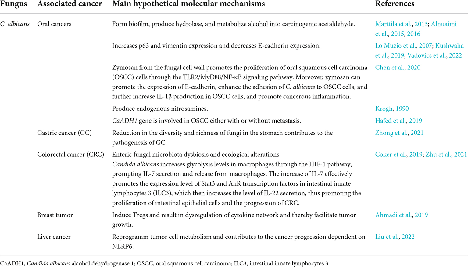
Candida albicans and oral cancerIn the observational study, a link between Candida infection induced oral leukoplakia and malignant tumors was found. Candida infection occurred in 13.5% of oral leukoplakia cases, and in 28.7% of the malignant tumor cases (Banoczy and Sugar, 1975; Warnakulasuriya and Ariyawardana, 2016; Di Cosola et al., 2021). Another study showed that 31% of 257 patients with oral leukoplakia were infected with Candida (Silverman et al., 1984). In the leukoplakia cancer group, 53% of patients were Candida positive before tumor formation (Perera et al., 2017). One study identified that C. albicans was the most frequently detected and abundant fungus in oral squamous cell carcinoma (OSCC) (Mäkinen et al., 2018). In addition, C. albicans was also the most commonly isolated Candida species from saliva samples of patients with oral cancer (Jain et al., 2016). From the current epidemiological studies, the detection rates of Candida in patients with oral cancer are increased, and C. albicans is the main one. Unfortunately, these are only epidemiological and descriptive studies, lacking effective experimental evidence. Therefore, the above studies can only show that C. albicans is associated with cancer development, but there is no definite causal relationship.
In order to prove the role of C. albicans infection in oral cancer, some researchers have carried out relevant experimental studies. It was found that the virulence attributes of Candida and the production capacity of ethanol derived acetaldehyde were related to the development of oral cancer (Alnuaimi et al., 2016). Furthermore, the high biofilm forming ability of Candida may ensure the long-term exposure of host tissue to fungal carcinogens such as acetaldehyde, and increased production of hydrolases may trigger chronic inflammatory responses in the host tissues (Alnuaimi et al., 2016). Chronic inflammation caused by microbial infection is one of the risk factors of tumor development (Karin and Greten, 2005). However, above study did not identified the correlation between the severity of inflammatory changes at cancer sites and the virulence of Candida isolated from these sites. Another study showed that zymosan from the cell wall of C. albicans promoted the proliferation of OSCC cells through TLR2/MyD88/NF-κB signaling pathway. In addition, zymosan could promote the expression of E-cadherin, thus enhancing the adhesion of C. albicans onto OSCC cells and further increasing IL-1β production (Chen et al., 2020). Meanwhile, zymosan was shown to participate in the IL-1β secretion by OSCC cells by regulating the NLRP3/IL-1β pathway. However, the secretion of proinflammatory cytokines (such as IL-1β) is also influenced by the microbiota or its cellular components, indicating a more complex interaction between cancer cells, immune cells, and the microbiota in the tumor microenvironment (TME) (Chen et al., 2020). NLRP3 inflammasome-induced IL-1β production promoted 5-FU resistance in OSCC both in vitro and in vivo (Feng et al., 2017). It can be inferred that the presence of C. albicans in oral cancer may influence the effect of chemotherapy by inducing IL-1β production, which was also a potential target for treating oral cancer. Some studies also found that C. albicans strains from oral precancerous lesions showed the highest nitrosation potential, while Candida tropicalis, Candida parapsilosis, and Torulopsis glabrata have lower nitrosation potential, which can induce epithelial carcinogenesis and promote tumor development (Krogh, 1990). Another study detected the prevalence of C. albicans alcohol dehydrogenase 1 (CaADH1) gene in oral dysplasia and OSCC and found that CaADH1 gene is associated with OSCC either with or without metastasis, indicating that it may be related to the progression and metastasis of OSCC (Hafed et al., 2019). However, they were unable to confirm whether the observed Candida infection was related to therapeutic interventions such as surgery or chemotherapy. Therefore, it is uncertain whether the development of OSCC is the primary or secondary effect of C. albicans infection. A further in vitro study found that the presence of living C. albicans promoted the progress of OSCC by stimulating the production of matrix metalloproteinases (MMPs), tumor metabolites, tumor promoting signal pathways. These results suggest that C. albicans actively participates in the complicated process of OSCC progression (Vadovics et al., 2022; Table 1). However, the specific roles of C. albicans in the development of oral cancer remains unclear.
TABLE 1
Table 1. The association of C. albicans and cancers.
Candida albicans and gastric cancerGastric cancer (GC) is the fifth most common cancer in the world and the third most common cause of cancer-related death (Smyth et al., 2020). After the continuous development of high-throughput sequencing technology, research on the correlation between gastric microbiome (other than Helicobacter pylori) and GC has gradually emerged. A study described the composition and ecological changes of fungi by analyzing the metagenomic sequences in cancer foci and adjacent non-cancer tissues of 45 GC patients. The results showed that GC related fungal biological community was unbalanced, which was characterized by changes in fungal composition and ecology, and suggested that C. albicans might be used as a fungal biomarker of GC. With the significant increase of C. albicans in GC, the abundance of Fusicolla acetilerea, Arcopilus aureus, and Fusicolla aquaeductuum were increased, while Candida glabrata, Aspergillus montevidensis, Saitozyma podzolica, and Penicillium arenicola were obviously decreased (Zhong et al., 2021). In addition, C. albicans reduced the diversity and abundance of fungi in the stomach, thus enhancing the development of GC (Zhong et al., 2021; Table 1). However, these studies did not further clarify the specific molecular mechanism by which C. albicans is involved in the progression of GC.
Candida albicans and colorectal cancerColorectal cancer (CRC) is the third most common cancer in the world and the second most common cause of cancer-related death (Siegel et al., 2021, 2022). It is well known that gut microbiota play critical roles in CRC development (Qing et al., 2022). Through intestinal microbiota biodiversity analysis, a study showed that fungal dysbiosis and altered fungal network might play an important role in the pathogenesis of CRC (Gao et al., 2017). Subsequently, another study reached the similar conclusion through the ecological analysis of gut microbiota (Coker et al., 2019). However, these studies did not clarify the specific role of symbiotic fungi in CRC. Recently, in fungal specific pattern recognition receptor Dectin-3 knockout mice (Dectin-3–/– mice), the deletion of Dectin-3 gene could lead to a significant increase in CRC development, and the fungal load in Dectin-3–/– mouse feces is significantly higher than that in wild-type (WT) mice. Interestingly, the proportion and abundance of C. albicans were significantly increased. The fecal fungus transplantation experiment further confirmed that the feces of Dectin-3–/– tumor bearing mice and C. albicans can promote the malignant process of CRC, and antifungal treatment can effectively alleviate the tumor load of Dectin-3–/– mice. In vivo and in vitro experiments also confirmed that Dectin-3 gene deletion can damage the ability of macrophages to scavenge C. albicans and increase the load of fungi. These studies reveal the molecular mechanism of C. albicans in regulating intestinal immunity and promoting the development of CRC (Zhu et al., 2021; Table 1).
Candida albicans and other cancersIt is well known that Aspergillus flavus, which produces aflatoxin, is closely related to liver cancer (Cai et al., 2020; Sun et al., 2021). C. albicans was also found to be closely related to liver cancer. A study found that the diversity of intestinal fungal community in patients with liver cancer decreased significantly, and the abundance of C. albicans increased. Abnormal colonization of C. albicans increased the size and weight of liver cancer, affected the cancer cell metabolism, thus promoting the progression of liver cancer dependent on nucleotide oligomerization domain-like receptor family pyrin domain containing 6 (NLRP6) (Liu et al., 2022). It indicates the harmful effect of C. albicans on liver cancer may be mediated by NLRP6, which provides a new target for the cancer treatment. Nevertheless, they have not identified the cell surface receptors that recognize C. albicans that can further activate NLRP6. Recently, another study showed that compared to the uninfected control group, C. albicans infection increased the number of Tregs in TME. In addition, compared to the tumor group, C. albicans infection increased tumor growth in the tumor/Candida infection group. It further shows that systemic infection of C. albicans could not only induce Tregs, but also lead to the imbalance of cytokine network, so as to promote the growth of tumor (Ahmadi et al., 2019; Table 1). Tregs have been shown to enhance tumor progression by inhibiting antitumor immune response (Deng et al., 2013; Lainé et al., 2021). However, the specific mechanism by which C. albicans promotes occurrence and development of breast cancer remains unknown. The fungal community of pancreatic ductal adenocarcinoma (PDA) was shown to be different from that of healthy controls. Furthermore, the fungal community of PDA was significantly enriched in Malassezia, which promoted tumor growth, while Candida could not accelerate tumor growth (Aykut et al., 2019). It indicates that C. albicans may not be involved in the progression of PDA. The role of fungal microorganisms in cancer development are gradually appreciated.
Candida albicans and other microorganisms during cancer developmentCandida albicans has synergy, symbiosis and antagonism with other microorganisms, which determines the role of the microbiota in which C. albicans is located. For example, the interaction of multiple microorganisms can improve the ability of C. albicans biofilm formation (Lohse et al., 2018), and then enhance the ability of C. albicans to invade the host. A study showed that the interaction between C. albicans and oral microorganisms might promote oral carcinogenesis (Arzmi et al., 2019). The metabolites of the polymicrobial membrane formed by C. albicans, Actinomyces naeslundii and Streptococcus mutans regulate the phenotype of cancer cells by increasing the adhesion of OSCC to extracellular matrix (ECM) and enhancing the expression of proinflammatory cytokines (Arzmi et al., 2019). However, C. albicans and S. mutans have antagonistic effects. S. mutans can inhibit the mycelial growth and biofilm formation of C. albicans and reduce its pathogenicity (Barbosa et al., 2016). Previous studies showed that C. albicans could inhibit S. mutans from producing extracellular polymeric substances (EPSs) and reduce its biofilm toxicity (Sztajer et al., 2014). It is also reported that oral actinomycetes (including A. naeslundii) also inhibit the proliferation, adhesion, metabolic enzyme activity, mycelial growth and biofilm formation of C. albicans (Guo et al., 2015). All these complicated interactions between multiple microorganisms may also explain the reduction of colonization on the surface of multiple microbial membranes and the differential regulation of oral cancer cell phenotypes, but not in single C. albicans and S. mutans.
Effect of cancer and anticancer therapy on Candida albicansIn cancer, microbial communities in cancer-related areas usually change, including fungal communities. A study investigated the steady-state changes of microbial community during the occurrence of GC, and found that GC was significantly related to the changes of fungal community in the stomach, including decreased biodiversity and richness the increased proportion of opportunistic fungi (Yang et al., 2022). Another study also showed that the abundance of C. albicans increased in GC and C. albicans promoted cancer progression by reducing the diversity of fungi in the stomach (Zhong et al., 2021). In CRC, there was also an imbalance of fungal community, with increased abundance of C. albicans (Wang et al., 2021a). C. albicans may promote the development of CRC through Dectin-1/Wnt pathway (Wang et al., 2021a). These studies suggest that cancer may promote the proliferation of C. albicans in fungal communities, and C. albicans may also promote the development of cancer. Unfortunately, the mechanism of the interaction between cancer and fungi are still largely unclear.
On the one hand, when cancer occurs, the mucosal barrier function of the host is often damaged, which may cause the invasion of the conditional pathogen C. albicans. On the other hand, cancer may suppress host immune function, which promotes invasion of C. albicans. Finally, anticancer treatment may damage the host immunity and further increase the infection of C. albicans. However, some studies also show that anticancer drugs can inhibit the formation of C. albicans biofilm and reduce its invasiveness (Wakharde et al., 2018). Another study showed that anticancer drugs and radiation could enhance the virulence of C. albicans and increase the risk of systemic candidiasis (Ueta et al., 2001). In addition, with the extension of anticancer treatment duration, C. albicans may produce a large number of phospholipases to enhance its invasion (Ramla et al., 2016). So far, these studies have not clearly clarified the specific mechanism of anticancer treatment on the invasiveness of C. albicans.
Possible carcinogenic mechanism of Candida albicansCurrent studies suggest that C. albicans may promote the development of cancer through various mechanisms such as damaging the mucosal epithelium, producing carcinogens, triggering chronic inflammation, and inducing Th17 immune responses. However, some of these mechanisms still lack strong and direct evidence and need to be further verified.
Candida albicans damages epithelial mucosal cells and invades the hostEpithelial mucosal cells are the first line of defense for the host protection against the invasion of pathogenic microorganisms. Adhesion of C. albicans to epithelial cells is the first step of fungal colonization and invasion. Subsequently, it invades epithelial cells by inducing endocytosis and active infiltration, which is a key step in the pathogenesis of Candida (Maza et al., 2017; Allert et al., 2018; Lipke, 2018). Among them, C. albicans invasive enzymes destroy the integrity of mucosal tissue structure and enhance its virulence; hemolytic factors help them obtain nutrients for its survival and reproduction; phenotypic transformation can help C. albicans adapt to the host tissue environment; adhesin can assist in its colonization and invasion of host cells (Zhu and Filler, 2010; Tao et al., 2014; Furlaneto et al., 2015; Noble et al., 2017). Once C. albicans invades epithelial mucosal cells, it may induce apoptosis and necrosis, destroy the host epithelial defense barrier, and finally leading to the structural changes of epithelial cells (Richardson et al., 2018; Mogavero and Hube, 2021). These are the preconditions for the cancer promoting C. albicans infection. Epithelial cells are damaged and the normal structure is changed, which results in abnormal proliferation and the formation of oral cancer (Engku Nasrullah Satiman et al., 2020). There is a significant positive correlation between C. albicans infection and oral mucosal epithelial dysplasia, and the deterioration of epithelial dysplasia induced by C. albicans infection occurs (Barrett et al., 1998). Multiple studies showed that C. albicans could induce epithelial dysplasia and further malignant tumor (McCullough et al., 2002; Dwivedi et al., 2009).
Candida albicans produces carcinogensThe substances such as nitrosamine (Mohd Bakri et al., 2010) and acetaldehyde (Stornetta et al., 2018) produced by C. albicans play a certain role in promoting cancer development. A study showed that C. albicans had higher nitrosation production potential than other fungi, and could convert N-benzylmethylamine (BMA) in vegetables, herring oil and freeze-dried coffee and nitrite produced by other microorganisms in the host’s mouth into N-nitroso-benzylmethylamine (NBMA), thus inducing the occurrence and development of OSCC (Krogh, 1990). However, the mechanism of direct carcinogenesis is still controversial. It may also due to the fact that the integrity of oral mucosal cell barrier and smoking and other risk factors enhance the virulence of C. albicans and jointly promote oral carcinogenesis (Ye et al., 2021). Recently, a study compared the ability of Candida isolated from oral cancer patients and matched oral healthy subjects to produce acetaldehyde. The results showed that Candida strains producing a large amount of acetaldehyde were more in patients with oral cancer than in healthy volunteers, further indicating the possible role of Candida in enhancing the occurrence of oral cancer (Alnuaimi et al., 2016). C. albicans may secrete alcohol dehydrogenase, which converts ethanol into acetaldehyde and participates in the carcinogenic process (Stornetta et al., 2018). Acetaldehyde can induce DNA adducts to interfere with DNA replication, resulting in point mutation and chromosome aberration. At the same time, it affects the enzymes involved in cytosine methylation and DNA repair, leading to protooncogene activation and cell cycle disorder, which may lead to tumor progression. In addition, acetaldehyde can also cause mitochondrial damage and promote apoptosis, which can be activated by NF-κB to offset. For example, in GC, NF-κB induced IL-6 upregulates the antiapoptotic gene MCL1 to inhibit DNA repair and apoptosis (Lin et al., 2001; Wang et al., 2021b). The combination of acetaldehyde and glutathione indirectly increases the production of reactive oxygen species (ROS), thus inducing DNA damage, which is conducive to the progress of cancer (Ramirez-Garcia et al., 2016; Mizumoto et al., 2017; Johnson et al., 2021). However, the correlation between the acetaldehyde concentration produced by C. albicans and the severity of cancer tissue is unknown, and the mechanism to promote cancer development is still unclear.
Candida albicans induces tumor microenvironmentStromal cells are composed of fibroblasts, vascular cells and inflammatory immune cells, which together constitute the TME. Both chronic disease induced inflammation and tumor induced inflammation have a great impact on the composition of TME, especially on the plasticity of tumor and stromal cells. Inflammatory substances released by immune cells in TME can directly affect precancerous cells and cancer cells by increasing the cell proliferation and their resistance to cell death and stress, so as to directly promote tumor progression (Greten and Grivennikov, 2019). Therefore, chronic inflammatory response plays an important role in the occurrence and development of tumors. Persistent Candida epithelial mucosal colonization and infection can also cause a chronic inflammatory state. C. albicans recognizes Toll like receptors (TLRs) and C-type lectin-like receptors (CLRs), and then activates the corresponding MAPK and NF-κB. Interferon and inflammatory signaling pathway promote the expression of a variety of related inflammatory genes and play a role in the connection between benign and malignant diseases. Studies have found that the expression of inflammatory factors prostaglandin E2 (PGE2) and MMPs (mainly MMP-9) increased after C. albicans infection in oral epithelial cells (Claveau et al., 2004; Nasry et al., 2018). In addition, prostaglandins, cyclooxygenase (COX) enzymes and MMPs can inhibit tumor suppressor genes through DNA methylation and post-translational modification, leading to the occurrence and development of cancer (Munn, 2017).
Studies have shown that PGE2 are overexpressed in a variety of cancer types, including breast cancer (Nandi et al., 2017), oral cancer (Tao et al., 2021), and CRC (Karpisheh et al., 2019). However, C. albicans can induce human peripheral blood mononuclear cells (PBMC) to produce PGE2 (Smeekens et al., 2010). PGE2 promotes tumorigenesis by producing ROS, stimulating carcinogenic transcription factors, inhibiting antitumor immune response (Liu et al., 2015), and enhancing angiogenesis (Mizuno et al., 2019). PGE2 inhibits the cytotoxic function of NK cells and prevents them from producing IFN-γ, and promotes malignant growth by avoiding type I interferon and T cell-mediated cell death. PGE2 promotes the inhibitory activity of Tregs, and contributes to the maturation of Tregs, thereby inhibiting antitumor immunity (Nasry et al., 2018). In addition, PGE2 increases MDSCs, and inhibits innate and adaptive antitumor immunity by downregulating cytokines of macrophages, inhibiting cytotoxicity of NK cells, blocking activation of cytotoxic T cells, and regulating the development of Tregs. PGE2 facilitates bone marrow mesenchymal stem cells migrate into the tumor environment and enables malignant cells to proliferate without interference from the host immune system (Nasry et al., 2018). PGE2 binds to PGE receptor (EP1) and activates protein kinase δ (protein kinase Cδ, PKCδ)/c-Src/AP-1 signaling pathway, upregulates intercellular adhesion molecule 1 (ICAM-1), and promotes the metastasis of cancer cells (Yang et al., 2010). However, the exact mechanism by which PGE2 produced by C. albicans in the process of chronic inflammation promotes cancer development is not clear.
Other studies have shown that MMP-9 is highly expressed in cancer tissues such as oral cancer (Xie et al., 2020), GC (Dong et al., 2020), CRC (Guo et al., 2020), breast cancer (Nazir et al., 2019), and cervical cancer (Azevedo Martins et al., 2020), and has been used as a potential marker of cancers (Huang, 2018). Some studies have shown that the high expression of MMP-9 leads to the enhancement of tumor invasion, which maybe because the effect on the transforming growth factor (TGF-β1) induced epithelial mesenchymal transition (EMT) process, which promotes the invasion and metastasis of cancer (Li et al., 2020a). At the same time, MMP-9 can degrade type IV collagen of basement membrane, destroy the integrity of basement membrane, and also contribute to the invasion and metastasis of tumor cells (Koontongkaew, 2013). In addition, MMP-9 degrades ECM components and activates angiogenic factor VEGF and TGF-β helps cancer angiogenesis, and cleavage of osteopontin (OPN) also contributes to cancer metastasis (Quintero-Fabián et al., 2019). These results suggest that MMP-9 has a role of cancer promoting mechanism. However, a study showed that epithelial origin MMP-9 exerts tumor inhibitory effect by activating MMP9-Notch1-ARF-p53 axis, resulting in increased apoptosis, and starts cell cycle arrest by activating p21WAF1/Cip1, and checks the damaged DNA until DNA repair. In addition, in colitis associated CRC, MMP-9 can prevent γH2AX reduced levels of genotoxicity, also plays a tumor suppressive role (Walter et al., 2017). Subsequently, another study found that the expression of MMP-9 was related to the decrease of ROS level, the decrease of DNA damage and the upregulation of mismatch repair pathway. This suggests that the expression of MMP-9 is a natural biological way to inhibit CRC by limiting ROS accumulation and colonic DNA damage. Therefore, inhibition of MMP-9 may be harmful to patients with CRC (Walter et al., 2020). These results indicate that MMP-9 has a protective host effect in CRC. Therefore, MMP-9 has both cancer promoting and inhibiting cancer effects. However, it is not clear whether MMP-9 produced by host with C. albicans infection plays a role in promoting or inhibiting cancer formation. Whether it is related to the site of C. albicans infection or the type of tumor formation has not been reported. These need to be further studied.
Candida albicans infection induces both host innate and adaptive immune responses, and the core of protection is often from adaptive Th1 and Th17 cell immune response, which is also considered to be the primary factor in the successful immune defense against C. albicans infection (Chen and Kolls, 2017). However, Th17 cells are found in various types of human tumors. Th17 cells and their effector molecules (such as IL-17 and IL-22) can regulate oncogene activated cancer cells themselves and adjacent normal epithelial cells, fibroblasts, endothelial cells, and other stromal cells (Chang, 2019). Current studies have found that IL-17 could promote tumor growth through IL-6-Stat3 signaling pathway (Li et al., 2020b), and also release IFN-γ and other cytokines through stimulating T cells, dendritic cells, NK cells and other immune cells, thus inhibiting tumor growth (Kryczek et al., 2009; Martin-Orozco et al., 2009). In addition to its direct effect on tumor, IL-17 can also reshape the TME by recruiting neutrophils and macrophages and promoting tumor occurrence, development, and metastasis (Rei et al., 2014; Liang and Ferrara, 2016). IL-17 driven antitumor immunity is attributed to its ability to recruit dendritic cells (You et al., 2018). These may be related to various tumor types or TME. Recently, some studies have found that C. albicans can induce the increase of glycolysis of macrophages through HIF-1 pathway and promote the secretion and release of IL-17 by macrophages. Increased IL-17 can effectively promote the expression of STAT3 and AHR transcription factors in intestinal innate lymphocyte 3 (ILC3), which in turn leads to the increase of IL-22 and promotes the proliferation of intestinal epithelial cells and the progress of CRC (Zhu et al., 2021). However, the exact mechanism of the immune response induced by C. albicans infection remains unclear.
ConclusionAt present, most of studies on interaction between C. albicans and cancer are epidemiological survey or descriptive studies. There are few molecular mechanistic studies in this field. Early researchers simply believe that cancer patients are prone to Candida infection. However, tumor is a disease caused by multiple factors. In the mouse model of oral carcinogenesis, a study showed that infection with C. albicans alone could not lead to oral cell dysplasia or OSCC, which requires pretreatment with epithelial carcinogenesis inducer (Dwivedi et al., 2009). C. albicans can promote the occurrence and development of cancer to a certain extent, which may be the result of synergy with the host’s own state and other factors. For example, defective host immunity provides opportunities for C. albicans infection; long-term smoking and drinking provide conditions for C. albicans to produce carcinogenic metabolites; chronic inflammation provides TME and other common factors for C. albicans to promote cancer, and promotes the occurrence and development of cancers.
In conclusion, various adverse factors cause compromised host immunity, leading to C. albicans infection. C. albicans infection increases the risk of cancer development and exacerbates cancer progression. Recent studies have shown that C. albicans infection may participate in the progression of cancer by damaging the epithelial mucosal barrier, producing carcinogenic metabolites, inducing chronic inflammation and Th17 immune response. The progression of cancer further aggravates C. albicans infection. The two promote each other and aggravate the malignant process of cancer development. Therefore, it seems that C. albicans infection may be accompanied by cancer development, and the two promote each other, which in turn aggravates the process of malignancy. It is hoped that these can provide direction for the study of the correlation between C. albicans and cancers, and also provide new ideas for the prevention, diagnosis, and treatment of cancers.
Author contributionsZL: original idea, planning, and editing. DY: writing and editing. Both authors read and agreed to the published version of the manuscript.
FundingThis work was supported by funds from the National Natural Science Foundation of China (31960163), Jianggang Scholar of Jiangxi Province (QD202205), the Project of Ganzhou Science and Technology Bureau (GSKF [2019] No. 60), and Inflammation and Immunology Research Team of Gannan Medical University (TD2021JC01) (to ZL).
Conflict of interestThe authors declare that the research was conducted in the absence of any commercial or financial relationships that could be construed as a potential conflict of interest.
Publisher’s noteAll claims expressed in this article are solely those of the authors and do not necessarily represent those of their affiliated organizations, or those of the publisher, the editors and the reviewers. Any product that may be evaluated in this article, or claim that may be made by its manufacturer, is not guaranteed or endorsed by the publisher.
ReferencesAhmadi, N., Ahmadi, A., Kheirali, E., Hossein Yadegari, M., Bayat, M., Shajiei, A., et al. (2019). Systemic infection with Candida albicans in breast tumor bearing mice: Cytokines dysregulation and induction of regulatory T cells. J. Mycol. Med. 29, 49–55. doi: 10.1016/j.mycmed.2018.10.006
PubMed Abstract | CrossRef Full Text | Google Scholar
Allert, S., Förster, T. M., Svensson, C. M., Richardson, J. P., Pawlik, T., Hebecker, B., et al. (2018). Candida albicans-Induced epithelial damage mediates translocation through intestinal barriers. mBio 9, e915–e918. doi: 10.1128/mBio.00915-18
PubMed Abstract | CrossRef Full Text | Google Scholar
Alnuaimi, A. D., Ramdzan, A. N., Wiesenfeld, D., O’Brien-Simpson, N. M., Kolev, S. D., Reynolds, E. C., et al. (2016). Candida virulence and ethanol-derived acetaldehyde production in oral cancer and non-cancer subjects. Oral Dis. 22, 805–814. doi: 10.1111/odi.12565
PubMed Abstract | CrossRef Full Text | Google Scholar
Alnuaimi, A. D., Wiesenfeld, D., O’Brien-Simpson, N. M., Reynolds, E. C., and McCullough, M. J. (2015). Oral Candida colonization in oral cancer patients and its relationship with traditional risk factors of oral cancer: a matched case-control study. Oral. Oncol. 51, 139–145. doi: 10.1016/j.oraloncology.2014.11.008
PubMed Abstract | CrossRef Full Text | Google Scholar
Arzmi, M. H., Cirillo, N., Lenzo, J. C., Catmull, D. V., O’Brien-Simpson, N., Reynolds, E. C., et al. (2019). Monospecies and polymicrobial biofilms differentially regulate the phenotype of genotype-specific oral cancer cells. Carcinogenesis 40, 184–193. doi: 10.1093/carcin/bgy137
PubMed Abstract | CrossRef Full Text | Google Scholar
Aykut, B., Pushalkar, S., Chen, R., Li, Q., Abengozar, R., Kim, J. I., et al. (2019). The fungal mycobiome promotes pancreatic oncogenesis via activation of MBL. Nature 574, 264–267. doi: 10.1038/s41586-019-1608-2
PubMed Abstract | CrossRef Full Text | Google Scholar
Azevedo Martins, J. M., Rabelo-Santos, S. H., do Amaral Westin, M. C., and Zeferino, L. C. (2020). Tumoral and stromal expression of MMP-2, MMP-9, MMP-14, TIMP-1, TIMP-2, and VEGF-A in cervical cancer patient survival: a competing risk analysis. BMC Cancer 20:660. doi: 10.1186/s12885-020-07150-3
PubMed Abstract | CrossRef Full Text | Google Scholar
Banoczy, J., and Sugar, L. (1975). Progressive and regressive changes in Hungarian oral leukoplakias in the course of longitudinal studies. Commun. Dent Oral Epidemiol. 3, 194–197. doi: 10.1111/j.1600-0528.1975.tb00307.x
PubMed Abstract | CrossRef Full Text | Google Scholar
Barbosa, J. O., Rossoni, R. D., Vilela, S. F., de Alvarenga, J. A., Velloso Mdos, S., Prata, M. C., et al. (2016). Streptococcus mutans Can Modulate Biofilm Formation and Attenuate the Virulence of Candida albicans. PLoS One 11:e0150457. doi: 10.1371/journal.pone.0150457
PubMed Abstract | CrossRef Full Text | Google Scholar
Barrett, A. W., Kingsmill, V. J., and Speight, P. M. (1998). The frequency of fungal infection in biopsies of oral mucosal lesions. Oral Dis. 4, 26–31. doi: 10.1111/j.1601-0825.1998.tb00251.x
PubMed Abstract | CrossRef Full Text | Google Scholar
Cai, P., Zheng, H., She, J., Feng, N., Zou, H., Gu, J., et al. (2020). Molecular Mechanism of Aflatoxin-Induced Hepatocellular Carcinoma Derived from a Bioinformatics Analysis. Toxins 12:203. doi: 10.3390/toxins12030203
PubMed Abstract | CrossRef Full Text | Google Scholar
Chen, X., Luo, Q., Ding, J., Yang, M., Zhang, R., and Chen, F. (2020). Zymosan promotes proliferation, Candida albicans adhesion and IL-1beta production of oral squamous cell carcinoma in vitro. Infect. Agent Cancer 15:51. doi: 10.1186/s13027-020-00315-6
PubMed Abstract | CrossRef Full Text | Google Scholar
Claveau, I., Mostefaoui, Y., and Rouabhia, M. (2004). Basement membrane protein and matrix metalloproteinase deregulation in engineered human oral mucosa following infection with Candida albicans. Matrix Biol. 23, 477–486. doi: 10.1016/j.matbio.2004.08.006
PubMed Abstract | CrossRef Full Text | Google Scholar
Coker, O. O., Nakatsu, G., Dai, R. Z., Wu, W. K. K., Wong, S. H., Ng, S. C., et al. (2019). Enteric fungal microbiota dysbiosis and ecological alterations in colorectal cancer. Gut 68, 654–662. doi: 10.1136/gutjnl-2018-317178
PubMed Abstract | CrossRef Full Text | Google Scholar
Deng, B., Zhu, J. M., Wang, Y., Liu, T. T., Ding, Y. B., Xiao, W. M., et al. (2013). Intratumor hypoxia promotes immune tolerance by inducing regulatory T cells via TGF-β1 in gastric cancer. PLoS One 8:e63777. doi: 10.1371/journal.pone.0063777
PubMed Abstract | CrossRef Full Text | Google Scholar
Di Cosola, M., Cazzolla, A. P., Charitos, I. A., Ballini, A., Inchingolo, F., and Santacroce, L. (2021). Candida albicans and oral carcinogenesis. a brief review. J. Fungi 7:476. doi: 10.3390/jof7060476
PubMed Abstract | CrossRef Full Text | Google Scholar
Dong, Z., Guo, S., Wang, Y., Zhang, J., Luo, H., Zheng, G., et al. (2020). USP19 Enhances MMP2/MMP9-Mediated Tumorigenesis in Gastric Cancer. Onco. Targets Ther. 13, 8495–8510. doi: 10.2147/OTT.S240543
PubMed Abstract | CrossRef Full Text | Google Scholar
Dwivedi, P. P., Mallya, S., and Dongari-Bagtzoglou, A. (2009). A novel immunocompetent murine model for Candida albicans-promoted oral epithelial dysplasia. Med. Mycol. 47, 157–167. doi: 10.1080/13693780802165797
PubMed Abstract | CrossRef Full Text | Google Scholar
Engku Nasrullah Satiman, E. A. F., Ahmad, H., Ramzi, A. B., Abdul Wahab, R., Kaderi, M. A., Wan Harun, W. H. A., et al. (2020). The role of Candida albicans candidalysin ECE1 gene in oral carcinogenesis. J. Oral Pathol. Med. 49, 835–841. doi: 10.1111/jop.13014
PubMed Abstract | CrossRef Full Text | Google Scholar
Feng, X., Luo, Q., Zhang, H., Wang, H., Chen, W., Meng, G., et al. (2017). The role of NLRP3 inflammasome in 5-fluorouracil resistance of oral squamous cell carcinoma. J. Exp. Clin. Cancer Res. 36:81. doi: 10.1186/s13046-017-0553-x
PubMed Abstract | CrossRef Full Text | Google Scholar
Furlaneto, M. C., Favero, D., França, E. J., and Furlaneto-Maia, L. (2015). Effects of human blood red cells on the haemolytic capability of clinical isolates of Candida tropicalis. J. Biomed. Sci. 22:13. doi: 10.1186/s12929-015-0120-8
PubMed Abstract | CrossRef Full Text | Google Scholar
Gao, R., Kong, C., Li, H., Huang, L., Qu, X., Qin, N., et al. (2017). Dysbiosis signature of mycobiota in colon polyp and colorectal cancer. Eur. J. Clin. Microbiol. Infect Dis. 36, 2457–2468. doi: 10.1007/s10096-017-3085-6
PubMed Abstract | CrossRef Full Text | Google Scholar
Guo, X., Liu, G., Xie, X., Li, J., Hou, Z., Gu, Y., et al. (2020). Expressions of CD23, IL-17 and MMP-9 in Patients with Colorectal Cancer. Iran J Public Health 49, 257–266. doi: 10.18502/ijph.v49i2.3088
CrossRef Full Text | Google Scholar
Guo, Y., Wei, C., Liu, C., Li, D., Sun, J., Huang, H., et al. (2015). Inhibitory effects of oral Actinomyces on the proliferation, virulence and biofilm formation of Candida albicans. Arch. Oral. Biol. 60, 1368–1374. doi: 10.1016/j.archoralbio.2015.06.015
PubMed Abstract | CrossRef Full Text | Google Scholar
Hafed, L., Farag, H., El-Rouby, D., Shaker, O., and Shabaan, H. A. (2019). Candida albicans alcohol dehydrogenase 1 gene in oral dysplasia and oral squamous cell carcinoma. Pol. J. Pathol. 70, 210–216. doi: 10.5114/pjp.2019.90398
PubMed Abstract | CrossRef Full Text | Google Scholar
Jain, M., Shah, R., Chandolia, B., Mathur, A., Chauhan, Y., Chawda, J., et al. (2016). The oral carriage of candida in oral cancer patients of indian origin undergoing radiotherapy and/or chemotherapy. J. Clin. Diagn. Res. 10, ZC17–ZC20. doi: 10.7860/JCDR/2016/15702.7180
PubMed Abstract | CrossRef Full Text | Google Scholar
Johnson, C. H., Golla, J. P., Dioletis, E., Singh, S., Ishii, M., and Charkoftaki, G. (2021). Molecular mechanisms of alcohol-induced colorectal carcinogenesis. Cancers 13:4404. doi: 10.3390/cancers13174404
PubMed Abstract | CrossRef Full Text | Google Scholar
Karpisheh, V., Nikkhoo, A., Hojjat-Farsangi, M., Namdar, A., Azizi, G., and Ghalamfarsa, G. (2019). Prostaglandin E2 as a potent therapeutic target for treatment of colon cancer. Prostaglandins Other Lipid Mediat. 144:106338. doi: 10.1016/j.prostaglandins.2019.106338
PubMed Abstract | CrossRef Full Text | Google Scholar
Klimesova, K., Jiraskova Zakostelska, Z., and Tlaskalova-Hogenova, H. (2018). Oral bacterial and fungal microbiome impacts colorectal carcinogenesis. Front. Microbiol. 9:710–774. doi: 10.3389/fmicb.2018.00774
PubMed Abstract | CrossRef Full Text | Google Scholar
Koontongkaew, S. (2013). The tumor microenvironment contribution to development, growth, invasion and metastasis of head and neck squamous cell carcinomas. J Cancer 4, 66–83. doi: 10.7150/jca.5112
PubMed Abstract | CrossRef Full Text | Google Scholar
Kryczek, I., Wei, S., Szeliga, W., Vatan, L., and Zou, W. (2009). Endogenous IL-17 contributes to reduced tumor growth and metastasis. Blood 114, 357–359. doi: 10.1182/blood-2008-09-177360
留言 (0)