
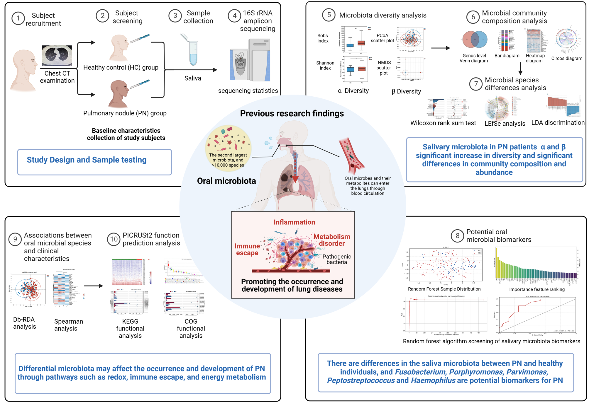
記住我


The isolate named Pseudomonas sp. MN1F was confirmed as belonging to Pseudomonas genus with proximity to Pseudomonas putida and Pseudomonas monteilii (Figure S1). Bacterial sequencing was performed using Illumina technology. After trimming and assembly, genome length was estimated at 6 Mbp, consistent with other Pseudomonas sequenced genomes [12, 13]. C + G content, number of coding sequences and number of tRNAs were 62,9%, 5.575 and 45, respectively. Global genomes statisitics are depicted in Table 1. Forty-eight insertion sequences (IS) were found, 34 of which were similar to Pseudomonas syringae sequences (Table S1).
Table 1 Global statistics of Pseudomonas MN1F genomeGene clusters of secondary metabolites were analyzed using the antiSMASH [14], indicating that MN1F has 31 genes related to pyoverdin synthesis, expression and transport. Genes related to antibiotic resistance beta-lactamase (1 gene) and efflux pumps (7 genes) were found. Numerous genes related to abiotic stresses have also been found, such as temperature stress (5 heat-shock proteins, 5 cold-shock proteins and 7 universal stress proteins), osmotic stress (1 Aquaporin Z, 7 proline/betaine transporter, 1 glycerol uptake protein, 1 osmotically inducible protein), oxidative stress (4 peroxidase, 1 metallothionein and 6 superoxide dismutase), amino acid and carbon starvation (VapBC toxin/anti-toxin system, 3 carbon starvation proteins). Our annotation revealed genes related to alginate synthesis (Alg8, Alg44, AlgK, AlgE, AlgJ, AlgF, AlgA, AlgL, AlgT and AlgX), which may contribute for exopolysachharides (EPS) formation. Genes for sigma fimbriae (usher protein, adhesion and chaperone), for type I (LapB, C, D, E, P, and RTX) and type VI (IcmF, B, G, A, VasA, F, B, D, I, VCA0109 and VgrG) secretion systems were also detected. In addition, five copies for rearrangement hotspot (rhs) repeat proteins were found.
In order to evaluate whether MN1F uses coffee molecules as energy sources, genes that are involved in the degradation of phytocompounds were searched. MN1F possess the genetic machinery required for the degradation of benzoate, quinate, cinnamate, and salicylate; phenolics commonly found in coffee plants [15,16,17]. The interconnection between these degradation systems culminates with the formation of acetyl-CoA, a precursor for citrate synthesis (Fig. 1). The degradation pathways of benzoate and salicylate converge on the conversion into catechol by BenD and SH enzymes, respectively. The quinate pathway converges on the benzoate pathway through the formation of 3-oxoadipate-enol-lactone, but the reverse may also occur when the benzoate pathway (via 4-hydroxy benzoate) converges to the quinate pathway. These three pathways end at acetyl-CoA. The cinnamate pathway, however, does not interconnect with any other and is incorporated into the citrate cycle, ending at fumarate rather than acetyl-CoA. HPLC experiments showed that MN1F can actually break the previous cited phytocompounds and incorporate them into the citrate cycle. Such incorporation was measured by the increasing of concentration of acetate by using the phytocompounds benzoate, cinnamate, quinate and salycilate as unique carbon source (Fig. 2).
Fig. 1
Metabolic map of pathway interconnections between quinate, benzoate, trans-cinnamate and salicylate catabolic systems. Precursors are indicated in red, enzymes are indicated in blue squares and intermediate compounds are indicated in small blue circles. Drawing based on Kyoto Encyclopedia of Genes and Genomes (KEGG) homepage (https://www.genome.jp/kegg)
Fig. 2
Measurement of phytocompounds incorporation into citrate cycle. Incorporation was measured by the decreasing concentration of phytocompounds (Coloured lines: cinnamate: red circle, benzoate: blue square, quinate: yellow triangle, salicylate: green rhombus) and concomitant acetate synthesis (Black lines: cinnamate: black circle, benzoate: black square, quinate: black triangle, salicylate: black rhombus)
EPS production and caffeine resistanceExopolysaccharides (EPS) are involved in regulating mechanisms that promote tolerance to desiccation in bacteria and fungi [18]. Based on the fact that leaf surface is an environment that is poor in water [19, 20], EPS production at increasing osmotic pressure was measured during MN1F growth at minimal media plus PEG. MN1F responded to induced drought stress by producing large amounts of EPS (Fig. 3; dark grey, > 10 g/L at high PEG concentrations).
Fig. 3
Production of water-soluble exopolysaccharides (EPS) by MN1F in response to PEG concentration. Bars represent water soluble EPS (g/L); dark gray bars: MN1f; light gray bars: E. Coli. Rhombus represent CFU (Log10 CFU/mL); dark blue rhombus: MN1F; light blue rhombus: E. coli. The asterisk indicates statistically significant differences (Tukey, α = 0.05)
After extensive genome annotation, no genes were found for caffeine degradation. To evaluate whether MN1F could incorporate caffeine by a non-canonical pathway, we grew bacteria in media with glucose (0—2 g) and NH4Cl (0—8 g) as carbon and nitrogen primary sources, respectively. The intention was to check whether MN1F induce cells to “replace” glucose and NH4Cl with caffeine. MN1F was not able to grow in media containing caffeine in low concentrations of caffeine (Fig. S2).
We evaluated MN1F resistance to high concentrations of caffeine (Fig. 4). Different caffeine concentrations were used (0.8, 2, 3, 6, 10, 15 g/mL) and measured bacterial growth after 8 h of incubation. MN1F was more resistant to caffeine than E. coli control (Fig. 4), indicating some degree of resistance to caffeine.
Fig. 4
Evaluation of caffeine resistance in MN1F. Dark gray bars: MN1F; Light gray bars: E. coli negative control. Bacteria growth was measured by CFU (Log10 CFU/mL). The asterisk indicates statistically significant differences (Tukey, α = 0.05)
Biocontrol against coffee leaf rustThe presence of Type I and VI secretion systems in MN1F gave us a hint that this bacteria could be used as a biocontrol agent. An initial experiment was performed to check whether MN1F had an antagonistic effect on H. vastatrix infection in C. arabica detached leaves (Figure S3). The application of MN1F decreased the severity of the infection (Fig. 5; treatment IV, V, VI). However, MN1F was less effective when applied after 24 h of H. vastatrix uredospores application (Fig. 5; treatment VI). It was verified whether the antagonism was due to extracellular metabolites secreted by MN1F. The supernatant was collected, filtered, and applied to the leaves with the uredospores. The severity of the infection did not differ between the application of the supernatant (Fig. 5; treatment III), the control treatments with saline (Fig. 5; treatment I) and culture medium (Fig. 5; treatment II).
Fig. 5
Severity of H. vastatrix infection after MN1F application. I- saline + uredospores applied simultaneously; II—King’s B liquid medium + uredospores applied simultaneously; III – MN1F supernatant filtered + uredospores applied simultaneously; IV—MN1F + uredospores applied simultaneously; V—MN1F + uredospores applied 24 h later; VI—Uredospores + MN1F applied 24 h later; VII—MN1F only; The asterisk indicates statistically significant differences (Dunnett, p < 0.05)
In vivo antagonism was verified by calculating the H. vastatrix incidence in response to MN1F application (Fig. 6a). The treatments with the fungicide azoxistrobin + cyproconazole (Fungi), the resistance inducer acibenzolar-S-methyl (ASM) and MN1F inoculant showed a positive effect in delaying the progress of infection, since all had a lower incidence than control plants. MN1F treatments ANTP (antagonism), INDP (induction) and INDP/ANTP (antagonism and induction) showed a final effect similar to the fungicide and ASM. However, after the final measurement, the application of MN1F few minutes before infection (ANTP) showed the best result of all treatment with an incidence of 12%, a better result than chemical treatments with 18% of incidence. Treatment with application two days and few minutes before infection (INDP/ANTP) proved to be inferior (around 18% of incidence) to ANTP alone. In addition. treatment with MN1F also reduced the average number of lesions per plant by about fourfold (Fig. 6b) compared to the untreated control, but there was no significant difference (p > 0.05) between treatment with MN1F and the other treatments (with BION and fungicide).
Fig. 6
In vivo antagonism of MN1F against H. vastatrix. a) Incidence of coffee rust in response to MN1F and other treatments. Control—Infected plants without treatment. BION (ASM)—plants sprayed with ASM (0.01 g/L) two days before infection; Fungi (fungicide Priori Xtra)—plants sprayed with Priori fungicide (50% v/v) two days before infection; INDP—plants sprinkled with MN1F two days before infection; ANTP—plants sprinkled with MN1F few minutes before infection; ANTP/INDP—plants that were sprinkled with MN1F two days before and a few minutes before infection; b) Severity, evaluated by the average number of lesions per plant in the same experiment; at 54 days after inoculation (Dunnett, p < 0.05)
The MN1F strain showed ability to inhibit 40% of uredospores germination, when applied as a solution in the same concentration of in vitro conditions (Fig. 7). Therefore, this effect over the germination, which is essential for the infection process, may be related with the control of the disease in the coffee seedlings in the treatment ANTP. However, because the coffee seedlings treated with MN1F showed lower incidences of coffee rust until the last evaluation (54 days after the inoculation), probably other mechanisms of control, such as induction of resistance, may also be acting in the biocontrol of the disease.
Fig. 7
Inhibition in uredospore germination promoted directly by MN1F contact. * The asterisk indicates statistically significant differences (Tukey, α = 0.05)
留言 (0)