What is the physical entity we call a wound? Wounds comprise not only the wound bed but also the wound edge and periwound skin. The wound bed is typically the open portion of the tissue or wound bed surface; it can vary in depth and may have tunneling or undermining that needs to be factored into the care plan. However, the wound encompasses not only the wound bed and edge but also the surrounding, periwound skin (Supplemental Figure, https://links.lww.com/NSW/A114).
For example, in moisture management, it is important not to let packing go above the wound surface and cause periwound maceration from leaked exudate. Exudate quantity may be influenced by edema control in the surrounding skin and the absorbency of the dressing applied. More research and expert consensus documents are needed to clarify the importance of the type of exudate (serous, sanguineous, pustular, or combinations), along with solutions for exudate management and the prevention of periwound maceration.
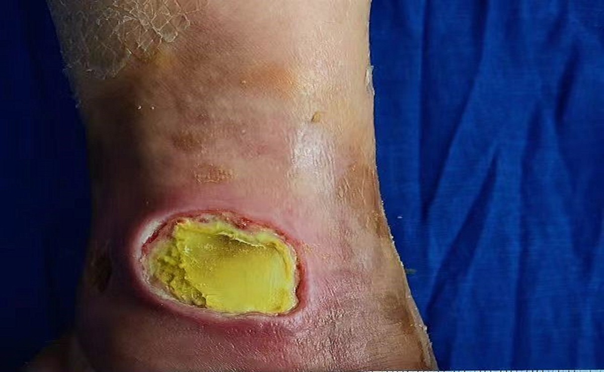
To identify infection, two mnemonics are useful: NERDS and STONEES. Woo and Sibbald1 validated NERDS (Non-healing, Exudate, Red friable, Debris, and Smell) in 2009 to help clinicians distinguish local infection from infection in the deep and surrounding compartment. Any three or more NERDS criteria indicate local infection and the need for a topical antimicrobial agent.
In contrast, the STONEES criteria relate to deep and surrounding infection.1 Three of the seven criteria indicate the need for systemic antimicrobial treatment. The criteria may include any three criteria from either the wound base (Os [Latin for bone], increased Exudate, or Smell) or the wound edge (Size increase, Temperature increase [periwound skin is ≥3 °F warmer than the opposite side of the body], New satellite periwound breakdown, Erythema/Edema [cellulitis], Exudate, and Smell). Both increased exudate and smell arise from the base of the wound and are present in the NERDS and STONEES criteria: an additional NERDS criterion is required for superficial infection, or an additional STONEES criterion is required for deep and surrounding infection. A wound base with exposed bone or probing to bone increases the likelihood of osteomyelitis,1–3 especially with gritty bone in the base of a foot ulcer.
The highest odds ratio for infection is increased temperature of the periwound skin, which is eight times more likely to be associated with deep and surrounding infection.1 However, clinicians must always take care to identify two additional criteria for deep and surrounding infection because the temperature change may also be associated with deep inflammation (eg, the Charcot foot) or unequal vascular supply.
Maceration of the wound edge from excess moisture escaping from the wound or indirectly from the surface dressing is also an issue. Protect periwound skin with surface barriers (eg, film-forming liquid acrylates, petrolatum, zinc oxide, or windowed adhesive dressings). Alternatively, dressings or superabsorbent products with fluid-lock technology may prevent this complication.
Deeper in the periwound skin, edema from venous disease or other etiologies may occur. In this situation, compression bandages for healing and compression stockings to prevent recurrence help optimize the periwound integrity. It is also important to prevent trauma to the surrounding skin: consider silicone dressings that have a lower tear force on removal compared with traditional acrylic adhesive dressings.
Local wound care can only be optimized with attention to the wound edge and the regional skin that must have adequate blood supply for compression therapy and a healthy epidermal barrier function to prevent moisture damage. Let's give periwound skin the respect and consideration it needs and deserves during wound bed preparation.
Elizabeth A. Ayello, PhD, MS, BSN, RN, CWON, ETN, MAPWCA, FAAN
R. Gary Sibbald, MD, DSc (Hons), MEd, BSc, FRCPC (Med Derm), FAAD, MAPWCA, JM
1. Woo KY, Sibbald RG. A cross-sectional validation study of using NERDS and STONEES to assess bacterial burden. Ostomy Wound Manage 2009;55(8):40–48.
2. Grayson ML, Gibbons GW, Balogh K, Levin E, Karchmer AW. Probing to bone in infected pedal ulcers. A clinical sign of underlying osteomyelitis in diabetic patients. JAMA 1995;273:721–3.
3. Lavery LA, Armstrong DG, Peters EJG, Lipsky BA. Probe-to-bone test for diagnosing diabetic foot osteomyelitis: reliable or relic?Diabetes Care 2007;30:270–4.


留言 (0)