
記住我


Non-dystrophic myotonias (NDMs) are rare genetic neuromuscular disorders caused by dysfunctional ion channels expressed in the skeletal muscle cell membrane and, depending on the ion channel involved, referred to as chloride or sodium channelopathies.
Myotonia congenita is a chloride channelopathy with autosomal dominant or recessive inheritance, leading to Thomsen's or Becker's disease, respectively. Chloride channels (ClC-1), encoded by the CLCN1 gene, play an essential role in restoring and maintaining the electrical stability of skeletal muscle cells. Up till now, more than 150 different CLCN1 mutations, resulting in a reduced conductance of the ClC-1, have been described (1–3). Sodium channelopathies are caused by mutations in the SCN4A gene, resulting in an impaired inactivation of the alpha-subunit of the skeletal muscle NaV1.4 channel. Two autosomal dominant inherited phenotypes are recognized, paramyotonia congenita (PMC) and sodium channel myotonia (SCM) (1, 3). Both ion channelopathies lead to hyperexcitability of the sarcolemma membrane, causing myotonia, characterized by delayed skeletal muscle relaxation after voluntary or evoked muscle contraction. The first symptoms usually appear in the first or second decade but may also emerge later in life. MC is characterized by muscle stiffness (myotonia) upon initiating movement, and alleviated by repeated muscle contractions, known as the “warm-up” phenomenon. Transient muscle weakness after initiating movements as well as muscle hypertrophy can be part of the phenotype. Symptoms in Becker's disease are often more severe (4). In contrast to SCN4A-associated myotonia, face and hand muscles are less involved in MC, and no or minimal cold sensitivity is reported. However, distinguishing SCM from MC can be challenging since there is some clinical overlap (1). Diagnosis is made by medical history, physical examination, neurophysiological tests, and genetic analysis. Most NDM patients present myotonic discharges on needle electromyography (nEMG). In addition, Fournier's short exercise test (SET) can be performed to differentiate sodium and chloride channelopathies based on postexercise changes in compound muscle action potential (CMAP) amplitude obtained by supramaximal nerve stimulation. In most MC cases, the CMAP amplitude decreases instantaneously after a short effort and quickly returns to baseline, classified as pattern II (5). However, in a minority of autosomal dominant MC cases, the CMAP amplitude is not altered by exercise, resembling the pattern III characteristics of SCM (5).
NDMs are skeletal muscle disorders without extramuscular manifestations. However, in 2016 our study group reported four families with coexisting Brugada syndrome (BrS) and NDM. Three families were diagnosed with genetically confirmed SCM and one with autosomal dominant MC (Thomsen disease) (6). BrS is another rare inherited channelopathy, due to alterations of ion currents at the level of the cardiac sarcolemma, predisposing to life-threatening ventricular arrhythmias and sudden cardiac death (SCD). The estimated prevalence is 1 in 2000 people worldwide. The diagnosis is confirmed on spontaneous or Ajmaline-induced ST segment elevation on the electrocardiogram (ECG) with a type I morphology of > 2 mm in more than one lead among the right precordial leads (V1-V3) (7, 8). Approximately 20–30% of BrS cases are attributed to a mutation in the SCN5A gene. Other genes are assumed to be causative in an additional 10% of cases. However, most cases remain genetically unsolved. We hereby present a second family with MC and coexisting BrS.
Case presentationThe index patient, a 52-year-old man (A1, Table 1), was referred to our neurology department. He complained of chronic painful muscle stiffness, mainly involving the shoulder and pelvic girdle and, to a lesser degree, the hands. He reported a warm-up phenomenon. Stress and preceding rest periods were exacerbating factors. Cold exposure, on the other hand, was not. He never experienced muscle weakness. These muscle complaints had been until now, attributed to fibromyalgia. Most importantly, there was a history of symptomatic BrS for which an ICD was implanted (Table 1). Upon physical examination diffuse hypertrophy was noted. When the patient tried to get up from a chair, initially we observed marked muscle stiffness of the lower limbs, which progressively improved upon repetition of the task. No other clinical myotonia was detected, including during the evaluation of hand grip, and eye closure. No percussion myotonia or lid lag could be elicited. Muscle strength was normal, and Gower's sign was negative. nEMG showed myotonic discharges mostly exceeding 1 second of duration in all muscles tested, without any myopathic features. The short exercise test was normal at room temperature and after cooling (pattern III). Genetic testing revealed that the patient was heterozygous carrier for a c.2287C>A, p.(Gln763Lys) variant in exon 19 of the CLCN1 gene, which is classified as a variant of unknown significance (VUS). Other genetic causes of myotonia, including myotonic dystrophy type 1 (DMPK) and 2 (ZNF9) and sodium channelopathies (SCN4A), were excluded. Subsequently we screened three relatives of our index patient, 2 of whom had BrS, for the presence of myotonia.
TABLE 1
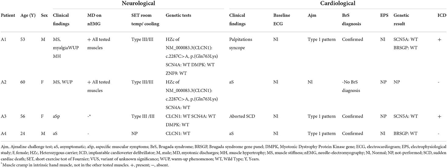
Table 1. Clinical and paraclinical features of the 4 relatives.
The eldest sister (A2) complained of muscle stiffness in the 4 limbs, especially after a period of rest, or after heavy exercise. There was no aggravation with exposure to cold. She had normal muscle strength and no signs of muscle hypertrophy. Although no clinical myotonia was detected, nEMG did reveal brief (100–200 msec duration) myotonic discharges in several muscles. The short exercise test was normal at room temperature and after cooling (pattern III). Sequencing the CLCN1 gene, the same heterozygous c.2287C>A, p.(Gln763Lys) variant in exon 19 was detected. No Brugada syndrome was diagnosed since she had a negative Ajmaline challenge test. The second sister (A3) had been diagnosed with Brugada Syndrome following a malignant cardiac event requiring an ICD implantation. She did not complain of muscle stiffness, myalgia, or delayed muscle relaxation. She mentioned sporadic muscle cramps, not preventing her from exercising. The neurological exam was normal, and no muscle hypertrophy nor clinical myotonia was present. During nEMG no myotonic discharges were observed. However, a slightly prolonged random insertional activity, was found in the intrinsic hand muscles and not in the other tested muscles, including the tibial anterior and biceps brachii. These findings were interpreted as a cramp and not as myotonic discharges. This was supported by the fact that at the same time, the patient experienced a slight painful cramping sensation at the site of the needle insertion. A short exercise test was also normal at room temperature and after cooling of the investigated muscle. Based on these clinical and electrophysiological features, no myotonic phenotype was retained. In the context of genetic screening of this family, the analysis of CLCN1, SCN4A, and DMPK genes was normal in this patient (A3). The son of our index patient A1 (A4) had a first negative Ajmaline challenge test at the age of 11 which turned positive when he was 19 years old, confirming the diagnosis of BrS. He never had any cardiac or neurological symptoms. No clinical or electrical abnormalities were detected during physical examination and nEMG. His genetic screening showed no variants in the CLCN1 gene. No SCN5A variant was found in this family. A primary cardiac arrhythmia gene panel was performed of the index patient (A1) and his son (A4). They both returned normal. The following genes were tested in this panel: ANK3, CACNA1c, CACNA2D1, CACNB2b, GPD1L, HCN4, KCNA3, KCNA4, KCNA7, KCNC4, KCND2, KCND3, KCNE1L (KCNE5), KCNE3, KCNH2, KCNJ8, RANGRF (MOG1), SCN1B, SCN2B, SCN3B, SCN5A, SCN7A (Nav.2.1), SGK1, SGK3, SLMAP, SNTG2, TRPM4. The clinical and paraclinical features of the 4 relatives are summarized in Table 1. The nEMG findings are shown in Figure 1.
FIGURE 1
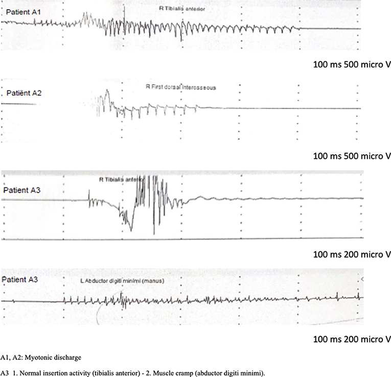
Figure 1. nEMG findings.
DiscussionWe report a family with a clinical and electrophysiological phenotype of NDM and the presence of a heterozygous c.2287C>A (p.Gln763Lys) CLCN1 gene variant. Although this variant was previously reported as likely benign by Brugnoni et al. we believe that in our family this variant is probably pathogenic (13). Indeed, Thomsen's disease is the most probable diagnosis based on the presence of cold-insensitive predominant lower limb muscle stiffness and associated warm-up phenomenon, muscle hypertrophy, myotonic discharges without myopathic features on nEMG, further supported by segregation analysis of this pedigree, and the absence of other gene mutations related to familial myotonic syndromes, including SCN4A, DMPK, and ZNF9 (14–16). Some family members of this pedigree were previously diagnosed with BrS. SCN5A sequencing and the primary cardiac arrhythmia panel analysis were normal. However, a causative role of the detected CLCN1 variant in relation to the arrythmia can be ruled out since two relatives (A3, A4) were diagnosed with BrS and did not carry this CLCN1 variant.
In 2016, our study group reported the first large family with Thomsen's disease due to a novel described CLCN1 mutation and coexisting BrS. Thirteen relatives with BrS were reported, of whom 11 carried the NM_198056.3(SCN5A): c.2632C>T, p.(Arg878Cys) variant of unknown significance (VUS), and five had a genetically confirmed MC due to the NM_000083.3(CLCN1): c.744+1G>A;[=] pathogenic variant. Remarkably, two relatives who showed a positive Ajmaline challenge test without a history of malignant cardiac events, carried the CLCN1 variant and not the SCN5A VUS (6).
Despite the absence of similar cases in the literature reporting the coexistence of BrS and MC, other cardiac arrhythmias and conduction disturbances have recently been described in patients with MC. Vereb et al. reported six MC patients who exhibited arrhythmias or conduction disorders, some of whom requiring a pacemaker (9). Wang et al. described nine autosomal dominant and recessive MC patients with cardiac arrhythmia resulting in minor cardiac symptoms. However, genetic analyses exploring primary cardiac arrhythmias were not specified in this article (10). Two additional case reports described the coexistence of MC and cardiac arrhythmia, including Wolff-Parkinson-White syndrome (11, 12). An overview of reported cases of cardiac arrhythmias in myotonia congenita patients is given in Table 2.
TABLE 2
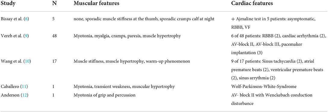
Table 2. Reported cases of cardiac arrhythmias in myotonia congenita patients.
It is unclear whether CLCN1 mutations may contribute to primary arrhythmias or conduction disorders. The ClC-1 channels are mainly expressed in the skeletal muscle, but low levels are also found in the kidney, heart, liver, smooth muscle, and the central nervous system (16–18). Other chloride channel isoforms, including ClC-2 and ClC-3 voltage-gated chloride channels, have been implicated in cardiac arrhythmias (19). By contrast, no clear link has been reported between cardiac ClC-1 channels and cardiac arrhythmias, including BrS. However, for several reasons our clinical data may support a potential role of CLCN1 in the pathogenesis of cardiac arrhythmia, especially BrS. First, since the likelihood of cardiac and muscular channelopathies occurring together in patients is extremely low, given their rare prevalence in the general population, a pathophysiological link between CLCN1 mutations and cardiac arrhythmias can be suspected. Second, we can assume that the prevalence of CLCN1-associated myotonia is underestimated in the BrS population, due to the rather unspecific symptoms, especially in mild cases. Patients may complain of chronic muscle stiffness and myalgia without muscle weakness. Because those symptoms are unspecific and common, NDM is rarely suspected, and diagnosis is often delayed. This is clearly illustrated by the history of our family cases A1 and A2, where in the former symptoms were attributed to an earlier fibromyalgia diagnosis, and, in the latter, mild muscular symptoms were not even reported. Thanks to increasing awareness of the presence of subtle muscular symptoms in myotonia, the rhythmology team referred the BrS patient (A1) to our neurology department. The NDM diagnosis would probably not yet have been made in other circumstances. Finally, the Mendelian model for BrS, characterized by single gene disease, has been recently challenged by an oligogenic model referring to a possible cumulative role of common and rare genetic variants (20). A possible explanation could be that CLCN1 variants contribute to the BrS phenotype. On the other hand, it cannot be excluded that it is a simple coexistence of two separate pathologies and that the gene responsible for Brugada syndrome in these patients is not yet known.
A parallel can be drawn with the SCN4A gene recently added to the list of minor genes associated with BrS (7, 8). In 2016, our study group reported 3 families with SCN4A-associated NDM and coexisting BrS, with this gene presumably acting as a modifier gene. Some of these patients had few muscle complaints, despite the presence of a deleterious variant in the SCN4A gene (6). The expression of the skeletal SCN4A isoform in cardiac muscle was also described (21, 22).
Even though current data are not sufficient to advocate a systematic cardiac work-up in patients with NDM, there is increasing evidence of an overlap between skeletal muscle and cardiac ion channelopathies. Therefore, we propose to perform at least a thorough cardiac history assessment and a 12-lead ECG in NDM patients. A history of palpitations, syncopes, conduction disorders, or a family history of sudden cardiac death should prompt an immediate referral to a cardiologist. Conversely, patients with primary cardiac arrhythmias, especially those with BrS, should be systematically asked about presence of muscle stiffness, myalgia, muscle weakness, and if indicated, be referred to the neurology department.
Sodium channel blockers are used in the symptomatic treatment of myotonia. Mexiletine, a class 1B antiarrhythmic drug, seems to be a safe anti-myotonic therapy in the presence of Brugada syndrome (23, 24). However, the use of Flecainide, a class 1C antiarrhythmic drug, should be started with caution, based on a case report in which BrS was unmasked after initiating this treatment in an SCM patient who had never experienced cardiac symptoms before (25). Considering this and the previously reported family with concomitant MC and BrS, the use of class 1C antiarrhythmic drug to alleviate myotonia seems less appropriate for MC patients as well (6).
Of course, our data need to be confirmed by new designed clinical and genome wide association studies in larger patient cohorts, as well as functional studies in order to further clarify the involvement of CLCN1 gene in cardiac arrhythmias.
Data availability statementThe datasets presented in this article are not readily available because of ethical and privacy restrictions. Requests to access the datasets should be directed to the corresponding author.
Ethics statementWritten informed consent was obtained from the individual(s) for the publication of any potentially identifiable images or data included in this article.
Author contributionsAC: data collection, review of the literature, and article writing. VB: review of literature, article writing, and article editing. AF, TR, AG, LP, CD, and GP: article editing. All authors contributed to the article and approved the submitted version.
Conflict of interestThe authors declare that the research was conducted in the absence of any commercial or financial relationships that could be construed as a potential conflict of interest.
Publisher's noteAll claims expressed in this article are solely those of the authors and do not necessarily represent those of their affiliated organizations, or those of the publisher, the editors and the reviewers. Any product that may be evaluated in this article, or claim that may be made by its manufacturer, is not guaranteed or endorsed by the publisher.
References2. Morales F, Pusch M. An up-to-date overview of the complexity of genotype-phenotype relationships in myotonic channelopathies. Front Neurol. (2019) 10:1404. doi: 10.3389/fneur.2019.01404
PubMed Abstract | CrossRef Full Text | Google Scholar
3. Vivekanandam V, Männikkö R, Mathew E, Hanna MG. Improving genetic diagnostics of skeletal muscle channelopathies. Expert Rev Mol Diagn. (2020) 20:725–36. doi: 10.1080/14737159.2020.1782195
PubMed Abstract | CrossRef Full Text | Google Scholar
4. Sun C, Tranebjaerg L, Torbergsen T, Homgren G, Van Ghelue M. Spectrum of CLCN1 mutations in patients with myotonia congenita in Northern Scandinavia. Eur J Hum Genet. (2001) 9:903–9. doi: 10.1038/sj.ejhg.5200736
PubMed Abstract | CrossRef Full Text | Google Scholar
5. Fournier E, Arzel M, Sternberg D, Vicart S, Laforet P, Eymard B, et al. Electromyography guides toward subgroups of mutations in muscle channelopathies. Ann Neurol. (2004) 56:650–61. doi: 10.1002/ana.20241
PubMed Abstract | CrossRef Full Text | Google Scholar
6. Bissay V, Van Malderen SCH, Keymolen K, Lissens W, Peeters U, Daneels D, et al. SCN4A variants and Brugada syndrome: phenotypic and genotypic overlap between cardiac and skeletal muscle sodium channelopathies. Eur J Hum Gene. (2016) 24:400–7. doi: 10.1038/ejhg.2015.125
PubMed Abstract | CrossRef Full Text | Google Scholar
7. Campuzano O, Sarquella-Brugada G, Cesar S, ARbelo E, Brugada J, Brugada R. Update on genetic basis of brugada syndrome: monogenic, polygenic or oligogenic? Int J Mol Sci. (2020) 21:7155 doi: 10.3390/ijms21197155
PubMed Abstract | CrossRef Full Text | Google Scholar
8. Campuzano O, Sarquella-Brugada G, Fernandez-Falgueras A, Cesar S, Coll M, Mates J, et al. Genetic interpretation and clinical translation of minor genes related to Brugada syndrome. Hum Mutat. (2019) 40:749–64. doi: 10.1002/humu.23730
PubMed Abstract | CrossRef Full Text | Google Scholar
9. Vereb N, Montagnese F, Gläser D, Schoser B. Non-dystrophic myotonias: clinical and mutation spectrum of 70 German patients. J Neurol. (2021) 268:1708–20. doi: 10.1007/s00415-020-10328-1
PubMed Abstract | CrossRef Full Text | Google Scholar
10. Wang Q, Zhao Z, Shen H, Bing Q, Li N, Hu J. The clinical, myopathological, and genetic analysis of 20 patients with non-dystrophic myotonia. Front Neurol. (2022) 13:830707. doi: 10.3389/fneur.2022.830707
PubMed Abstract | CrossRef Full Text | Google Scholar
13. Brugnoni R, Kapetis D, Imbrici P, Pessia M, Canioni E, Colleoni L, et al. A large cohort of myotonia congenita probands: novel mutations and a high-frequency mutation region in exons 4 and 5 of the CLCN1 gene. J Hum Genet. (2013) 589:581–7. doi: 10.1038/jhg.2013.58
PubMed Abstract | CrossRef Full Text | Google Scholar
15. Matthews E, Fialho D, Tan SV, Venance SL, Cannon SC, Sternberg D, et al. The non-dystrophic myotonias: molecular pathogenesis diagnosis and treatment. Brain. (2010) 133:9–22. doi: 10.1093/brain/awp294
PubMed Abstract | CrossRef Full Text | Google Scholar
16. Imbrici P, Altamura C, Pessia M, Mantegazza R, Desaphy JF, Conte Camerino D. ClC-1 chloride channels: state-of-the-art research and future challenges. Front Cell Neurosc. (2015) 9:156. doi: 10.3389/fncel.2015.00156
PubMed Abstract | CrossRef Full Text | Google Scholar
17. Steinmeyer K, Ortland C, Jentsch TJ. Primary structure and functional expression of a developmentally regulated skeletal muscle chloride channel. Nature. (1991) 354:301–4. doi: 10.1038/354301a0
PubMed Abstract | CrossRef Full Text | Google Scholar
18. Chen TT, Klassen TL, Goldman AM, Marini C, Guerrini R, Noebels JL. Novel brain expression of ClC-1 chloride channels and enrichment of CLCN1 variants in epilepsy. Neurology. (2013) 80:1078–85. doi: 10.1212/WNL.0b013e31828868e7
PubMed Abstract | CrossRef Full Text | Google Scholar
19. Duan D. Phenomics of cardiac chloride channels: the systematic study of chloride channel function in the heart. J Physiol. (2009) 587:2163–77. doi: 10.1113/jphysiol.2008.165860
PubMed Abstract | CrossRef Full Text | Google Scholar
21. Pereon Y, Lande G, Demolombe S, Nguyen The Tich S, Sternberg D, Le Marec H, et al. Paramyotonia congenita with an SCN4A mutation affecting cardiac repolarization. Neurology. (2003) 60:340–2. doi: 10.1212/01.WNL.0000042093.96309.5A
PubMed Abstract | CrossRef Full Text | Google Scholar
22. Kaufmann SG, Westenbroeck RE, Maass AH, Lange V, Renner A, Wischmeyer E, et al. Distribution and function of sodium channel subtypes in human atrial myocardium. J Mol Cell Cardiol. (2013) 61:133–41. doi: 10.1016/j.yjmcc.2013.05.006
PubMed Abstract | CrossRef Full Text | Google Scholar
23. Vicart S, Franques J, Bouhour F, Magot A, Péréon Péréon Y, Sacconi S, et al. Efficacy and safety of mexiletine in non-dystrophic myotonias: a randomised, double-blind, placebo-controlled, cross-over study. Neuromuscul Disord. (2021) 31:1124–35. doi: 10.1016/j.nmd.2021.06.010
PubMed Abstract | CrossRef Full Text | Google Scholar
24. Modoni A, D'Amico A, Primiano G, Capozzoli F, Desaphy JF, Lo Monaco M. Long-term safety and usefulness of mexiletine in a large cohort of patients affected by non-dystrophic myotonias. Front Neurol. (2020) 11:300. doi: 10.3389/fneur.2020.00300
PubMed Abstract | CrossRef Full Text | Google Scholar
25. Cavalli M, Fossati B, Vitale R, Brigonzi E, Ricigliano V, Saraceno L, et al. Flecainide-induced Brugada syndrome in a patient with skeletal muscle sodium channelopathy: a case report with critical therapeutical implications and review of the literature. Front Neurol. (2018) 9:385. doi: 10.3389/fneur.2018.00385
留言 (0)