Study participants and dietary challenge
Study participants were from the USDA Nutritional Phenotyping Study which included healthy men and women, aged 18–66 y with a normal to obese BMI of 18–44 kg/m2 living near Davis, California beginning in May 2015. Details of study recruitment and participation are contained in separate reports [17, 18]. Briefly, the study included two visits to the United States Department of Agriculture – Agricultural Research Service – Western Human Nutrition Research Center (WHNRC) scheduled within a period of 10–14 days. On visit 1 subjects were provided informed consent and screened to ensure the volunteers fell within designed ranges for the study. Visit 2 was the challenge meal test day. The night before visit 2, subjects were provided a high carbohydrate meal (17% kcal from fat, 77% kcal from carbohydrate, and 7.5% kcal from protein) and instructed to consume it before 19:00 h. Subjects arrived fasted (12 h) the next morning and fasting blood was collected before ingestion of a high-fat liquid challenge meal (60% kcal from fat, 25% kcal from carbohydrates, and 15% kcal from protein). Additional details of the standardized high carbohydrate dinner and high-fat challenge meal are contained in separate reports [19, 20]. Postprandial blood was drawn at 3 and 6 h after consumption of the challenge meal. Ethnicity was self-reported by subjects using a demographic questionnaire and is presented in Supplemental Table 1. Subjects were grouped White or Caucasian (n = 214), Hispanic or Latino/a (n = 45), Asian (n = 41), Multi-racial (n = 22), Black or African-American (n = 16), Middle Eastern (n = 5), Native Hawaiian or other Pacific Islander (n = 2), American Indian or Alaska Native (n = 1), or declined to respond (n = 3). The “Asian” group was comprised of subjects who responded as Asian, East Asian, South Asian, or Southeast Asian; Multi-racial subjects (“Multi”), identified as more than one ethnic group.
The study was registered at clinicaltrials.gov (identifier: NCT02367287) and received ethical approval from the University of California, Davis, Institutional Review Board. All participants provided written informed consent and received monetary compensation for their participation. Data were stored using the Research Electronic Data Capture (REDCap) application hosted by the University of California Davis Health System Clinical and Translational Science Center.
Analysis of monocyte frequencies
Monocyte frequencies were quantified using complete blood count (CBC) with differential. During the four-year recruitment period from June 2015 through July 2019 the CBC analyses were performed using whole blood (treated with EDTA as an anticoagulant) in the UC Davis Health, Department of Pathology and Laboratory Medicine Clinical Laboratory using a Beckman Coulter LH750/780 (prior to October 2016) or a Beckman Coulter DxH800 automated hematology analyzer, with the exception that twelve samples early in the study (prior to August 14, 2015) were analyzed on an Abbott Cell-Dyn 322 analyzer at the WHNRC.
Clinical parameters
Fasting and postprandial blood was collected and serum or plasma was obtained by centrifugation at 1300 × g at 4 °C for 10 min. Lipid-related markers including triglycerides, total cholesterol, HDL-cholesterol (HDL-C), and LDL-cholesterol (LDL-C) were measured using a Cobas Integra 400/800 kit (Roche, Indianapolis, IN), a Cobas CHOL2 kit (Roche), a Cobas HDL-C plus 3rd generation kit (Roche), and a Cobas LDLC3 kit (Roche), respectively. All assays were completed on an auto-analyzer, Cobas Integra 400 + instrument (Roche). Glucose concentrations in plasma samples were measured using Glucose HK Gen.3 kits (Roche) conducted on the Cobas Integra 400 + instrument (Roche). Serum insulin levels were determined by Elecsys Insulin kits running on a Cobas e411 analyzer (Roche).
Plasma immune markers measured by ELISA
Neopterin concentration (nmol/L) was measured using undiluted sodium heparin plasma using a commercial, competitive enzyme immunoassay (Alpco, BRAHMS GmbH, Salem, NH, USA) according to the manufacturer’s instructions. Myeloperoxidase concentrations (µg/L) were measured using sodium heparin plasma (1:10 dilution) using a commercial ELISA kit (Alpco Immunodiagnostik, Salem, NH, USA) according to the manufacturer’s instructions. Soluble CD14 (sCD14) concentrations (µg/L) were measured in duplicate using sodium heparin plasma (1:400 or 1:600 dilution) using a commercial ELISA kit (Bio-Techne R&D Duoset Systems, Minneapolis, MN USA) according to the manufacturer’s instructions. Plates for these three assays were read on an Agilent BioTek Synergy reader (Santa Clara, CA USA) and data analyzed using the BioTek Gen5 software.
Plasma immune markers measured by multiplexed MSD assay
The concentrations of plasma proteins (µg/L) were measured in plasma using MSD assay kits and the MSD sector imager 2400 (MESO Scale Discovery). EDTA plasma was used for proteins (CRP, SAA, ICAM-1, VCAM-1 using the Vplex Vascular Injury Panel 1 with samples diluted 1:1,000; CCL2 using the Vplex Custom Human Biomarker Chemokine Panel 1 with samples diluted 1:4; TNF, IL-1β, IL-6, IL-8 and IL-10 using the Vplex Custom Human Biomarker Proinflammatory Panel 1 with samples diluted 1:2). Three levels of lyophilized controls were used on each plate to assess plate-to-plate variation. Mean concentrations (µg/L) of duplicate wells were used for analysis.
Analysis of monocyte subsets by flow cytometry
Monocyte subsets at fasting and postprandial time points were analyzed using 100 µl of whole blood collected into EDTA-treated tubes mixed with pre-titrated volumes of the following antibodies in BD Brilliant stain buffer (catalog # 563,794 BD Biosciences): CD45-BV786 (catalog # 563,716 BD Biosciences), CD91-PE (catalog # 550,497 BD Biosciences), CD14-BUV395 (catalog # 563,561 BD Biosciences), and CD16-BV421 (catalog # 562,878 BD Biosciences). Following a 20-min incubation at room temperature 1X BD FACS Lysing Solution (catalog# 349,202 BD Biosciences) was added to whole blood/antibody mixture and incubated at room temperature for an additional 10 min. Cells were washed twice with cold wash/stain buffer (containing 0.1% BSA (w/v), 0.05% NaN3 (w/v) in PBS) then analyzed using an LSR Fortessa flow cytometer (BD Biosciences) configured with blue (488 nm), red (640 nm), violet (405 nm) and UV lasers (355 nm). Data were collected using FACSDiva and analyzed using FlowJo version 10.6.1 software (BD Biosciences).
Statistical analysis
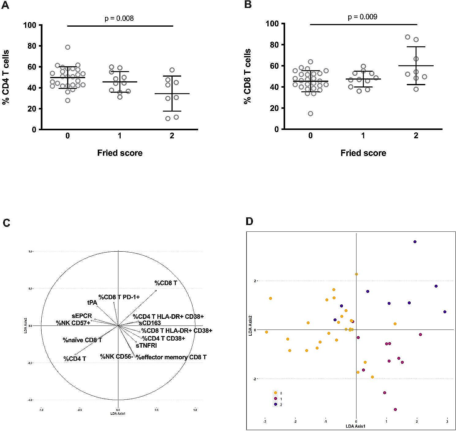
All statistical analyses were performed in GraphPad Prism 9 (Version 9.3.1). Unless noted otherwise, graphical data are presented in violin plots as means with individual data points displayed. Data normality was assessed using the Anderson–Darling test. Cohort characteristics, experimental measurements, as well as monocyte frequencies and subsets at fasting were compared between age groups using Kruskal–Wallis non-parametric one-way ANOVA with Dunn’s multiple comparisons test. Paired monocyte frequencies and subsets were compared between metabolic state (fasting and at 3 and 6 h after meal consumption) using Friedman non-parametric, repeated measures one-way ANOVA with Dunn’s multiple comparisons test. Differences were considered significant when P < 0.05 (*P < 0.05; **P < 0.01; ***P < 0.001; ns = not significant). Correlation analysis was performed using non-parametric Spearman’s rank-order correlation. Correlations were considered significant when P < 0.05 (*P < 0.05; **P < 0.01; ***P < 0.001). Final assembly and preparation of all figures was done using CorelDRAW 2021 (Corel Corporation, Ottawa, Canada).

留言 (0)