
記住我


Ankle osteoarthritis (OA) is a chronic disease.[1] The causes include trauma, degenerative changes, rheumatic diseases, hemophilia, hemochromatosis, gout, avascular necrosis, and post-infectious states. End-stage ankle arthritis often manifests as severe weight-bearing pain, dysfunction, and impaired mobility.[1] Currently, common surgical procedures for the treatment of end-stage ankle arthritis include ankle arthrodesis (AA) and total ankle replacement (TAR). Previous studies have confirmed that AA can effectively relieve pain and restore limited function, but this operation sacrifices ankle joint mobility and may cause or aggravate arthritis in adjacent joints.[2] TAR can relieve pain and improve function while preserving the range of motion of the ankle joint, and its clinical outcome has gradually been confirmed.[2–9] It has been considered that AA is the gold standard for the treatment of end-stage ankle arthritis before TAR. In recent years, with the continuous improvement of surgical techniques and prosthetic products, the application of TAR has become more common, and it has become a common treatment for end-stage ankle OA.[2–6]
There have been many reports of the use of different types of TAR prostheses for the treatment of end-stage ankle OA, and the satisfaction rate, function, and pain improvement are significant.[7–9] However, the clinical and radiographic results of using INBONE-II (Wright Medical Technology, Arlington, TN, USA), prosthesis were rarely reported. INBONE-II is a third-generation, 2-component TAR prosthesis, which allows for improved biomechanical stability over the previous design, particularly in the coronal plane.[10] In 2016, the INBONE-II prosthesis was put into the market in China. It is the only TAR prosthesis on the market in our country for a period of time, but there is still a lack of relevant large-sample clinical studies of whether it is suitable for ankle OA caused by common domestic causes and its clinical efficacy. Therefore, this study aimed to explore the medium-term and short-term clinical outcomes, radiographic findings, complications, and prosthetic survival of single-center, large-sample TAR surgery using INBONE-II prosthesis.
Methods Ethical approvalThis study was approved by the Ethics Committee of Beijing Jishuitan Hospital (No. 202204-16-01). In this retrospective study, informed written consent was obtained from all patients before their enrollment in this study. All patients allowed researchers to analyze their data and produce academic results.
Inclusion and exclusion criteriaInclusion criteria: (1) INBONE-II prosthesis use for TAR; (2) Age ≥18 years; (3) Patient informed consent obtained; (4) Negative past history of TAR or AA surgery; and (5) Post-operative follow-up time ≥2 years.
Exclusion criteria: (1) Active infection or previous infection in the foot and ankle; (2) Neurogenic arthropathy (Charcot joint); (3) Severe benign joint hypermobility syndrome; (4) Full-length non-digital lower limb x-rays with abnormal alignment in knee joints or hip joints; (5) Uncontrolled diabetes; (6) Tumors, peripheral neurovascular diseases, etc.; and (7) Incomplete medical records.
Observation indicatorsFrom August 1, 2016 to June 31, 2019, a total of 69 patients underwent TAR surgery using the INBONE-II prosthesis, all of which were performed by the same surgeon. According to the above inclusion and exclusion criteria, a total of 64 patients were included in the study.
We recorded various demographic data from the hospital medical record system, including age, gender, height, weight, etiology, history of smoking, history of diabetes, and history of previous ankle surgery. We also recorded surgery-related information, including the amount of bleeding, the length of the operation, etc. According to the medical record system and post-operative follow-up, the types and numbers of intra-operative and post-operative complications were recorded. The grade of complications was classified according to the Glazebrook classification system.[11]
We used the American Orthopedic Foot and Ankle Society (AOFAS) ankle hindfoot scale, visual analog scale (VAS), and the Short Form-36 (SF-36) to evaluate patients pre-operatively and at the final follow-up. Using the hospital imaging system, on the weight-bearing X-ray images, we used the tibial anterior surface angle (TAS), tibial lateral surface angle (TLS), and talar tilt angle (TT) to measure the radiographs of patients with pre-operative and follow-up time of >1 year after surgery [Figure 1]. TAS is defined as the angle between the articular surface of the distal tibia and the axis of the tibia on the anterior-posterior view, TT is defined as the angle between the articular surface of the distal tibia and the articular surface of the talus, TLS is defined as the angle between the articular surface of the distal tibia and the axis of the tibia on the lateral view. The range of motion of the patient's ankle dorsiflexion and plantar flexion was recorded before the operation and at the final follow-up.
 Figure 1:
 Measurement of tibial anterior surface angle (TAS), talar tilt (TT) on anterior-posterior view (A), and tibial lateral surface angle (TLS) on lateral view (B). a: TAS; b: TT; c: TLS.
Surgical methodsThe patient was brought into the operating room and was placed in a supine position with a thigh tourniquet applied to the operative lower extremity. An ipsilateral hip bump was placed to internally rotate the leg. The foot and ankle were prepped to the level of the knee in the standard fashion by pre-operative fluoroscopy. A central anterior incision or arc incision of the ankle joint was made to expose the joints from the interval of tibialis anterior and the extensor hallucis longus. The osteophytes and hyperplastic synovium were resected, and the soft tissue released. After reducing the ankle to the anatomically corrected position, K-wire was used to cross fix the tibiotalar joint. The lower extremity was then placed into the external fixation jig with the help of manufacturer's guidelines and technique for the INBONE-II TAR. Intraoperative fluoroscopy was used for the alignment of our extra-medullary alignment and sizing for our tibial and talar cuts under standard manufacturer's guidelines and techniques. A 6 mm drill bit was used to drill through the calcaneus into the talus, about 5 to 6 cm deep into the medullary cavity of the tibia. An appropriate size osteotomy guide was selected, and fixed to the front of the fixator, after making sure the size of the osteotomy guide was appropriate (just within the ankle joint). Using a pendulum saw to cut the bone, the front guide plate was removed, the bone block taken out and rinsed with the flushing gun, and finally the anteroposterior size of the tibial tray was tested. The tibial stem and tibial tray were installed, and fluoroscopy was performed. The talus stem and talus dome were then installed, and the lateral view checked through fluoroscopy. Finally, the polyethylene component was inserted and the position of the prosthesis was checked through fluoroscopy. The mobility of the ankle joint was also checked [Figure 2]. Achilles lengthening was also performed as needed. In this study, one patient underwent additional Achilles tendon lengthening.
 Figure 2:
 Intra-operative photographs (A–E) of the ankle joint and immediate post-operative X-rays (F,G). (A) Shows the interval between the tibialis anterior and the extensor hallucis longus; (B) shows the joint capsule is opened, and osteophyte proliferation is obvious, the articular surface is severely worn, and the cartilage damage area exceeds 2/3; (C) shows the osteotomy after removing the osteophyte and synovium; (D) shows the removed osteophytes and bone obtained from osteotomy; (E) shows the implantation of the prosthesis; (F) and (G) show the X-ray on AP and lateral view immediately after the operation, when the prosthesis is in a good position.
Statistical methodsIBM SPSS 25.0 (SPSS Inc., Chicago, IL, USA) software was used for statistical analysis. For continuous variables, Shapiro– Wilk's test for normality detection was first used, then mean ± standard deviation (SD) for normal distribution. Two independent sample t test was used for independent sample comparison between the two groups, and paired sample t test was used for pre-operative and post-operative sample comparison. Those that did not conform to the normal distribution were expressed by median (P25, P75), and the Wilcoxon signed-rank test was used for comparison between the two groups. The difference was considered to be statistically significant when P <0.05.
ResultsA total of 64 patients (33 males, 31 females) were available for the final follow-up in this study, with an average age of 59.9 ± 10.5 years (39–79 years) at the time of surgery. In terms of etiology, traumatic arthritis, primary arthritis, and arthritis due to other causes (all rheumatic) accounted for 47, 14, and 3 cases of arthritis, respectively. Among all 64 patients, twenty of them had a history of smoking, nine had type 2 diabetes, and 19 patients had a history of ankle surgery. The operating time was 164.2 ± 33.4 minutes, and the amount of bleeding was 186.3 ± 90.9 ml.
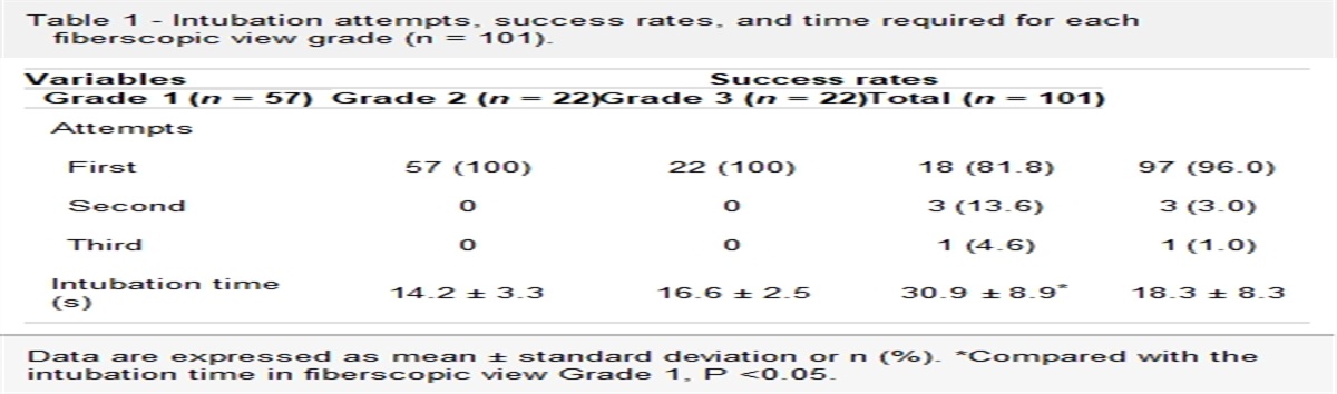
The average follow-up time was 37.9 months (24.0–59.0 months). During this time, the post-operative AOFAS score increased from 47.1 to 83.6, the VAS score decreased from 5.0 to 1.0, and the SF-36 score increased from 94.0 to 127.4. The differences of pre-operative and post-operative clinical results were statistically significant (P < 0.01); the ankle dorsiflexion range increased from 7.6° to 13.2°; and the plantar flexion range was restored from 15.1° to 22.2°. These were statistically significant differences (P < 0.01). The average follow-up time of the patient's imaging results was 22.8 months (12.0–59.0 months), the last follow-up TAS was 87.8 ± 2.4°, TLS was 87.4 ± 2.3°, and TT was 1.3 ± 1.3°. There were significant differences of TLS and TT before and after surgery (P < 0.01) [Table 1]. At the last follow-up, none of the patients had any failure of the prosthesis, and the survival rate of the prosthesis was 100% (64/64).
Table 1 - Pre-operative and final follow-up clinical outcomes and radiographic findings. Items Pre-operative Post-operative Statistics P value AOFAS 47.1 ± 14.9 83.6 ± 10.8 16.14∗ <0.01 VAS 5.0 (4.0, 6.0) 1.0 (0.0, 2.0) 270.00† <0.01 SF-36 94.0 ± 11.7 127.4 ± 9.5 9.56∗ <0.01 Ankle range of motion (°) 22.8 ± 7.5 35.2 ± 10.9 8.01∗ <0.01 Ankle dorsiflexion range (°) 7.6 ± 4.8 13.2 ± 5.0 6.32∗ <0.01 Ankle plantar flexion range (°) 15.1 ± 4.6 22.2 ± 7.4 7.38∗ <0.01 TAS (°) 84.9 ± 12.8 87.8 ± 2.4 1.52∗ 0.14 TLS (°) 80.4 ± 7.7 87.4 ± 2.3 5.94∗ <0.01 TT (°) 4.7 ± 4.3 1.3 ± 1.3 8.16∗ <0.01Values are presented as mean ± standard deviation or median (P25, P75).AOFAS: American Orthopedic Foot and Ankle Society; SF-36: Short Form-36; TAS: Tibial anterior surface angle; TLS: Tibial lateral surface angle; TT: Talar tilt; VAS: Visual analog scale.
∗Paired t test.
†Wilcoxon signed-rank test.
A total of 10 patients had surgery-related complications, and the complication rate was 15.6%. Among the 64 patients, three cases (4.7%) had medial malleolus fractures during operation, 4 (6.3%) had wound healing problems after operation, two (3.1%) had toe numbness, and one (1.6%) had wound infection, all of which were mildly complicated. The symptoms were detected on time and treated accordingly, and all the cases turned out to be good [Table 2]. No serious complications occurred in all patients.
Table 2 - Treatment and outcome of 10 complications. Patient No. Complication Time of occurrence Treatment Outcome 1 Numbness of the toes Post-operation Neurotrophic drug therapy Got better, still a little numb 2 Medial malleolus fracture During operation Intra-operative reduction, fixation with one screw Healed 3 Medial malleolus fracture During operation Intra-operative reduction, fixation with one screw Healed 4 Medial malleolus fracture During operation Intra-operative reduction, fixation with two screws Healed 5 Delayed wound healing Post-operation Change the dressing regularly Healed 12 weeks after surgery 6 Wound dehiscence Post-operation Rehospitalized at 1 month after surgery for local flap transfer Healed 4 months after surgery 7 Delayed wound healing Post-operation Change the dressing regularly Healed 12 weeks after surgery 8 Delayed wound healing Post-operation Change the dressing regularly Healed 4 months after surgery 9 Numbness of the toes Post-operation Neurotrophic drug therapy Got better, still a little numb 10 Wound infection Post-operation Rehospitalized at 1 month after surgery for debridement Healed 2 months after surgeryAmong all the patients, 47 patients (73.4%) were very satisfied with the operation, 11 (17.2%) were relatively satisfied, and four (6.3%) considered they only got little effect. The overall satisfaction rate reached 90.6%. There were two patients (3.1%) who were not satisfied with the operation due to persistent pain in the ankle joint and persistent numbness in the toes [Table 3]. Figure 3 shows a typical case of TAR using INBONE-II prothesis.
Table 3 - Six patients evaluated as not ideal or unsatisfied. Patient No. Evaluation Reasons for dissatisfaction Treatment Outcome 1 Not ideal Numbness of the toes Neurotrophic drug therapy Got better, still a little numb 2 Not ideal Ankle pain Drug therapy, rehabilitation exercise Improved, still pain when walking with weight-bearing 3 Not ideal Ankle pain and swelling Drug therapy, rehabilitation exercise The swelling is gone, still pain when walking with weight-bearing 4 Not ideal Limping, difficult to turn when walking Drug therapy, rehabilitation exercise Got better 5 Unsatisfied Ankle pain Drug therapy, rehabilitation exercise Got better, still having resting pain 6 Unsatisfied Numbness of the toes Neurotrophic drug therapy Got better, still a little numb Figure 3:
 The male patient, 47 years old, with traumatic ankle arthritis. (A-C) The pre-operative X-ray showed that the ankle joint was seriously damaged, osteophyte hyperplasia is obvious, the joint space disappeared, and the distal tibia and the top of the talus had sclerosis; (D-F) 2 years after TAR, X-ray showed that the position of the prosthesis was good, the alignment was neutral, and there was no obvious prosthesis displacement and heterotopic ossification. TAR: Total ankle replacement.
DiscussionIn this study, we performed TAR using INBONE-II prosthesis. The post-operative clinical results, radiographic findings, and ankle range of motion were significantly improved. The incidence of complications was low (15.6%, 10/64), and there was no case need revision. Only 3.1% (2/64) were not satisfied with the operation. It is proved that the use of INBONE-II prosthesis in the treatment of end-stage ankle arthritis can significantly release pain, improve function, and bring good imaging performance.
There have been literature reports on the post-operative efficacy and imaging of other prostheses for TAR surgery, most of which shown that they produce reliable and significant improvement, and that their imaging performance is good.[7–10] However, there are still a few clinical research reports on the use of INBONE-II prosthesis for TAR [Table 4]. Adams et al[12] first published the early results of 194 cases of INBONE-II prosthesis for initial replacement in 2014, and the results showed that the clinical score had significantly improved 3.7 years after surgery (P < 0.003). A retrospective study of 59 cases of INBONE type I and type II prostheses showed that at a follow-up time of 2 years, the AOFAS score in the INBONE-II group averaged 90.1 points, the average VAS score was 1.3 points, and the average ankle joint range of motion was 39.7°.[13] Another study showed that 56 cases of INBONE-II prosthesis replacement had a good clinical outcome 2 years after surgery; the proportion of post-operative prostheses in a neutral position in the coronal position was 96.4%, and this proved that the use of INBONE-II prosthesis replacement has a good corrective effect on the TT.[14] Research by Rushing et al[15] showed that 6 weeks after INBONE-II prosthesis replacement, the coronal and sagittal alignments were satisfactory, and it was observed that the alignment remained good at the 7-year follow-up (P = 0.684, P = 0.837). Our study showed that the post-operative AOFAS score, VAS score, SF-36 score, and ankle range of motion significantly improved compared to their pre-operative values; TAS, TLS, and TT showed that the alignment of post-operative ankle joints can be improved (Table 1). Therefore, we believe that the use of INBONE-II prosthesis for TAR has a reliable, obvious therapeutic effect, and satisfactory radiographic performance. In addition, the pre-operative TT of the patients in this study was 4.7 ± 4.3°, and eight patients had TT ≥ 10°. Previous studies have shown that excessive pre-operative coronal deformity may be a risk factor for early failure of TAR. As the degree of deformity increases, it becomes difficult to restore the neutral position of the ankle joint alignment. Poor alignment is likely to be left behind in the ankle joint after surgery, which may lead to prosthesis wear, joint instability and dislocation, and lead to failure of the operation.[16,17] However, recent studies have shown that if the ankle joint is stable and in a neutral position after TAR, the pre-operative deformity will not reduce the clinical outcome.[17–23] In our study, all the patients were satisfied with post-operative TT correction (TT = 1.3 ± 1.3°), and the clinical effect was good, indicating that the use of INBONE-II prosthesis for TAR can also make patients with severe ankle coronal deformity obtain good clinical results and imaging performance.
Table 4 - Relevant literature on the use of INBONE-II prosthesis for total ankle arthroplasty. Article Type of study No. Follow-up time (years) Clinical outcomes Radiographic findings Implant survival rate (%) Complications (%) Hsu and Haddad[13] Retrospective study 31 2.9 AOFAS: 90.1 ± 11.9VAS: 1.3 ± 2.1Ankle range of motion: 39.7 ± 10.4° – 100 25.8 Lewis et al [14] Single-center prospective controlled study 56 2.1 SF-36 averaged: 78.7VAS averaged: 0.9AOFAS averaged: 82.5 Neutral alignment: 96.4%TT: 1.6° (preoperative varus deformity)TT: −1.0° (preoperative valgus deformity) 97.4 8.9 Rushing et al [15] Case series study 15 7.1 – TT: 2.6 ± 2.2°Tibial axis–talar ratio (TTR) averaged: 36%Rate of heterotopic ossification: 66.7% 93.7 33.3 Berlet et al [24] Case series study 121 2.4 – – 95.0 24.8AOFAS: American Orthopedic Foot and Ankle Society; SF-36: Short Form-36; TT: Talar tilt; VAS: Visual analog scale; –: Not applicable.
The complication rate in this study was low (15.6%, 10/64) compared to previous studies, of which three cases (4.7%) had medial malleolus fractures during operation, four of 64 (6.3%) had wound healing problems after operation, 2 (3.1%) had toe numbness, and one had (1.6%) wound infection, but all were mild complications. They were promptly detected and treated accordingly, and they all recovered well. No serious complications occurred in our cohort group, and this result is close to previous studies[13–15,24]. Among them, three patients with medial malleolus fractures all had osteoporosis, two were post-menopausal women, and one was a man with traumatic arthritis and had disuse osteoporosis due to long-term crutches. Therefore, for patients with osteoporosis, intra-operative manipulation should be carried out more carefully to avoid fracture. There were four patients had wound healing problems, and three of them had diabetes. This reminds us that for patients with diabetes, special attention should be paid to peri-operative blood glucose control. In this study, the survival rate of the prosthesis was 100%. In the previous literature on the use of INBONE-II prosthesis for total ankle arthroplasty, the study by Hsu and Haddad[13] showed that the survival rate of INBONE-II prosthesis was 100% at a follow-up time of 2 years. Research by Lewis et al[14] showed that the reoperation rate of 56 cases of INBONE-II prosthesis after 2 years was 15.9%, and the failure rate was 2.6%. Research by Rushing et al[15] showed that 7 years after INBONE-II prosthesis replacement, the prosthesis survival rate reached 93.7%, and the complication rate was 33.3%. The case series of Berlet et al[24] showed that the 2.4-year implant survival rate after INBONE-II prosthesis replacement was 95.0%, and the complication rate was 24.8%. Therefore, we believe that the short-term complication rate of TAR with INBONE-II prosthesis is relatively low, and the survival rate of the prosthesis is relatively high. On the one hand, the surgeons in this study have expertise in using other TAR prostheses; but the medium-term and long-term complication rate and revision rate of the TAR using INBONE-II prosthesis need further follow-up.
In this study, the satisfaction rate of patients who underwent TAR using INBONE-II was 90.6%, which is similar to the results of previous literature[7–15,24]. Dissatisfaction in this group of cases mainly manifested as numbness around the wound and distal toes, difficulty in wound healing, and pain around the ankle joint. The operation of TAR surgery cannot dislocate and fully expose the joints like knee and hip replacements. Instead, the prosthesis needs to be implanted in a relatively small space. In patients with varus deformity, it is necessary to perform a thorough soft tissue release on the medial ankle, and special attention should be paid to the protection of soft tissues during the operation. Excessive peeling and stretching will affect the blood supply of the skin and damage the cutaneous nerves, resulting in difficulty in wound healing, swelling, and numbness around the wound. Post-operative pain around the ankle joint is relatively common and can occur in the early post-operative period, such as within 3 months. This may be related to insufficient clearance of the abnormal synovial membrane during the operation and soft tissue swelling and compression. The pain in the later stage may be caused by inadequate osteotomy, heterotopic ossification, and slight changes in the position of the prosthesis. This requires the operation to carefully clean up the osteophytes formed by long-term deformities after osteotomy according to the osteotomy guide plate, especially in the areas where post-operative impact may occur, such as the posterior medial ankle point and the posterior malleolus. The bone debris should be rinsed from multiple osteotomies in the wound promptly to prevent premature heterotopic ossification. After the operation, a brace should be used to restrict activities or walking boots be worn to control weight-bearing for a month; these measures are conducive for soft tissue recovery and reduce heterotopic ossification.
The limitations of this study are that, first, the follow-up time of patients is relatively short (24–59 months), and a small number of patients do not have imaging follow-up data for >2 years, which may affect the results. Second, the imaging findings of this study mainly focused on the observation of the angles on the coronal and sagittal planes of the ankle. In fact, the post-operative anteroposterior position of the talus prosthesis relative to the tibia may have an impact on the clinical efficacy which has been obtained, and clinical biomechanical experiments of Barg et al[25], Wood et al,[26] and Tochigi et al[27] had confirmed that. Thus, we need to accumulate cases for risk factor analysis in future research.
In summary, the short-term and medium-term clinical results, imaging findings, complications and prosthetic survival rate after TAR with INBONE-II prosthesis is good. It is a good choice for the treatment of end-stage ankle OA.
FundingThis study was support by a grant from the Capital's Funds for Health Improvement and Research (No. CFH 2022-2-1122).
Conflicts of interestNone.
References 1. Thomas RH, Daniels TR. Ankle arthritis. J Bone Joint Surg Am 2003; 85:923–936. doi: 10.2106/00004623-200305000-00026. 2. Haddad SL, Coetzee JC, Estok R, Fahrbach K, Banel D, Nalysnyk L. Intermediate and long-term outcomes of total ankle arthroplasty and ankle arthrodesis. A systematic review of the literature. J Bone Joint Surg Am 2007; 89:1899–1905. doi: 10.2106/JBJS.F.01149. 3. Easley ME, Adams SB Jr, Hembree WC, DeOrio JK. Results of total ankle arthroplasty. J Bone Joint Surg Am 2011; 93:1455–1468. doi: 10.2106/JBJS.J.00126. 4. Anderson T, Montgomery F, Carlsson A. Uncemented STAR total ankle prostheses. J Bone Joint Surg Am 2004; 86-A: (Suppl 1 Pt 2): 103–111. doi: 10.2106/00004623-200409001-00001. 5. Singh JA, Ramachandran R. Time trends in total ankle arthroplasty in the USA: a study of the national inpatient sample. Clin Rheumatol 2016; 35:239–245. doi: 10.1007/s10067-014-2703-2. 6. Mulligan RP, Parekh SG. Safety of outpatient total ankle arthroplasty vs traditional inpatient admission or overnight observation. Foot Ankle Int 2017; 38:825–831. doi: 10.1177/1071100717709568. 7. Kofoed H. Scandinavian Total Ankle Replacement (STAR). Clin Orthop Relat Res 2004; 424:73–79. doi: 10.1097/01.blo.0000132414.41124.06. 8. Zhao H, Yang Y, Yu G, Zhou J. A systematic review of outcome and failure rate of uncemented Scandinavian total ankle replacement. Int Orthop 2011; 35:1751–1758. doi: 10.1007/s00264-011-1339-y. 9. Zaidi R, Cro S, Gurusamy K, Siva N, Macgregor A, Henricson A, et al. The outcome of total ankle replacement: a systematic review and meta-analysis. Bone Joint J 2013; 95-B:1500–1507. doi: 10.1302/0301-620X.95B11.31633. 10. Scott RT, Witt BL, Hyer CF. Design comparison of the INBONE I versus INBONE II total ankle system. Foot Ankle Spec 2013; 6:137–140. doi: 10.1177/1938640012473148. 11. Glazebrook MA, Arsenault K, Dunbar M. Evidence-based classification of complications in total ankle arthroplasty. Foot Ankle Int 2009; 30:945–949. doi: 10.3113/FAI.2009.0945. 12. Adams SB Jr, Demetracopoulos CA, Queen RM, Easley ME, DeOrio JK, Nunley JA. Early to mid-term results of fixed-bearing total ankle arthroplasty with a modular intramedullary tibial component. J Bone Joint Surg Am 2014; 96:1983–1989. doi: 10.2106/JBJS.M.01386. 13. Hsu AR, Haddad SL. Early clinical and radiographic outcomes of intramedullary-fixation total ankle arthroplasty. J Bone Joint Surg Am 2015; 97:194–200. doi: 10.2106/JBJS.N.00227. 14. Lewis JS Jr, Green CL, Adams SB Jr, Easley ME, DeOrio JK, Nunley JA. Comparison of first- and second-generation fixed-bearing total ankle arthroplasty using a modular intramedullary tibial component. Foot Ankle Int 2015; 36:881–890. doi: 10.1177/1071100715576568. 15. Rushing CJ, Mckenna BJ, Zulauf EA, Hyer CF, Berlet GC. Intermediate-term outcomes of a third-generation, 2-component total ankle prosthesis. Foot Ankle Int 2021; 42:935–943. doi: 10.1177/1071100720986114. 16. Hintermann B, Valderrabano V, Dereymaeker G, Dick W. The HINTEGRA ankle: rationale and short-term results of 122 consecutive ankles. Clin Orthop Relat Res 2004; 424:57–68. doi: 10.1097/01.blo.0000132462.72843.e8. 17. Trincat S, Kouyoumdjian P, Asencio G. Total ankle arthroplasty and coronal plane deformities. Orthop Traumatol Surg Res 2012; 98:75–84. doi: 10.1016/j.otsr.2011.10.007. 18. Hobson SA, Karantana A, Dhar S. Total ankle replacement in patients with significant pre-operative deformity of the hindfoot. J Bone Joint Surg Br 2009; 91:481–486. doi: 10.1302/0301-620X.91B4.20855. 19. Kim BS, Choi WJ, Kim YS, Lee JW. Total ankle replacement in moderate to severe varus deformity of the ankle. J Bone Joint Surg Br 2009; 91:1183–1190. doi: 10.1302/0301-620X.91B9.22411. 20. Shock RP, Christensen JC, Schuberth JM. Total ankle replacement in the varus ankle. J Foot Ankle Surg 2011; 50:5–10. doi: 10.1053/j.jfas.2010.08.016. 21. Trajkovski T, Pinsker E, Cadden A, Daniels T. Outcomes of ankle arthroplasty with preoperative coronal-plane varus deformity of 10° or greater. J Bone Joint Surg Am 2013; 95:1382–1388. doi: 10.2106/JBJS.L.00797. 22. Joo SD, Lee KB. Comparison of the outcome of total ankle arthroplasty for osteoarthritis with moderate and severe varus malalignment and that with neutral alignment. Bone Joint J 2017; 99-B:1335–1342. doi: 10.1302/0301-620X.99B10.BJJ-2016-1275.R1. 23. Lee GW, Wang SH, Lee KB. Comparison of intermediate to long-term outcomes of total ankle arthroplasty in ankles with preoperative varus, valgus, and neutral alignment. J Bone Joint Surg Am 2018; 100:835–842. doi: 10.2106/JBJS.17.00703. 24. Berlet GC, Brandão RA, Consul D, Ebaugh P, Hyer CF. Short- to midterm follow-up of cemented total ankle replacement using the INBONE II: a retrospective chart review. Foot Ankle Spec 2021; 14:302–311. doi: 10.1177/1938640020913126. 25. Barg A, Elsner A, Anderson AE, Hintermann B. The effect of three-component total ankle replacement malalignment on clinical outcome: pain relief and functional outcome in 317 consecutive patients. J Bone Joint Surg Am 2011; 93:1969–1978. doi: 10.2106/JBJS.J.01415. 26. Wood PL, Prem H, Sutton C. Total ankle replacement: medium-term results in 200 Scandinavian total ankle replacements. J Bone Joint Surg Br 2008; 90:605–609. doi: 10.1302/0301-620X.90B5.19677. 27. Tochigi Y, Rudert MJ, Brown TD, McIff TE, Saltzman CL. The effect of accuracy of implantation on range of movement of the Scandinavian total ankle replacement. J Bone Joint Surg Br 2005; 87:736–740. doi: 10.1302/0301-620X.87B5.14872.
留言 (0)