
記住我


We collected 7 commercially available spore-forming oral suspension products of the Bacillus clausii strain marketed in India. These products are available with the following brand names: BACIPRO®, ENTEROGERMINA®, β-LOCK®, BENEGUT®, PROCILLUS®, PROALANA-B®, and TUFPRO®. The manufacture/supplier information and batch number of the selected products are presented in Table 1. To rule out the batch-specific variation, we randomly chose 3 different batches of each product. However, due to the unavailability of different batches, only one lot of β-LOCK®, BENEGUT®, PROCILLUS®, and PROALANA-B® products were selected. We obtained 10 vials of each brand and stored them at a temperature not exceeding 30 °C.
Table 1 Details of commercial Bacillus clausii spore suspension probioticsViable spore count and isolation of bacteriaThe isolation and enumeration of bacteria were performed by the pour-plate method as described earlier [22]. In brief, isolation, and cultivation of bacteria were done in brain heart infusion (BHI) media and BHI agar. Typically, 5 mL of oral suspension was diluted with an equal volume of saline and vortexed. This stock solution was then serially diluted to obtain 106, 107, and 108 dilutions of each product. Before plating, spores of the different samples were heat-killed at 75 °C for 25 min, to ensure the absence of any residual vegetative cells or germinated spores. Next, spores were plated and allowed to incubate at 44 °C for 48 h. Post-incubation visible colonies were counted and expressed as CFU.
The spread plate procedure was performed as described previously [25]. The same dilutions made for the pour-plate method were used for this procedure. Briefly, 100 μL of the sample was transferred aseptically to BHI 2% agar plate, and then the sample was uniformly spread using a Z glass rod. The plates were incubated at 37 °C for 24 h. The bacterial counting was manually done by two independent researchers using the microbiological plating method.
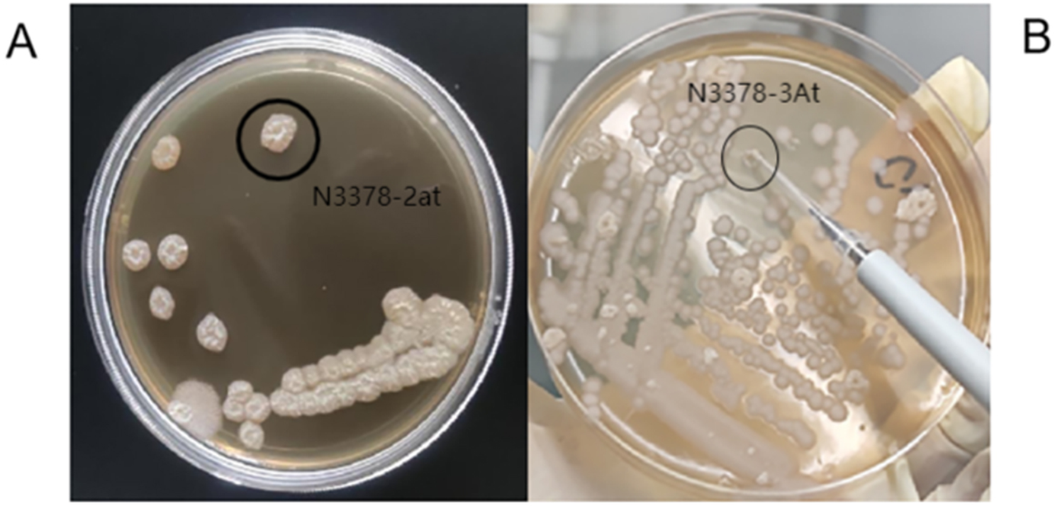
Molecular characterization: Colony fingerprinting PCRThe bacterial colony was isolated using the streak plate method. The bacterial DNA was extracted by phenol:chloroform:isomyl method as reported earlier [26] and DNA purity and integrity was confirmed by agarose gel electrophoresis (Fig. 1). DNA templates were amplified using 2 μL of forward and reverse primers, 20 μL of master mix (Taq polymerase, 10x buffer, Mg2+ ions, disH2O, Green Taq color), and RNASE free water to achieve the final reaction mixture of 40 μL. A thermal cycler was used to perform PCR amplification, which included an initial denaturation phase (95 °C for 7 min), 30 cycles of denaturation (90 °C for 30 s), annealing (40 °C for 1 min), extension (65 °C for 8 min), and a single final extension step (65 °C for 16 min). The PCR products were electrophoresed in 8% (w/v) agarose gel and the resulting fingerprints were compared directly with 1.5 kb DNA ladder under UV transilluminator after staining with ethidium bromide (Fig. 2).
Fig. 1
SDS-PAGE of the genomic DNA (gDNA): the agarose gel electrophoresis image represents the integrity of the isolated gDNA
Fig. 2
PCR gel: the agarose gel electrophoresis image indicates the integrity of amplified PCR products
Antibiotic susceptibility of probiotic productsThe disc diffusion methodology was used to perform the antimicrobial susceptibility test as reported earlier [27]. Thirty-one commercially available paper antibiotic discs with a defined concentration were employed. The results were categorized as susceptible when zone of inhibition diameter was 10 mm or more, and no inhibition zone diameter was considered as resistant [28]. The composition of the employed antibiotics is mentioned in Table 2.
Table 2 Composition of antibioticsThe enriched BHI broth was prepared and one colony from the previous streaked plate of a sample was inoculated in the broth. This broth was incubated at 37 °C ± 2 °C for 24 h. In the culture inoculated agar plate, 31 commercially manufactured paper antibiotic discs of various doses were placed. The diameter of the zone (mm) indicates the measure of the susceptibility of the isolate and the amount of drug diffused through the agar medium. Each batch of individual products was tested independently using microbiological plating and molecular techniques to determine whether the strain was resistant to antibiotics (Bacillus clausii).
Hemolytic activityThe test for hemolytic activity was performed as per the previously mentioned method [9]. Briefly, bacterial cells were cultured on Columbia blood agar base (Oxoid, Thermo Fisher Scientific, USA), supplemented with 5% (v/v) sheep blood to test their potential to cause distinct forms of hemolysis. Plates were incubated in an aerobic incubator at 37 °C. The observations were made based on the type of hemolysis, and labeled as alpha, beta, and gamma after 24 h and 72 h of incubation periods. A bacterial colony growing on agar is bordered by a greenish discoloration known as alpha hemolysis. Beta hemolysis is the complete breakdown of red blood cells hemoglobin in the presence of the bacterial colony. Gamma hemolysis is indicated by the lack of hemolysis in the area surrounding the bacterial colony. The brownish color of the blood agar plate indicates gamma hemolysis.
Species identification by 16S rRNA gene sequencing methodOne representative isolate from each fingerprinting pattern was selected for PCR amplification of the 16S rRNA gene to identify various bacterial species. The primers 27F (5′-AGAGTTTGATCCTGGCTCAG-3′) and 1492R (5′-TACGGYTACCTTGTTACGACTT-3′) were used to amplify DNA fragments of around 1 kb (equivalent to the size of the 16S rRNA gene). Template DNA, 2 μM primer concentration, and 20 μL Megamix were used to make the reaction mixture (40 μL). An initial denaturation step (94 °C, 5 min), 30 cycles of denaturation (94 °C for 30 s), annealing (58 °C for 30 s), extension (72 °C for 1 min), and a final extension phase (72 °C for 7 min) were performed in a thermal cycler. The PCR products were run on 0.8% (w/v) agarose gels, purified with genomic gel and PCR clean-up and quantified using the gene ruler marker molecular weight standard. The samples were then sent for 16S rRNA gene sequencing analysis as described earlier [22, 29].
The collected rRNA sequences were converted to FastQ format. The FastQ files containing information on the sequences of the studied areas of the 16S rRNA gene, as well as information on the reliability of reading each nucleotide, were generated as a result of the sequencing. The preliminary bioinformatic processing was performed by combining forward and backward reads, filtering sequences with low individual nucleotide readings and chimeric sequences, distributing reads based on barcode sequences, and removing technical sequences using ChromasPro version 2.1.10. The taxonomic verification was done by running the processed sequences using BLASTn [30] against the NT library. The purity of formulation for bacillus was obtained by comparing the % similarity. Phylogenetic analysis (supplementary graphs) was performed using the neighbor-joining method according to the best model identified by MEGA11 version 11.0.11 using the bootstrap test with 1000 replicates [31].
留言 (0)