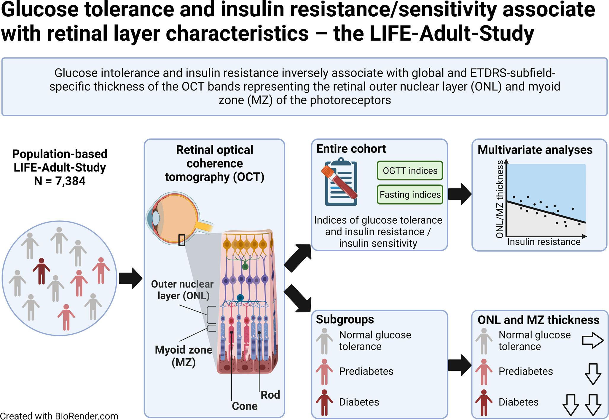
As far as we are aware, this is the first large-scale study to assess the systematic difference in retinal vessel measurements by diabetes status and the modifying effect of diabetes status on the associations between retinal vessel morphometry and key cardiometabolic risk factors. We confirm that those with diabetes tend to have wider arterioles and more tortuous venules. A key finding from this study was that BP associations with arteriolar diameter were attenuated among those with diabetes but associations of LDL-cholesterol with arteriolar diameter were stronger compared with those without diabetes, while venular tortuosity associations with TFMI, CRP, WCC and granulocyte count were stronger, and associations with HbA1c were weaker, among those with diabetes compared with those without diabetes.
Retinal vessel tortuosity and diameters by diabetes status
In the present study, those with diabetes had more tortuous venules compared with those without diabetes, and this difference was not explained by adjustment for key cardiometabolic risk factors (Table 2). Previous literature has been limited on this point, and the results have been inconsistent, with some studies showing more tortuous venules among those with diabetes [11, 35], while another study showed no difference in venular tortuosity [36] among those with and without diabetes. In agreement with the current study, the European Prospective Investigation into Cancer (EPIC) study of 5942 participants (including 238 with diabetes) showed that those with diabetes had more tortuous venules compared with those without diabetes [11]. Consistent with this, a small clinic-based study by Sasongko et al, which included 327 participants (224 with diabetes and 103 without diabetes) aged ≥18 years, also showed that those with diabetes had more tortuous arterioles and venules [35]. Our analyses adjusted for a wide range of CVD risk factors (age, sex, BP, BMI, cholesterol, triacylglycerols, anti-hypertensive and lipid-lowering medications). In contrast, a study of people of Asian Malay descent by Cheung et al showed that those with diabetes (n=594) were more likely to have straighter (less tortuous) arterioles, with no difference in venular tortuosity compared with those without diabetes (n=2141) [36]. They also adjusted for a range of CVD risk factors (sex, mean arterial BP, BMI, total cholesterol and current smoking). The discrepancies in findings may be due to the smaller sample sizes in the previous studies or to characteristics of the population (i.e. differences in diabetes duration, ethnicity, and risk factor profile, including diabetes management and history of CVD events). In the present study once those on hypertension treatment or with a history of a heart attack or stroke were removed, differences in venular tortuosity in those with compared with without diabetes were less pronounced. This is because those with advanced metabolic disease have been removed from the analysis, suggesting these changes were associated with advanced disease and events. The study differences may relate to the disease time course and duration of elevated cardiometabolic risk factors, which may differ between populations. Research has consistently shown that retinal blood flow is reduced among those with diabetes of short duration and then increases over time, possibly to maintain the microvascular integrity [37, 38]. Increased venular tortuosity has been associated with hyperglycaemia-mediated changes [14], with several studies having shown a disturbance in blood flow and loss of endothelial cells and pericytes from the vessel walls [39]. These changes may lead to loss of autoregulatory function and loss of capacity to accommodate fluctuations in hydrostatic pressure, leading to the development of diabetes-related complications [14]. This would fit with the hypothesis that mechanical instability and remodelling may be the mechanisms for the initiation and development of tortuous vessels [40].
The difference in arteriolar diameters by diabetes status observed in the present study supports the previous literature, which has consistently shown those with diabetes have wider arteriolar diameters compared with those without diabetes [36, 41,42,43]. These changes in arteriolar diameter may occur early in the disease course, as the changes have been consistently shown across studies regardless of the duration of diabetes. It is thought that hyperglycaemia and hypoxia may lead to vasodilation and early vascular changes [14]. While BP was higher, lipids were lower among those with diabetes, but arteriolar diameter remained wider in those with diabetes compared with those without diabetes after adjustment for these factors or exclusion of those on hypertension treatment or with a history of a heart attack or stroke (Table 2). Further adjustment for lipid-lowering therapy did not materially alter the findings. It may be that the full impact of this metabolic cascade cannot adequately be determined by the measurements adjusted for. The effects of LDL-cholesterol and activation of CD36/oxidised LDL receptor warrant further investigation, as it has been shown that CD36 mediates multiple pathways associated with the early pathogenesis and progression of diabetes-related complications in general [44]. In the present study, we did not observe any systematic differences in venular diameter between those with or without a diabetes. This is in contrast to previous research; for example, in the study by Cheung et al [36], those with diabetes had significantly wider venular diameters, as also observed in the study by Nguyen et al [42]. While we have adjusted for ethnicity in the present study, the majority of our participants were of European descent, while the study by Cheung et al comprised an Asian population, and that by Nguyen et al comprised a more diverse population, including white, black, Hispanic and Chinese people. Systematic differences between ethnic groups and medication use may be partially confounding these observations, but it was not possible to explore these interactions here as the number of participants of non-European descent in the present study is limited.
Retinal vessel morphology associations with cardiometabolic risk markers are modified by diabetes status
In this study, we have confirmed established patterns between retinal vessel diameter and BP, and shown that these associations were attenuated in those with diabetes compared with those without diabetes (Table 4 and electronic supplementary material [ESM] Table 1). Individuals with diabetes may have had pre-existing vascular disease (e.g. vascular sclerosis) or a breakdown in the blood/retinal barriers, resulting in defective autoregulation, which may have limited the change in arteriolar diameter in response to higher levels of BP [45]. Retinal venular tortuosity was overall negatively associated with blood lipids (total cholesterol, LDL-cholesterol and triacylglycerols) but positively associated with HbA1c, haematological indices (CRP and granulocyte count), BP (systolic BP, diastolic BP and MAP), and body composition (TFMI) (ESM Table 2), with several associations also being evident for arteriolar tortuosity. However, the associations of venular tortuosity with HbA1c, WCC, granulocyte count, CRP and TFMI in the present study were modified by diabetes status, meaning that the slopes were different for those with and without diabetes (Table 3). These differences in associations remained after exclusion of those with a history of heart attack, stroke or on medication for hypertension. Once diabetes is diagnosed, there is a higher likelihood that other comorbidities will be identified and treated. Therefore, the fact that the interactions are still seen among those not treated for hypertension implies that prompter or stricter control of high BP among those with diabetes cannot explain the effect modifications observed. As far as we are aware, this is the first study to assess the modifying effect of diabetes on a range of associations of cardiometabolic risk factors with retinal tortuosity. The fundamental message of the HbA1c venular tortuosity interaction by diabetes status may be twofold. The greater tortuosity among those with diabetes than among those without diabetes and the change in slope are indicative of a loss of autoregulation [14], and the changes in inflammatory marker associations are supportive of this. The strong positive correlation of tortuosity with HbA1c among those without diabetes suggests that the venular changes occur along a continuum, with higher HbA1c within the normal and prediabetes range being associated with increased retinal tortuosity. This linear association is consistent with the known association between higher glucose levels and the development of diabetic retinopathy. In our previous work on the diagnostic criteria for type 2 diabetes, we showed that the association of each glucose measure with retinopathy was linear in several populations, with no evidence of a threshold [46, 47]. Given that changes in the retinal microvascular architecture predict the development of diabetic retinopathy, it is not surprising to find evidence of this linear association among those without diabetes [48]. Once you have diabetes, it appears that the degree of glycaemic control does not influence venular tortuosity much; perhaps the venular tortuosity damage has already been done by the time of clinical diagnosis. In contrast, for venular diameter, the association with HbA1c continues right through the range of glucose values.
Strengths and limitations
As far as we are aware, the present study is the largest to assess the impact of diabetes on retinal vessel morphometry. Although the present study identified novel associations with retinal tortuosity and confirmed the associations for diameters between those with and without diabetes in terms of absolute differences and CVD risk marker interactions, it is a cross-sectional study. Further research is needed at scale using longitudinal data to determine whether these associations can be replicated, particularly given the mixed findings between cross-sectional and longitudinal studies to date [14]. In particular, research on a large scale to determine the longitudinal impact of trajectories of cardiometabolic risk factors (risk factors that are also key risk factors for diabetes-related complications) on retinal morphology and the development of diabetes-related complications is required if we are to develop risk prediction tools to identify those at high risk of developing diabetes-related microvascular complications within a 5-year window, allowing time for a suitable intervention to be implemented. The QUARTZ software is fully automated, incorporates convolutional neural network (CNN) technology and uses information from all vessels extracted within an image, providing precise measurement. Previous grading systems have only used a section of the retinal image for grading; however, given that our findings are consistent with previous literature, this is unlikely to be a major issue.
Conclusion
We provide clear evidence of the modifying effect of diabetes on the retinal microvasculature. These observations are indicative of preclinical disease processes, and may be a sign of impaired autoregulation due to hyperglycaemia, changes that have been suggested to play a pivotal role in the development of diabetes-related microvascular complications. Longitudinal investigation on a large scale to determine the usefulness of these non-invasive measures as predictors of diabetes-related complications is warranted.


留言 (0)