Study design
This was a single-center, retrospective study conducted at the Xinqiao Hospital, Army Medical University (Third Military Medical University) in Chongqing, China. We consecutively enrolled patients diagnosed with DDVT by compression ultrasonography (CUS), from January 1st to December 31st, 2020. The inclusion criteria were: 1) hospitalized patients with age ≥ 18 years; 2) diagnosed with IDDVT by CUS; 3) with at least one CUS follow-up examination. Patients were excluded if they presented concomitant PDVT or PE, without follow-up CUS, outpatients, significant bleeding at admission, indication for long-term anticoagulation for other reasons, and lost to follow-up. The present study complied with the Declaration of Helsinki and was approved by the ethics committee of the Xinqiao Hospital, Army Medical University (Third Military Medical University). Written consents were waived for its retrospective design by the committee. Patients were divided into two groups, those who received any kind of anticoagulation drugs [anticoagulation (AC) group], and those managed without anticoagulation drugs [non-anticoagulation (non-AC) group].
Compression ultrasonography (CUS)
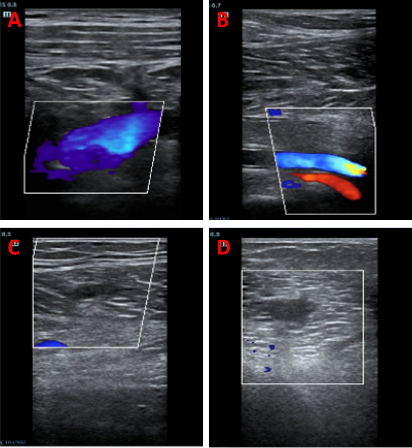
Baseline and follow-up ultrasounds were performed at the diagnostic unit or bedside by trained sonographers according to a standardized protocol that included a complete whole-leg ultrasonography [18]. Briefly, patients first laid in the supine position, and the proximal deep veins in the lower extremities were continuously imaged along the veins’ course in the transverse plane with a linear probe (5–10 HZ), including the common femoral, superficial femoral, deep femoral, and popliteal veins. Then, the patients took a seated position with the lower legs hanging down, and the distal deep veins were sequentially examined in this position, including the posterior tibial, peroneal, gastrocnemius, and soleus veins. Anterior tibial veins were not imaged since they were difficult to be compressed and partially obscured by the interosseous membranes and bones. If patients could not change between body positions, CUS was performed just in the supine position. DVT was diagnosed when a filling defect and any lack of compressibility of the deep venous segments were detected. Complete thrombosis resolution during follow-up was defined as no filling defect and complete compressibility restored in segments that were initially involved in the thrombosis by CUS.
Data collection
The demographic information, medical history, and examination results of participants were collected from electronic medical records. The following data were retrieved: diagnosis date, examination frequencies, and CUS follow-up results. Risk factors for DVT were also documented, including bedridden for more than three days, recent surgery or trauma, pregnancy, family history of venous thromboembolism, congestive heart failure (CHF), stroke, paraplegia, and malignancy history. Other data, such as hypertension, diabetes mellitus (DM), renal insufficiency (RI), hepatic insufficiency (HI), intensive care unit admission (ICU), sepsis, acute myocardial infarction (AMI), and chemo-radiotherapy history, were also collected. The first results of white blood cell (WBC) count, hemoglobin, platelet (PLT) count, serum creatine, and D-dimers at admission were recorded as the baseline data. Meanwhile, the status, drugs, dosage, and duration of anticoagulation therapy during hospitalization and after discharge were also collected.
Endpoints and follow-up
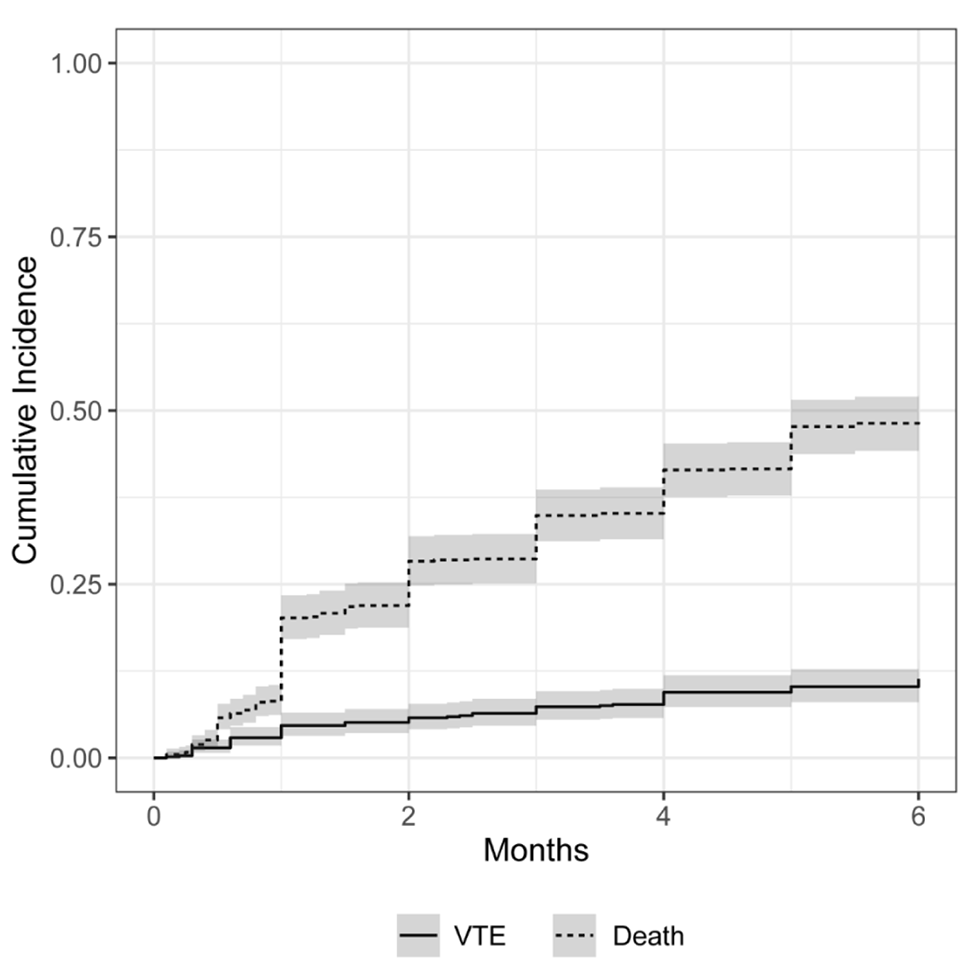
The primary endpoints of this study were: 1) thrombosis extension into PDVT/PE, and 2) all-cause mortality. Thrombosis extension was defined as imaged-confirmed thrombosis extension to any of the popliteal, femoral, iliac, and cava veins, regardless of whether the patients had clinical symptoms or not. PE was diagnosed according to a recent guideline [19]. The secondary endpoints were: 1) complete thrombosis resolution, and 2) bleeding, including major or minor bleeding events. Participants were followed up through telephone, clinic interviews, and medical records. We also collected CUS data and computed tomography pulmonary angiography from the medical records at other institutions or hospitals, including results during admission, and outpatient visits.
Statistical analyses
The sample size was calculated using PASS 15 Software (NCSS, LLC. Kaysville, Utah, USA). Referring to the published reports, we assumed the incidence of primary outcome (thrombosis progression to PDVT or PE) is 10% and 3% in IDDVT patients without and with anticoagulation, respectively [5, 7, 20]. The sample size was estimated to be 404 (with 135 in non-AC group and 269 in AC group, respectively), achieving 80% power to detect a difference between the group proportions with significant two-sided level of 0.05.
In unmatched cohorts, continuous variables with normal distribution are presented as means ± standard deviations or medians with interquartile ranges. Comparisons between two samples were performed using t-tests or Wilcoxon rank-sum tests. Categorical variables are presented as frequencies and percentages and were compared with χ2 or Fisher’s exact tests.
Propensity-score matching (PSM) was performed according to the method to control the measured confounders between the non-AC and AC groups [21]. Briefly, a propensity score was generated for each patient using multivariable binary logistic regression analysis. Variables with initial clinical relevance and those with statistical significance in the univariate analysis (p < 0.10) were included. The non-AC group was matched with the AC group based on the propensity score in a 1:1 ratio, with the Nearest Neighbor Matching algorithm and a caliper of 0.05. After PSM, distribution differences of baseline covariates were assessed through the method proposed by Peter C. Austin and were described as standardized differences [22]. A cutoff < 0.10 indicated well matching between treatment groups.
The time-to-event rates for each group were estimated using the Kaplan–Meier method and compared with log-rank tests in matched cohorts. Moreover, both in unmatched and matched cohorts, a multivariable Cox proportional hazards model analysis was performed to calculate the hazard ratio (HR) and 95% confidence intervals (CIs) to explore the association of anticoagulation and other risks factors with the primary endpoints. Variables considered to be clinically relevant or that showed statistical significance in the univariable analysis (p < 0.10) with the primary endpoints were included in the multivariable regression model. A p < 0.05 indicated significant differences between measurements. All analyses were performed using SPSS version 22.0 (IBM Corp., Armonk, NY, USA), and the PSM also required the R software (v. 2.15.3, R Foundation for Statistical Computing), SPSS R plug-in (SPSS Statistics Essentials for R 22.0.0, IBM, USA), and PS Matching in SPSS (version 3.04, SourceForge, San Diego, USA).


留言 (0)