
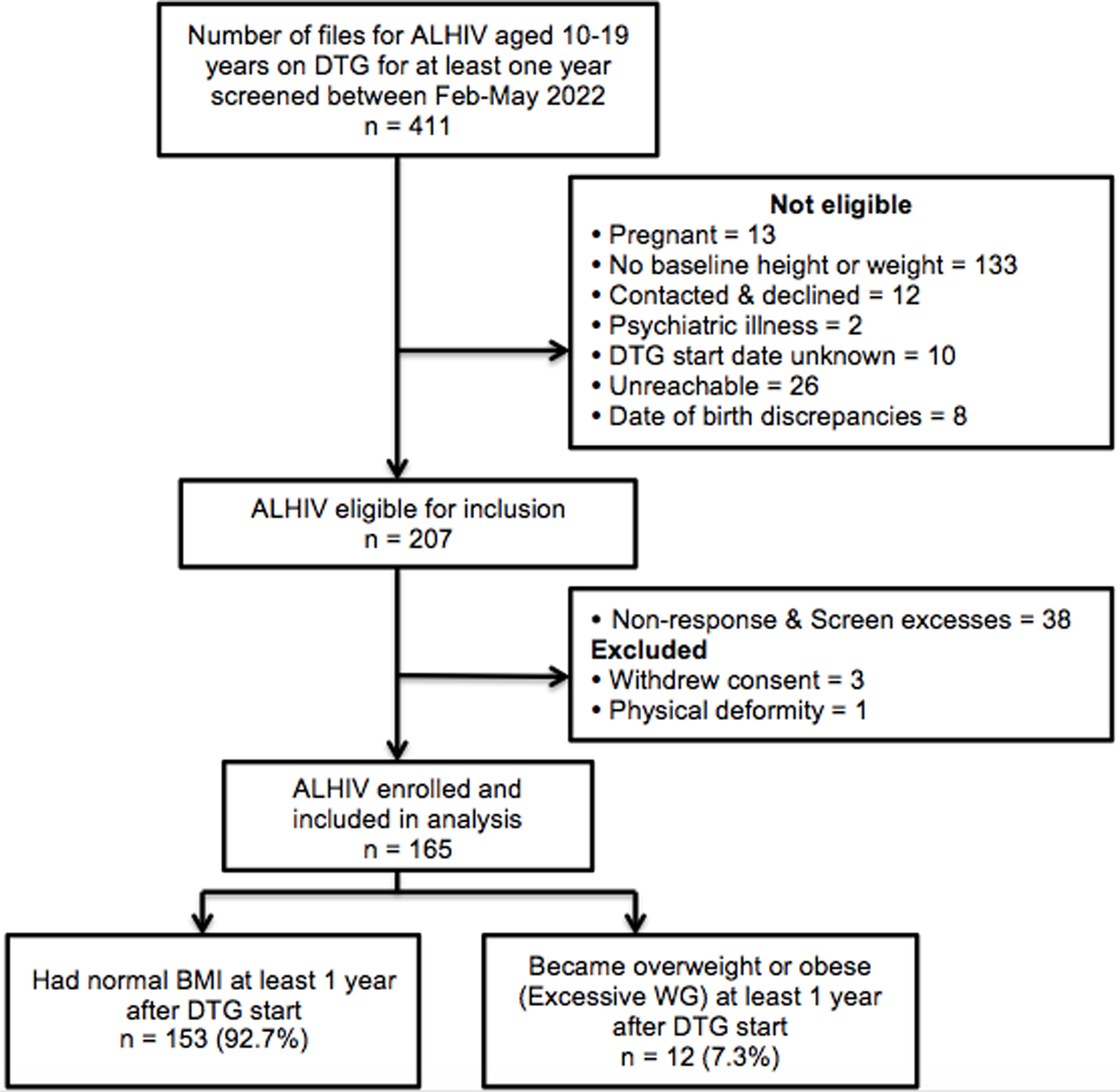
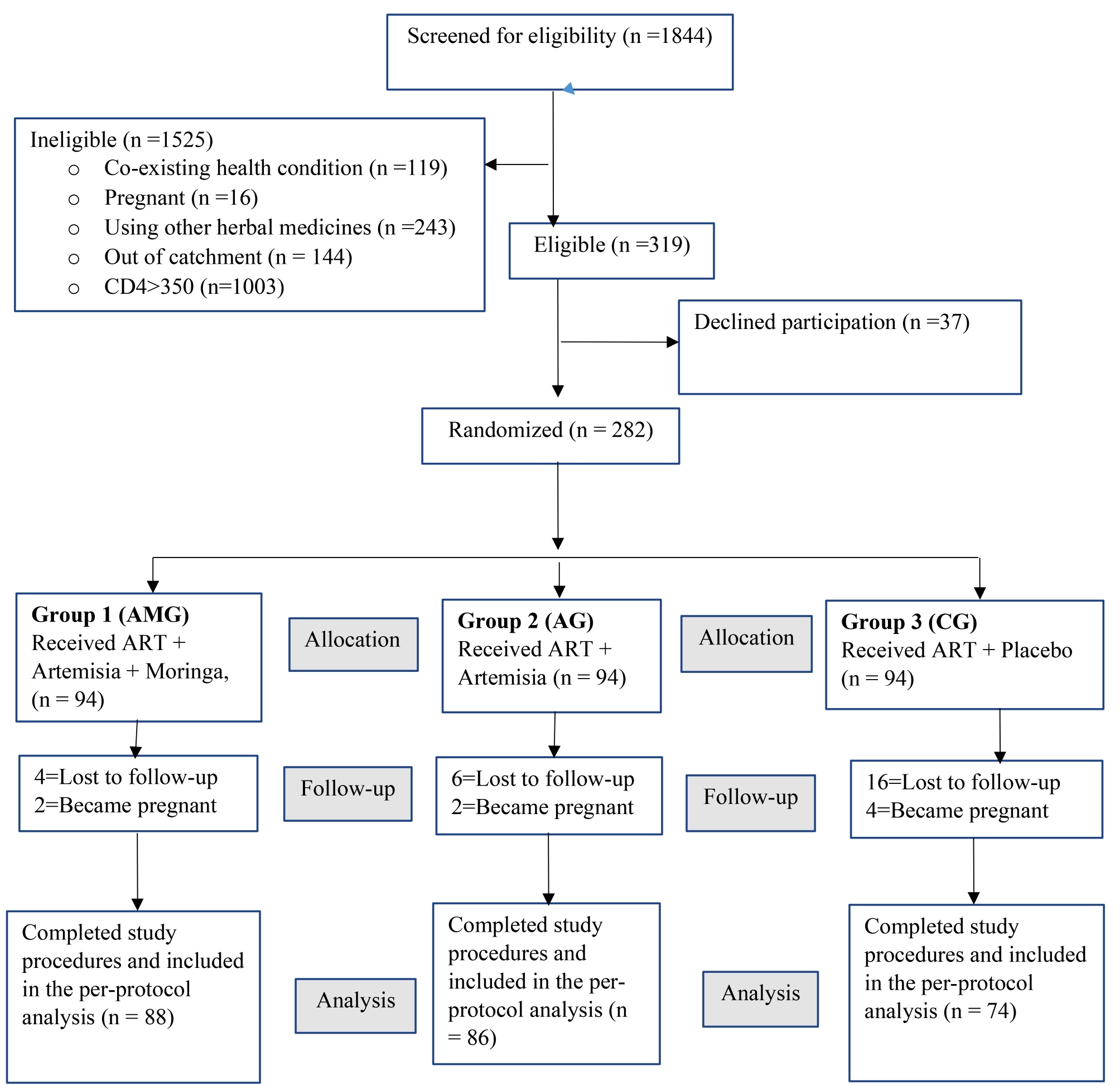
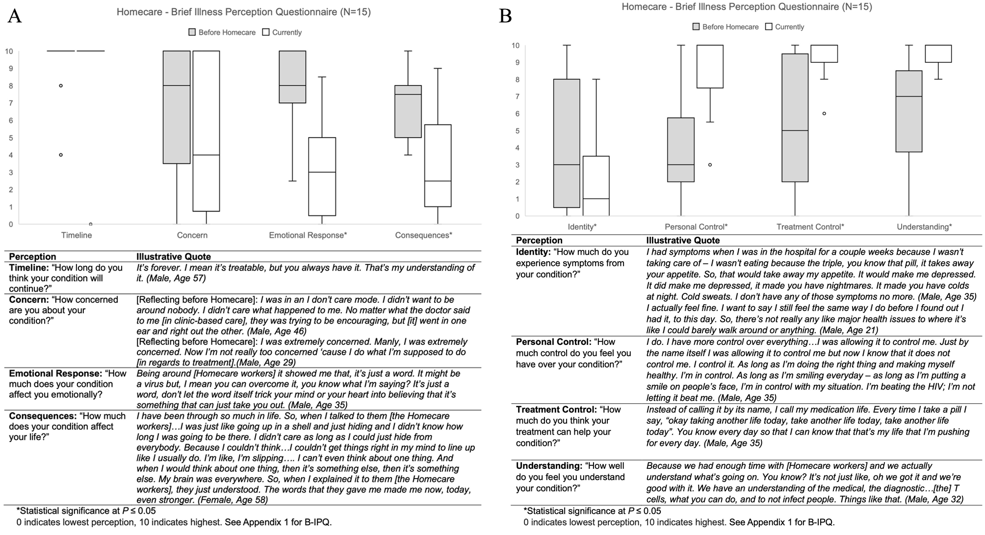
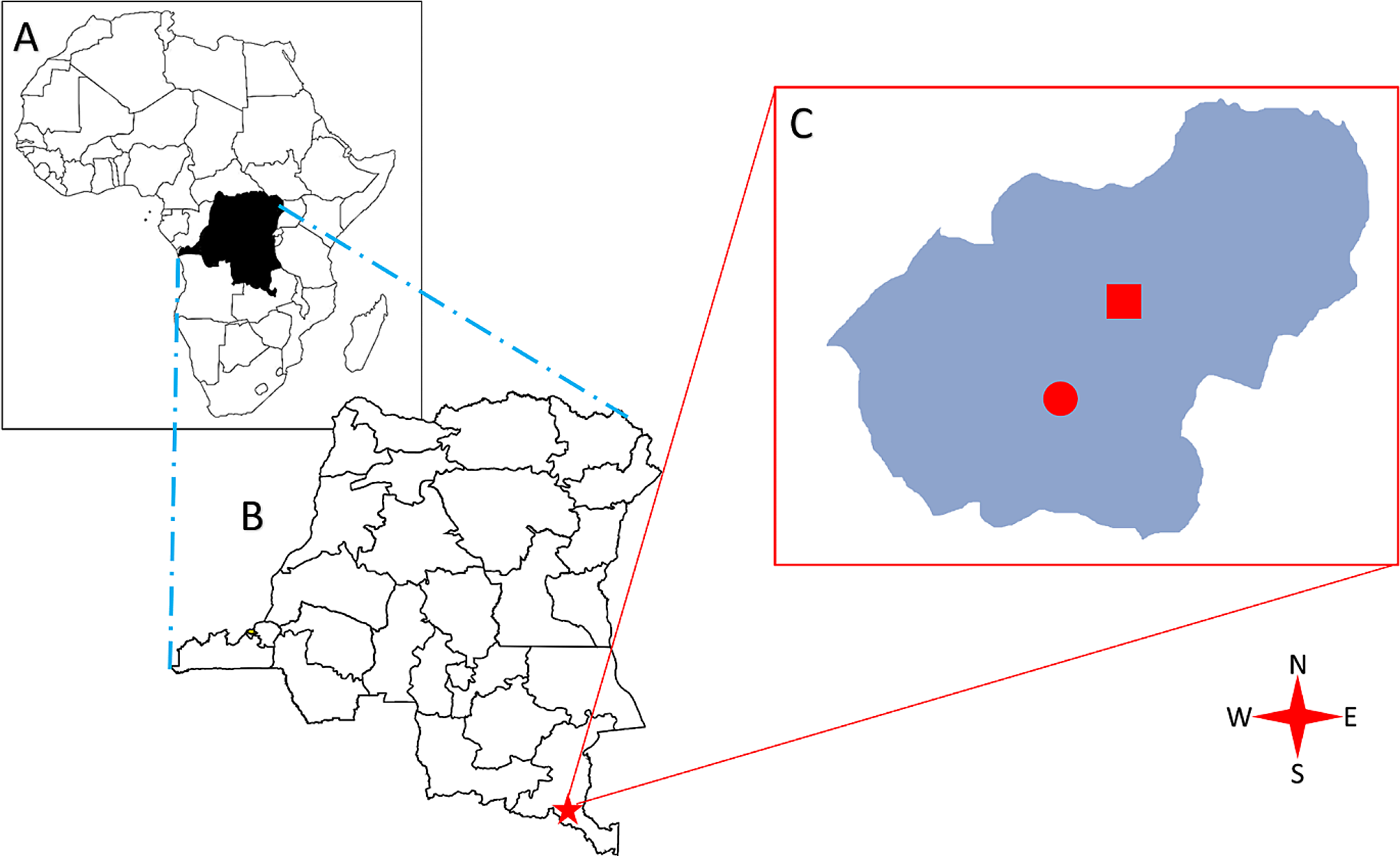
記住我

The patient is a 47-year-old, married, male construction worker. The patient was treated at a hospital in Chongqing on May 10, 2021 due to cough, shortness of breath, and other discomfort and was accordingly diagnosed with pneumocystis carinii pneumonia (PCP) and AIDS. His baseline CD4+ T-cell count and HIV-RNA level were unknown. After anti-PCP treatment (with compound sulfamethoxazole tablets combined with prednisone), his symptoms were relieved. Thereafter, antiretroviral therapy (ART), including lamivudine (3TC), tenofovir (TDF), and efavirenz (EFV) was initiated. He took the ART and compound sulfamethoxazole tablets regularly.
He was hospitalized on August 19, 2021, due to ghosting vision for > 2 months and anhidrosis on the left head and face for the past 20 days. Over 2 months before hospital admission, the patient experienced bilateral ghosting vision and limited rotation of the left eye without any obvious induction. Moreover, he had no fever, dizziness, headache, tinnitus, decreased hearing and vision, visual rotation, syncope, disturbance of consciousness, nausea, vomiting, incontinence, abdominal pain, diarrhea, or other discomforts. Accordingly, the patient was treated at a hospital in Chongqing. His cranial MRI revealed multiple intracranial abnormal enhancement signals with edema. Furthermore, routine examination and biochemical detection of the cerebrospinal fluid indicated minimal abnormalities. He was diagnosed with TE and AIDS. After treatment with azithromycin combined with compound sulfamethoxazole tablets against Toxoplasma gondii for 2 weeks, the patient reported slight improvement in his ghosting vision. Then, he was discharged without any imaging re-examination and was continued on the original anti-Toxoplasma gondii treatment after discharge. Then, 20 days before his readmission (the patient had been receiving continuous anti-Toxoplasma gondii treatment until his hospitalization in our hospital), the patient still had anhidrosis on the left head and face. In addition, his bilateral ghosting vision due to unknown causes had worsened, accompanied by limited rotation of the left eye, slight cough, and less sputum production. He had no tinnitus, decreased hearing and vision, visual rotation, blackness, epilepsy, disturbance of consciousness, or other discomforts. His brain MRI re-examination in other hospitals indicated that the intracranial lesions had increased significantly. The patient was subsequently transferred to our hospital for further treatment.
The results of the patient’s physical examination on admission were as follows: normal vital signs and a clear mind, he answered to the point; the bilateral pupils were equal in size and circular-shaped, showing sensitivity to light reflection; the left eye had external rotation disorder, but the right eye had normal activity; his lungs had clear respiratory sound, without rhonchus and moist rales; no abnormalities were detected upon cardiac and abdominal physical examination; the muscle strength and tension of the limbs were normal; pain and temperature sensation were normal, and bilateral pathological signs were negative. The results of his laboratory examination were as follows: cranial MRI showed patchy mixed long T1 and T2 signals in the bilateral frontal lobes and mixed high signals in flair, surrounded by massive edema. Enhanced scanning revealed that multiple intracranial lesions were circular, nodal, and enhanced, and the intracranial pia mater was slightly thickened. His chest CT indicated that the lungs were scattered with patchy, strip-shaped, nodule-like, acinar nodular increased shadows with uneven density and some unclear boundaries. His routine blood test results were as follows: white blood cells 3.08 × 10⁹/L, neutrophils 2.29 × 10⁹/L, and cerebrospinal fluid: pressure 140 mmHg, routine examination of the cerebrospinal fluid revealed a positive result for Pandy’s test. His cerebrospinal fluid biochemistry was as follows: total protein 498.77 mg/L, microalbumin 360.7 mg/L, β2 microglobulin 5.26 mg/L, glucose 3.41 mmol/L, and chloride 128.8 mmol/L. The following test results were obtained: CD4+ T-cell count 57 cells/µL, CD8+ T-cell count 322 cells/µL, CD4/CD8: 0.18; Toxoplasma gondii IgG antibody in the blood: positive, Toxoplasma gondii IgM antibody and DNA: negative, galactomannan (GM) test in the blood, (1,3)-β-d glucan (BG), Talaromyces marneffei antigen, Cryptococcus neoformans antigen, latent tuberculosis infection, and cytomegalovirus DNA: negative. Cerebrospinal fluid staining with India ink, cryptococcal antigen, cytomegalovirus DNA, tuberculosis fluorescence staining, tuberculosis sandwich cup, bacterial and fungal cultures: were all negative. The primary diagnosis was: (1) TE(?), and (2) AIDS. The patient continued to receive 3TC/TDF/EFV anti-human immunodeficiency virus (anti-HIV) treatment and azithromycin combined with compound sulfamethoxazole against Toxoplasma gondii. After 1 week of treatment, the symptoms of ghosting vision and facial anhidrosis did not improve. Per a past report, patients with AIDS complicated with TE have shown significant curative effects after regular treatment for 2–4 weeks [7]. Therefore, we wondered whether TE was erroneously diagnosed. For precise diagnoses, the cerebrospinal fluid was collected for mNGS examination (RDP-seq®, Guangzhou Sagene Biotechnology Co., Ltd.), and the results indicated no possible pathogenic bacteria, such as Toxoplasma gondii, CMV, and Cryptococcus. Then, tNGS examination was performed on the cerebrospinal fluid sampled previously (Guangzhou Sagene Biotechnology Co., Ltd.). The results indicated Aspergillus fumigatus (number of sequences: 1). CNSAG was not diagnosed immediately as the number of t-NGS sequences in the cerebrospinal fluid was extremely low. Considering that Aspergillus fumigatus usually invades the lungs and that lesions were present in the patient’s lungs, fiberoptic bronchoscopy was immediately performed. The bronchoalveolar lavage fluid (BALF) was collected for mNGS examination (RDP-seq®, Guangzhou Sagene Biotechnology Co., Ltd.), and the results showed a large number of Aspergillus fumigatus (number of sequences: 5842, the degree of confidence: 99%) (Fig. 1).
Fig. 1
The mNGS test results for bronchofiberscope alveolar lavage fluid
In addition, the BALF-GM test result was positive. Based on the patient’s symptoms, signs, laboratory tests, and past treatment reactions, the diagnosis was corrected to CNSAG, invasive pulmonary aspergillosis (IPA), and AIDS. Voriconazole (intravenous drips of 6 mg/kg/day q12-h on the first day, followed by intravenous drip of 4 mg/kg/day q12-h) was administered for antifungal treatment. Due to the interaction between voriconazole and EFV, the antiviral treatment regimen was revised to 3TC, TDF, and dolutegravir (DTG). After 2 weeks of this treatment, the patient’s ghosting vision and facial anhidrosis were relieved. The routine examination of cerebrospinal fluid revealed normal biochemistry. His cranial MRI indicated that the lesions in the frontal lobe were absorbed, and the surrounding edema was alleviated (Fig. 2). The patient was discharged and continued on oral voriconazole treatment (200 mg/kg/day). After 6 weeks of antifungal treatment, the patient's ghosting vision and facial anhidrosis were significantly relieved. Re-examination of his cranial MRI indicated that the lesions in the frontal lobe were significantly absorbed and improved, and the surrounding edema was alleviated (Fig. 2). His chest CT showed focal absorption (Fig. 3).
Fig. 2
Enhanced scanning of cranial MRI A before treatment: multiple intracranial circular and small-nodular enhanced lesions, as indicated by the arrow, B two weeks after treatment C six weeks after treatment, B and C indicate that the intracranial lesions were absorbed and improved compared with those before treatment
Fig. 3
Chest CT A Before treatment, the lung tissues seemed scattered with patchy, strip-shaped shadows, nodule-like and increased acinar nodular shadows with uneven density and some unclear boundaries were noted. B After 6 weeks of treatment, chest CT showed that the lesions were resolved
留言 (0)