The endometrium is a unique mucosal microenvironment under the control of pregnancy-associated hormones. The fate of the endometrium is to prepare the uterus for embryo implantation and support pregnancy when the embryo arrives; otherwise, menstruation occurs (Critchley et al., 2020). The endometrium can only accept embryos within a limited period called the window of implantation (WOI), which occurs in the mid-secretory phase (Zhang et al., 2013). On arrival at the appropriate implantation site, the blastocyst begins to adhere and initiates communication with the endometrium, establishing the first maternal-embryonic interface (Ye, 2020). Decidualization is the landmark event for embryo implantation, transforming endometrial stromal cells into specialized decidual cells that provide a nutritive and immune-privileged matrix for trophoblast syncytialization and placental development (Gellersen and Brosens, 2014).
The infiltrated immune cells in the endometrium show a synergistic dynamic pattern during the menstrual cycle (Wang et al., 2020), of which macrophages play an essential role in rebuilding the endometrium in the nonpregnant cycle (Critchley et al., 2020). During embryo implantation, macrophages coordinate with decidualization (Erlebacher, 2013). The role of decidual macrophages during pregnancy, including the functions in aiding in remodeling of the spiral arteries and trophoblast invasion, polarization to maintain the maternal-fetal tolerance, interactions with other cell types, the balance of immune tolerance and infection prevention, related pregnancy complications and possible therapeutic targets have been comprehensively reviewed elsewhere (Faas and De Vos, 2017, Ander et al., 2019, Jena et al., 2019, Yao et al., 2019). However, a review of the dynamics of endometrial macrophages before pregnancy and the possible ripple effect this may have on subsequent pregnancies is still imperative. Here, we summarize the evidence on how endometrial macrophages change during the menstrual cycle, how they contribute to the pregnancy, the transition of endometrial macrophages at the very beginning of pregnancy, and the endometrial macrophages involved in pregnancy complications, as well as pregnancy-related diseases.
In human endometrium, CD68+ or CD163+ macrophages constitute 1–2% of total endometrial cells in the proliferative phase (Salamonsen et al., 2002, Russell et al., 2011), and then 1–5% in early-to-mid secretory phase (Salamonsen et al., 2002, Russell et al., 2011, Diao et al., 2020, Zhao et al., 2020), next dramatically up to 7% in late secretory phase (Russell et al., 2011), and finally 6–15% pre-menstrual phase (Salamonsen et al., 2002, Russell et al., 2011). In addition, macrophages can polarize into M1 (classically activated macrophages) and M2 (alternatively activated macrophages) depending on the microenvironment. The M1 macrophages are usually identified by the markers nitric oxide synthase 2 (NOS2/iNOS), suppressor of cytokine signaling 3 (SOCS3), indoleamine 2,3-dioxygenase 1 (IDO1), CD80 or CD86. Conversely, the M2 macrophages are identified by CD163, mannose receptor C-type 1 (MRC1, CD206), or interleukin 10 (IL10). In addition to the M1/M2 polarization, more subpopulations of macrophages are characterized by other grouping methods (Mosser and Edwards, 2008). The frequency of IL-10-iNOS+CD68+/CD68+ M1 macrophage is significantly higher in the proliferative phase than in the secretory phase, while M2 is vice versa (Tsao et al., 2018) (Fig. 1).
Endometrial macrophage is likely to play an essential role during the menstrual cycle, especially in the menstrual context of tissue degradation, which requires regulated repair, regeneration, and phagocytic clearance of endometrial tissue debris to reestablish tissue integrity in preparation for pregnancy. The significance of macrophages in the menstrual cycle for the human endometrium has been elaborated by several elegant works in the literature (Salamonsen and Lathbury, 2000, Salamonsen et al., 2002, Thiruchelvam et al., 2013, Critchley et al., 2020). This review only focuses on the pregnancy-related function and regulation of endometrial macrophages and their interactions with other cells in the endometrium for pregnancy preparation.
During the proliferative phase, the development of the endometrial glands lays the foundation for subsequent pregnancy. In women under 35 years of age with idiopathic infertility, inflammation-related cytokine profiles of uterine perfusions in the proliferative phase were significantly elevated compared to those of fertile women (Fitzgerald et al., 2016). Improvement of the inflammatory endometrial microenvironment involving proliferative macrophages is a therapeutic target. Endometrial regeneration and fertility restoration are expected to be achieved by exosome-filled scaffold design, where exosome-induced macrophage polarization reduces inflammation and increases anti-inflammatory response (Xin et al., 2020).
However, controversy is still ongoing regarding the determination of excessive inflammation and whether anti-inflammatory treatment contributes to a successful pregnancy. For example, an injury-induced inflammation in the proliferative phase favors pregnancy outcomes in IVF patients. A prospective study showed that endometrial biopsy on both proliferative and mid-secretory phases in a spontaneous menstrual cycle before IVF treatment doubled the chance for a take-home baby (Barash et al., 2003). Biopsies from proliferative phase of the same menstrual cycle increased the percentages of HLA-DR+CD11c+ macrophages/dendritic cells in leukocytes in the secretory phase, as well as the levels of the pro-inflammatory cytokines TNF-α, growth-regulated oncogene α (GROα) also name CXCL1, IL-15, and macrophage inflammatory protein 1B (MIP-1β), in favor of pregnancy outcomes (Gnainsky et al., 2010) (Fig. 2). These HLA-DR+CD11c+ cells were a mix of dendritic cells and macrophages, and it was not sure whether all these macrophages were M1 polarized since both HLA-DR and CD11c were also found expressed on M2 macrophages (Shaul et al., 2010, Buchacher et al., 2015). However, the changes in total macrophages and their M1/M2 polarization ratios after biopsies remain unknown. In vitro study showed that TNF-α stimulated primary endometrial stromal cells to express monocyte-attracting cytokines to induce the differentiation of monocytes into macrophages and DCs (Gnainsky et al., 2015). In freshly isolated proliferative endometrial stromal and epithelial cells, a conditioned medium of monocyte-derived macrophages upregulates implantation-associated genes, including carbohydrate sulfotransferase 2 (CHST2), CCL4 (MIP1B), and CXCL1 (GROα) (Gnainsky et al., 2015).
Macrophages may also contribute to stemness or be influenced by stromal cell senescence associated with implantation outcomes. Women with implantation failure exhibited a significantly higher proportion of senescent cells, higher senescence-associated secretory gene expression, and lower endometrial stromal cell stemness during the proliferative phase than the successful group (Tomari et al., 2020). The ability of macrophages to regulate stromal cells has been reported in other fields. Macrophages promote bone regeneration through crosstalk with mesenchymal stem cells in bone healing (Pajarinen et al., 2019). They are recruited to support the stemness of the cancer cells during tumorigenesis (Fang et al., 2017).
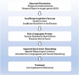
Prediction of implantation outcomes or pregnancy outcomes by endometrial examination during the secretory phase is considered feasible and reasonable (Craciunas et al., 2019). The commercial products Endometrial Receptivity Analysis (ERA) and Endometrial Function Test (EFT) are based on the mid-secretory phase endometrial examination results. They have been used for decision-making before embryo transfer for years (Díaz-Gimeno et al., 2011, Kliman and Frankfurter, 2019). A recent clinical study shows that the percentage of CD68+ and CD163+ macrophages in the mid-secretory endometrium and embryo transfer strategy is significantly associated with the first IVF-ET treatment (Diao et al., 2020). A similar study showed that the ratio of CD206+ M2-like macrophages to CD68+ pan-macrophages in endometrial biopsies was reduced considerably, and TNF-α mRNA expression was significantly increased in the pregnancy failure group compared with the pregnancy success group (Ono et al., 2020). These data suggested that the decreased M2-like macrophage ratio and increased total macrophages are harmful for embryo implantation. Dysregulated macrophages are associated with miscarriage. CD68+ macrophage density and the clustering level between CD68+ macrophages and CD56+ NK cells were significantly increased in mid-secretory endometrium of recurrent miscarriage women compared with fertile women (Zhao et al., 2020) (Fig. 2).
Seven macrophage chemoattractants are among the highly abundant chemokines in the endometrium. They are upregulated in the mid-secretory phase and maintained until early pregnancy. Their localization indicates a complex spatio-temporal regulation on endometrial macrophages preparing for endometrial receptivity and the coming pregnancy (Jones et al., 2004). Macrophage colony-stimulating factor (CSF-1) is expressed by both endometrial stromal cells and endometrial epithelial cells during the menstrual cycle and pregnancy (Hatayama et al., 1994). IL34 and CSF-1 were shown to share a common receptor, CSF-1R. IL-34 favors the differentiation of circulating monocytes to the M2 subtype instead of M1, whereas CSF-1 mainly promotes M1 differentiation (Muñoz-Garcia et al., 2021). In the mid-secretory phase, human uterine endometrial tissues exhibited higher N-glycosylation than in the proliferative phase, particularly in the epithelium. N-glycosylation of endometrial cells contributed to the successful implantation in mouse experiments. N-glycosylation of LIF receptor (LIFR) played a crucial role in LIF‐induced receptivity (Yu et al., 2020). The endometrial RNA expression of LIF during embryo implantation in successful IVF patients was 12.25 folds higher than those in failed (Camargo-Díaz et al., 2017). Inhibition of N-glycosylation decreased M2 polarization and related chemokine CCL22 in bone marrow cells (Jha et al., 2015), which suggested a possible regulation of macrophages for pregnancy.
Beyond the cytokines, some macrophage-related molecules expressed in the secretory phase are closely related to pregnancy. SPP1 is the most highly upregulated endometrial gene during the mid-secretory phase in humans (Carson et al., 2002). In mice, Spp1 is also a marker of endometrial receptivity (Casals et al., 2010). Endometrial macrophages might pave the way for implantation via the expression of Spp1. It is expressed by a subpopulation of macrophages embedded within the stroma of both cyclic and pregnant mice (White et al., 2006). The induction of monocyte chemoattractant protein-1 (MCP-1) and MIP-1β mediated this effect in the context of clinical rheumatoid arthritis (Zheng et al., 2009). Relaxin is a significant endometrial factor in preparing the endometrium for early pregnancy and may favor implantation via endometrial macrophage migration or differentiation regulation. It selectively increases the concentration of local immune cells required for implantation, including CD68+ macrophages (Goldsmith and Weiss, 2009). Relaxin is secreted predominantly by endometrial epithelial cells during the menstrual cycle. Relaxin and its receptor RXFP1 (LGR7) are likely to peak during the secretory phase, responsible for endometrial vascular remodeling and response to steroid hormones (Marshall et al., 2017). Relaxin in tumors can suppress macrophage migration and regulate M1/M2 cytokine profiles (Figueiredo et al., 2009).


留言 (0)