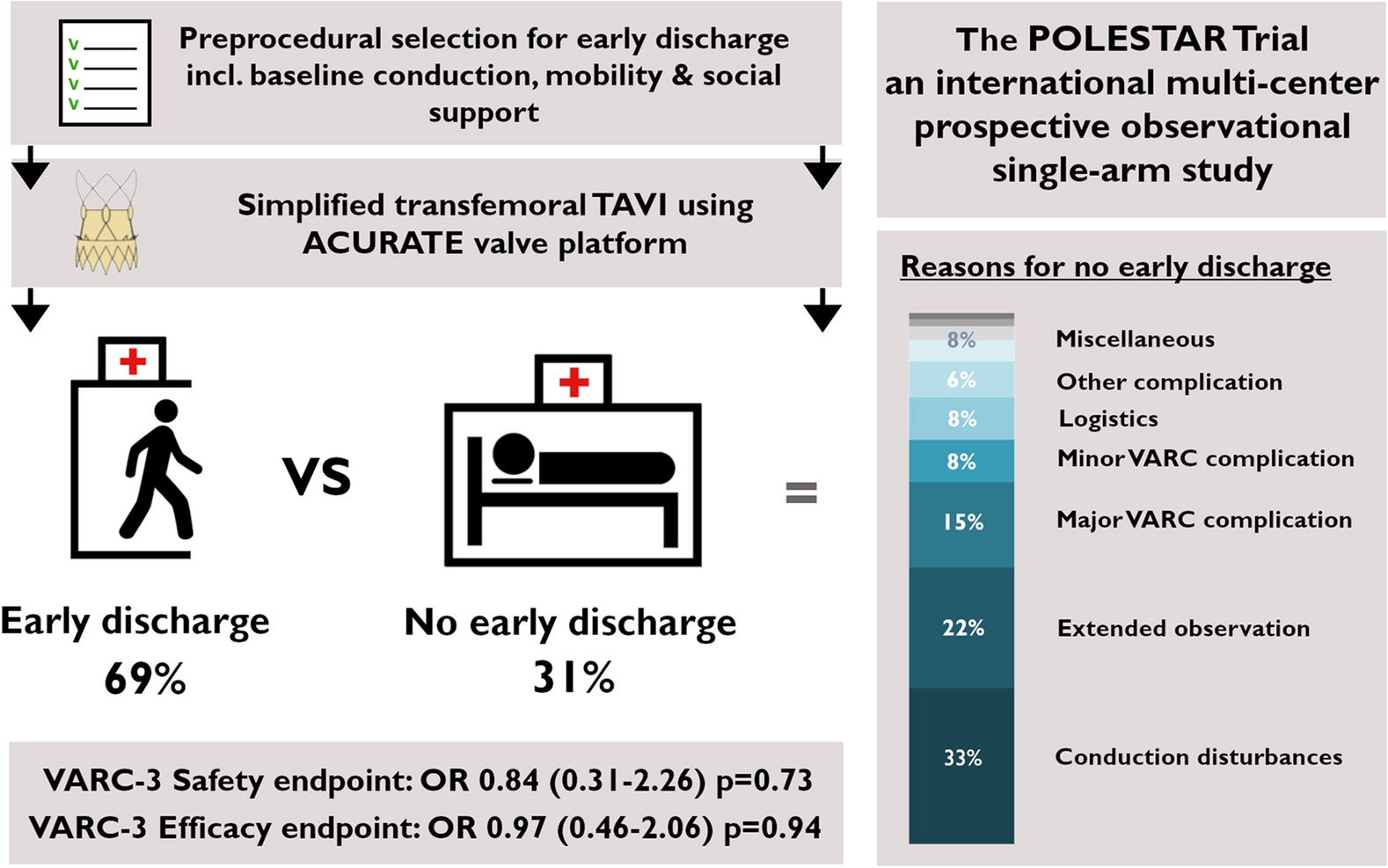
This study evaluated the incidence of vascular access site complications following transradial coronary angiography in a large cohort of unselected patients including a significant proportion of patients undergoing complex coronary intervention with large-bore access. Key finding was that the contemporary radial occlusion rate was with 4.6% lower as compared to many previously reported results [8, 11, 15]. Another key finding was that the incidence of other relevant complications was similarly low and, in particular, relevant peripheral neurologic or major bleeding even did not occur. These excellent results might be explained by the use of modern hydrophilic sheaths, improved puncture techniques or operator experience, and a standardized radial compression procedure.
Radial patency was evaluated on the day following coronary angiography. This early point of time was chosen since the majority of patients were discharged on this day. In previous studies, radial artery patency was evaluated from 24 h up of 30 days following the procedure [8, 13]. Even if direct comparison of different cohorts has major limitations, it appears if the incidence of radial occlusion might have been higher in studies with early testing as compared to studies with a later evaluation of this endpoint [10]. The overcome this potential limitation in the present study, patients diagnosed with radial artery occlusion were scheduled for a clinical follow-up demonstrating that the occlusion persisted in two of three patients despite intensified anticoagulation. This could indicate that rather traumatic than thrombotic factors might be the key mechanism leading to postprocedural radial occlusion.
In the present study, all patents were screened by duplex ultrasonography regardless of symptoms. This is an important feature of this study and might explain differences as compared to previous data. Earlier studies have infrequently reported even lower radial occlusion rates of less than 4%. However, these were mainly retrospective analyses with lower sample sizes. In some analyses, only symptomatic patients were apparently sent for ultrasound examination [16]. Even recent randomized trials demonstrating the superiority of the radial approach did only focus on clinical overt complications and did not screen for this complication [6]. As demonstrated in the present study, the majority of patients with radial occlusion were asymptomatic and patients with or without palpable pulse were found in both groups with or without radial occlusion. Thus, some asymptomatic patients with probably persistent occlusion might have been missed in previous studies. Apart from the routine ultrasound-based screening, further key features of the present study are the large sample size, which is one of the largest cohorts for this setting published so far, the consecutive enrollment of patients, which reduces the potential for selection bias, as well as the analysis of multiple variables that might impact on the incidence of radial occlusion. Given the enrollment in a tertiary care center, patients representing the whole spectrum of coronary diagnostics and interventions could be enrolled starting from the simple diagnostic angiography of young patients with planed valvular surgery to very complex and time-consuming coronary interventions needing large-bore access.
Overall, patients diagnosed with radial occlusion were more often female, had a lower prevalence of arterial hypertension and less frequently a history of coronary heart disease, which might be explained by their younger age. However, these patients were more often active smokers, demonstrated less frequently an obstructive coronary artery disease needing revascularization and had higher creatine kinase levels, which might point to a higher level of physical activity.
Young age has already previously been identified as predictor for radial artery occlusion [10, 11]. This might be explained by the higher sympathetic reactivity in younger individuals leading to a higher risk for vascular spasm as compared to elderly individuals with more atherosclerotic changes of the vascular wall and decreased sympathetic tone. A recent study assumed that higher age might be associated with ischemic pre-conditioning leading to larger artery diameter, based on the finding that patient’s age and body height was associated with a significant positive correlation with the radial artery diameter [15]. This finding is also in line with the present study demonstrating that low body height and low body weight were also predictors for radial occlusion. However, in multivariable models, neither body height nor body weight were independent predictors of radial occlusion.
Another analysis of 1706 patients undergoing transradial catheterization found a correlation of radial artery diameter with female gender, but not with body mass index [17]. This might explain why female sex emerged as strongest independent predictor for radial occlusion in almost all analyses of our cohort. It is well known that female patients trend to have smaller radial artery diameters as compared to male patients [11, 17]. The mismatch of vascular diameter and sheath size could have led to mechanical vascular damage. This potential pathomechanism is also underscored by the finding that the combination of female sex and use of a larger sheath size were associated with a more than four-fold increased risk for radial occlusion. Apart from mechanical vascular damage, vascular spasm might be one of the key mechanisms leading to radial vascular occlusion. Previous data demonstrated that female sex, younger age and lower body mass index are independent predictors of radial artery spasm [18]. Another study found only female sex as independent predictor for radial spasm, which was present in 10.3% of female patients [19]. However, procedural and fluoroscopy time showed also a trend to higher incidence of radial spasm in this analysis.
The last key predictor of radial occlusion in the present cohort was active smoking, which can be also associated with vascular spasm and has previously been reported to be associated with radial occlusion [20]. However, this association was in the present cohort only be seen for diagnostic angiography indicating that in patients undergoing coronary intervention, other variables such as sheath size might play a more important role.
Thus, pathogenesis of radial artery occlusion appears to be multifactorial. Sheath insertion and a mismatch between radial artery diameter and sheath size can lead to vascular damage and create a pro-thrombotic environment [21]. The evolution in techniques for radial access such as the use of hydrophilic sheaths might have reduced these effects given the lower incidence of radial occlusion in our cohorts as compared to previous reports. However, it is uncertain if these advances can also reduce the other key mechanism, which might be vascular spasm.
The results of the present analysis can certainly help to identify patients at risk for radial occlusion. Most cases of postprocedural radial occlusion occurred in patients undergoing only diagnostic coronary angiography (69 out of 93 in total) and in particular in female patients (n = 43) with an incidence of 8.3%. Another key subgroup of patients with only diagnostic angiography were active smokers with an incidence of radial occlusion of 11.7% (n = 23). Thus, one potential approach for risk reduction in these subgroups could be non-invasive imaging, which appears feasible in particular for younger patients with a lower prevalence of vascular calcifications. Current guidelines recommend coronary computed tomography angiography in selected cohorts due to the excellent outcomes demonstrated in patients presenting to the emergency department with low-to-intermediate pre-test probability for ACS. In addition, computed tomography imaging can exclude other causes of acute chest pain [22]. Other potential approaches for prevention of radial occlusion include a more careful vascular compression following angiography as well as the use of a far more distal vascular access [9, 23, 24].
In patients needing coronary intervention, key for reduction of radial occlusion appears to be the correct selection of sheath size. Overall, the use of large-bore sheaths was clinically safe in the present cohort with not any major bleeding or neurologic event, which is in line with previous clinical data [6]. However, similar as in other cohorts the incidence of radial occlusion increased with rising sheath size. In the Leipzig registry, an incidence of radial occlusion of 30.5% was seen if 6 F sheaths were used [8]. Another study using only 6 F sheaths demonstrated a radial artery diameter ≤ 2.5 mm and radial artery to sheath ratio < 1 as independent predictors for postprocedural occlusion of radial artery [11]. In the present study, the effect of rising sheath size was significantly dependent on sex with only a limited increase of incidence in male patients but a dramatic rise in female patients. Women receiving a 7 French sheath had an incidence of radial occlusion of 25%. Thus, aiming for smaller sheath sizes and probably pre-interventional sizing of the radial artery for sheath size selection might be potential approaches to reduce this vascular complication.
The optimal treatment for radial artery occlusion is still not well defined. Mechanical interventions such as an approach with transient ulnar compression has been suggested [12]. The most commonly described antithrombotic approach is the use of low-molecular-weight heparin for 7–30 days [25, 26]. The present study evaluated the use of oral anticoagulation, which represents the local standard of care. Even if the reperfusion rate appeared to be higher with oral anticoagulation as compared to no anticoagulation, the clinical success rate with oral anticoagulation of less than 1/3 was limited. This finding suggests that the local trauma might be more important than thrombotic mechanism. The missing association of antiplatelet or anticoagulatory treatment on admission with radial artery occlusion supports this theory. It is difficult to compare these results with previous data since in the majority of these studies, only symptomatic patients were treated. This might explain the higher success rates with low-molecular-weight heparin of about 87% [26].
Limitations
This is a monocentric study with all adherent limitations. The majority of patients received a 6 F sheath and only about 10% of patients a 5 F or 7 F sheath limiting the informative value for these less frequently used sizes.
Radial occlusion was examined one day following coronary angiography given that most patients were discharged on this day. It cannot be excluded that evaluation of the primary endpoint at a later point of time would have shown different result. Data on treatment of radial occlusion were only observative and the majority of patients received oral anticoagulation, which was not standardized. Thus, this study cannot determine the natural course of radial occlusion or if other types of treatment would have had a higher success rate. Even if there was a standard for dosing of periinterventional drugs such as heparin or nitroglycerine, preferences of the operator or clinical circumstances have certainly caused some interindividual variations that might bias our results. Other potential reasons for bias are undetected differences in operator experience or radial vasomotility or spasm.


留言 (0)