In this study, we demonstrated that resilience is accompanied by changes in the expression levels of IEGs and ERGs mainly in the prefrontal cortex and by the ability of the ventral hippocampus to face a novel challenge, a function that is instead impaired in the subpopulation of animals showing the depressive-like behavior.
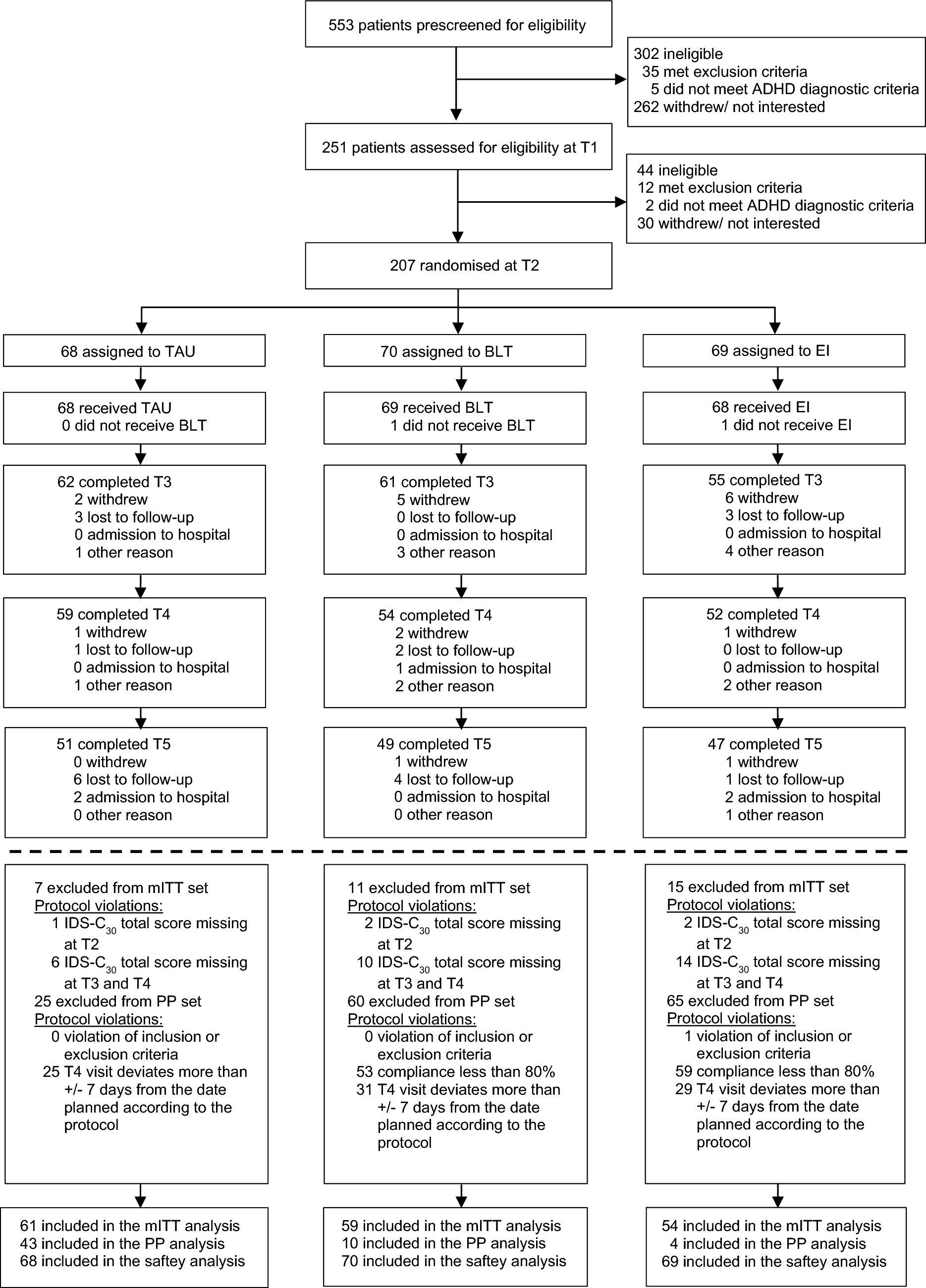
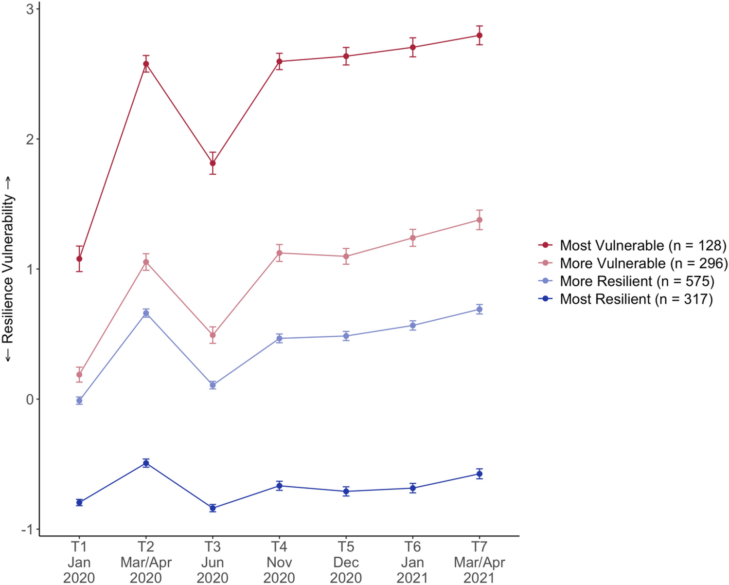
According to our previous results [5, 19], here, we confirmed that anhedonia, a core symptom of depression in humans [23], was developed only in a part of animals exposed to the CMS paradigm. Indeed, approximately 30% of stressed animals displayed resilience to the stress protocol.
Moreover, in line with the finding that spontaneous recovery from the depressive-like behavior induced by 6 weeks of CMS was achieved after 4 weeks of rest [24], we showed that vulnerable animals took 3 weeks of washout to completely recover the pathological phenotype.
After the behavioral characterization in terms of hedonic phenotype, we investigated the response of vulnerable and resilient animals to a novel acute stressor by focusing on neuronal activation to explore whether previous stress exposure may have affected their ability to deal with a new challenge.
As of IEGs, we found that, regardless of stress exposure, ARS enhanced the expression of Arc and Cfos in all the limbic structures investigated, implicating that CMS does not interfere with the activation of these brain structures and suggesting that the responsiveness of these regions was retained, independently from the hedonic phenotype, to promote neuronal mechanisms following external stimuli. In line, we previously found that both ARS and acute forced swim stress exposure [10, 25,26,27], as well as cognitive demanding task [19], led to an overexpression of the IEGs in unstressed animals in different brain regions; notably, in the Pfc, such modulation was still present after a period of washout following 4 weeks of chronic restraint stress [27].
The hallmark of the stress response is the activation of the HPA axis and the effects of acute and chronic stressors on the axis activity are well described by the inverted U-shaped dose–response curve of glucocorticoids [9]. In particular, activation of the HPA axis and the consequent increase of hormone release are associated with both single and prolonged stress, whereas the outcomes are very different with short and moderate stress being “positive” while chronic stimuli resulting in maladaptive consequences. On these bases, the elevated levels of CORT, already observed in our previous study [5], due to chronic stress have a “negative” impact and are related to the vulnerability to CMS, whereas the transient increase observed following the ARS in no stress and resilient animals may be related to the physiological activation of the HPA axis. Moreover, even if, after the recovery period, the hormone levels almost return to the basal levels, the expected activation induced by ARS exposure is completely absent indicating that CMS induced long-lasting effects.
Afterward, we measured the expression of a series of genes, Gadd45β, Sgk1, Dusp1, and Nr4a1, which could be clustered as early response genes. Indeed, the ERGs are rapidly transcribed following acute challenges and respond to HPA-axis activity [12,13,14,15].
In the vHip, we found that Gadd45β, Sgk1, Dusp1 were enhanced by ARS not only in the control group but also in animals that were resilient to CMS. By contrast, we did not observe this upregulation in CMS-vul group, whereas the vulnerable rats that recovered from the pathological phenotype were still able to mount such response.
On the contrary, in line with the notion that Nr4a1 is highly expressed when the levels of circulating corticosterone are elevated [13], its expression was enhanced by ARS in all the experimental groups, including the vulnerable ones.
Moreover, the physiological recovery from CMS at behavioral level was paralleled by a restored ability to deal with challenging conditions measured as proper response in terms of ERGs following the novel acute stressor. Nevertheless, as mentioned, ARS did not enhance circulating levels of CORT in vulnerable animals following the recovery period, suggesting that the regulation of ERGs expression is complex and involves different and independent systems, which will be further investigated in future studies.
The Z-score activation following ARS supports all these results, by highlighting the direction of genes’ regulation in the brain areas considered and strengthening the finding that chronic stress induced functional alterations in vHip in our experimental settings.
The fact that resilient rats exhibited a control-like response to ARS, together with evidence highlighting the importance of neurogenesis [28, 29], neuroplasticity [5], and also the mitochondrial activity [6], further supports the concept that the ventral subregion of the hippocampus is extremely involved in the response to CMS.
Accordingly, we have recently demonstrated that the novel antidepressant drug vortioxetine targeted specifically the vHip by modulating neuronal plasticity following an acute stressor [26].
As observed for the IEGs, we found a different pattern in dHip, Amy, and Pfc, with ARS increased mRNA levels of almost all the genes investigated, regardless of the behavioral susceptibility to stress, pointing out that these limbic structures were not impaired in their ability to be promptly activated following novel stimuli.
Of note, in Pfc, we found an upregulation of the IEGs and Gadd45β, Dusp1, Nr4a1 in resilient/No ARS animals in comparison to the unstressed/No ARS counterpart, possibly suggesting that the overexpression of these genes may contribute to promote the mechanisms of resilience. Consistent with this finding, it has been shown that mice resilient to the chronic predator or social defeat stress had a greater degree of Cfos expression in the glutamatergic neurons of the Pfc [30, 31], thus indicating that the enhanced neuronal activation in No ARS animals might represent a pro-resilience adaptation. In line, the Pfc has been associated with stressor resistance [32] and it has been shown that this brain region, through the enhancement of neuroplastic mechanisms, is involved in stress-coping strategies to promote resilience [33].
A potential limitation of our findings derives by the evidence that we primarily rely on mRNA data to infer changes in function, i.e., the ability to cope with a further challenge. However, we need to consider that the fast coping to the acute challenge needs to rely on evaluating changes in gene expression as initial responses, whereas changes in the related protein levels would take much longer. Furthermore, as our findings have been collected from male rats, we cannot say whether these results can also be generalized to female rats.
In summary, our results confirm the key role played by the vHip, the hippocampal subregion mainly involved in stress response and in the management of the HPA-axis activity [34], not only in the development of the anhedonic phenotype but also in promoting the mechanisms of resilience both in basal conditions and in response to a novel stress exposure.

留言 (0)