Mucinous Proliferations of the Uterine Corpus: Comprehensive Appraisal of an Evolving Spectrum of Neoplasms
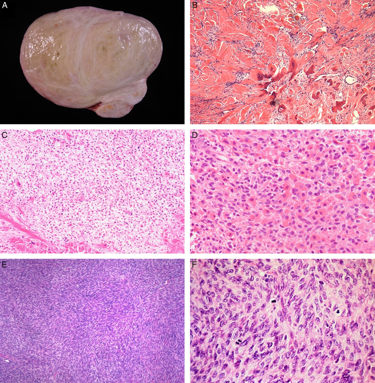
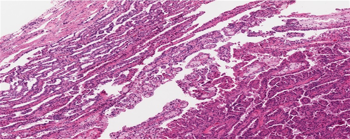
A variety of endometrial lesions may contain mucinous cells. Herein, the author reviews the literature on the classification and clinicopathologic significance of uterine corpus proliferations with a significant mucinous component, assesses the 2020 World Health Organization classification of such lesions, and presents a diagnostic framework. The key epithelial mucinous lesions include mucinous metaplasia, atypical mucinous glandular proliferation and mucinous carcinoma. Each of these categories are classifiable into “usual” and gastrointestinal subtypes, the latter being indicative of intestinal (presence of goblet cells) and/or gastric-type (abundant, pale eosinophilic or clear cytoplasm and well-defined cell borders) morphology. It has been proposed that at least focal expression of gastrointestinal immunohistochemical markers be required for all gastrointestinal type lesions, and for gastrointestinal type atypical mucinous glandular proliferation and carcinoma, minimality or absence of estrogen receptor expression, and the absence of an endometrioid component. Mucinous carcinomas of the usual type, in which >50% of the tumor is comprised of a mucinous component, are the most common. Morphologic subtypes include mucinous carcinoma with microglandular features and mucinous carcinoma with signet rings (signet ring carcinoma). Endometrioid carcinomas with a less than a 50% mucinous component are classified as endometrioid carcinoma with mucinous differentiation. Several studies have directly compared endometrioid and mucinous carcinomas, the latter presumably of the usual type, with respect to patient outcomes after treatment. All have found no difference in overall and disease free survival between these groups. However, three major studies have found mucinous carcinomas to be associated with a higher risk of lymph node metastases. Nineteen cases of mucinous carcinoma of the gastrointestinal type have been reported, and the limited data on their follow-up after primary treatment suggests that this subtype is more clinically aggressive and should accordingly be classified separately from mucinous carcinomas of the usual type. The morphologic spectrum of mucinous carcinoma of the gastrointestinal type is unclear and continues to evolve. Mucinous change, which may sometimes be extensive, may also be associated with papillary proliferation of the endometrium, adenomyoma of the endocervical type, atypical, and typical adenomyomas. In a curettage or biopsy, intestinal type mucinous epithelium may be indicative of any of the gastrointestinal lesions mentioned above, but may also represent samplings of uterine teratomas, yolk sac tumors, genital and extragenital adenocarcinomas with intestinal differentiation, or low-grade appendiceal mucinous neoplasms that secondarily involve the endometrium.

留言 (0)