
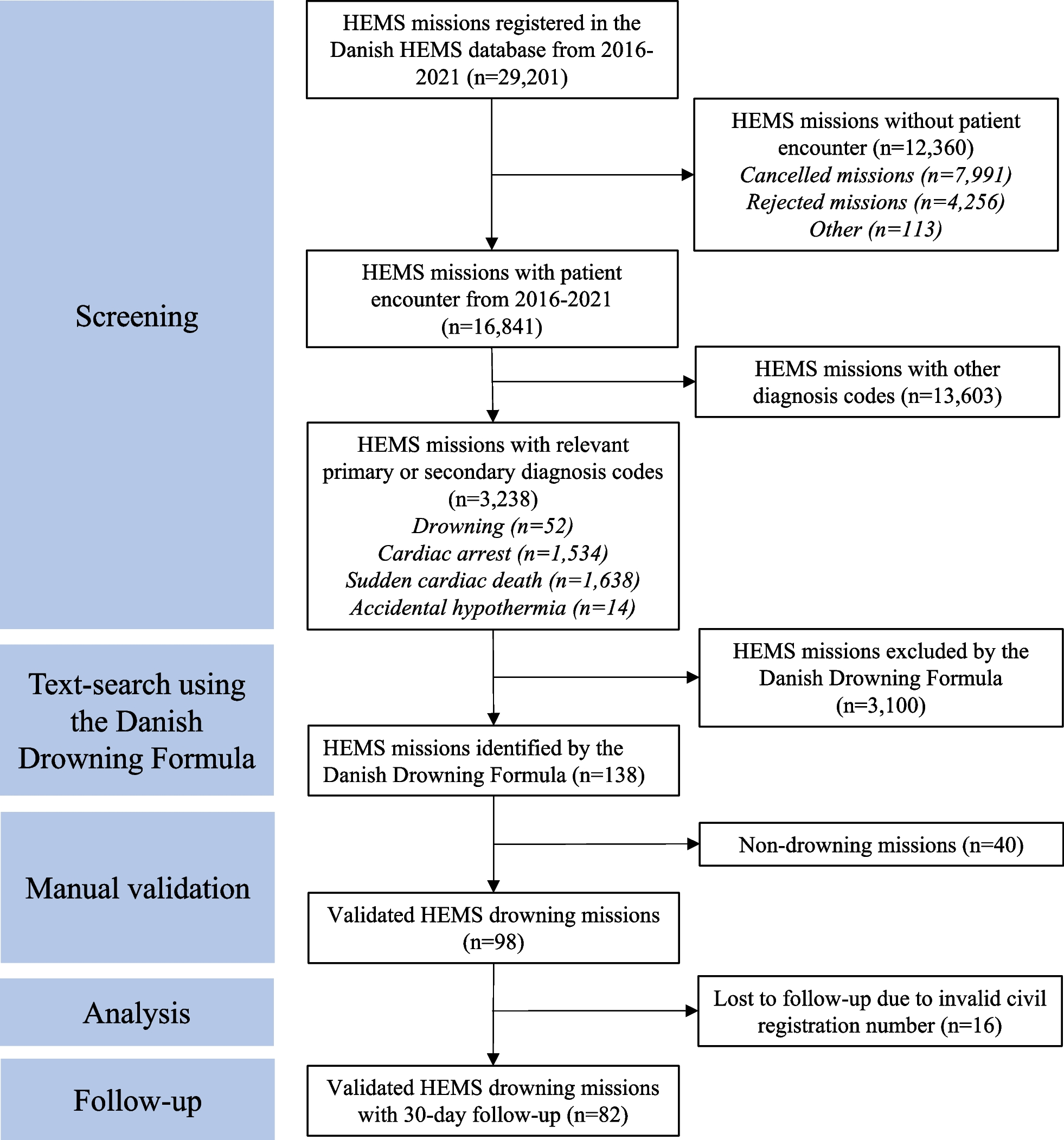
記住我
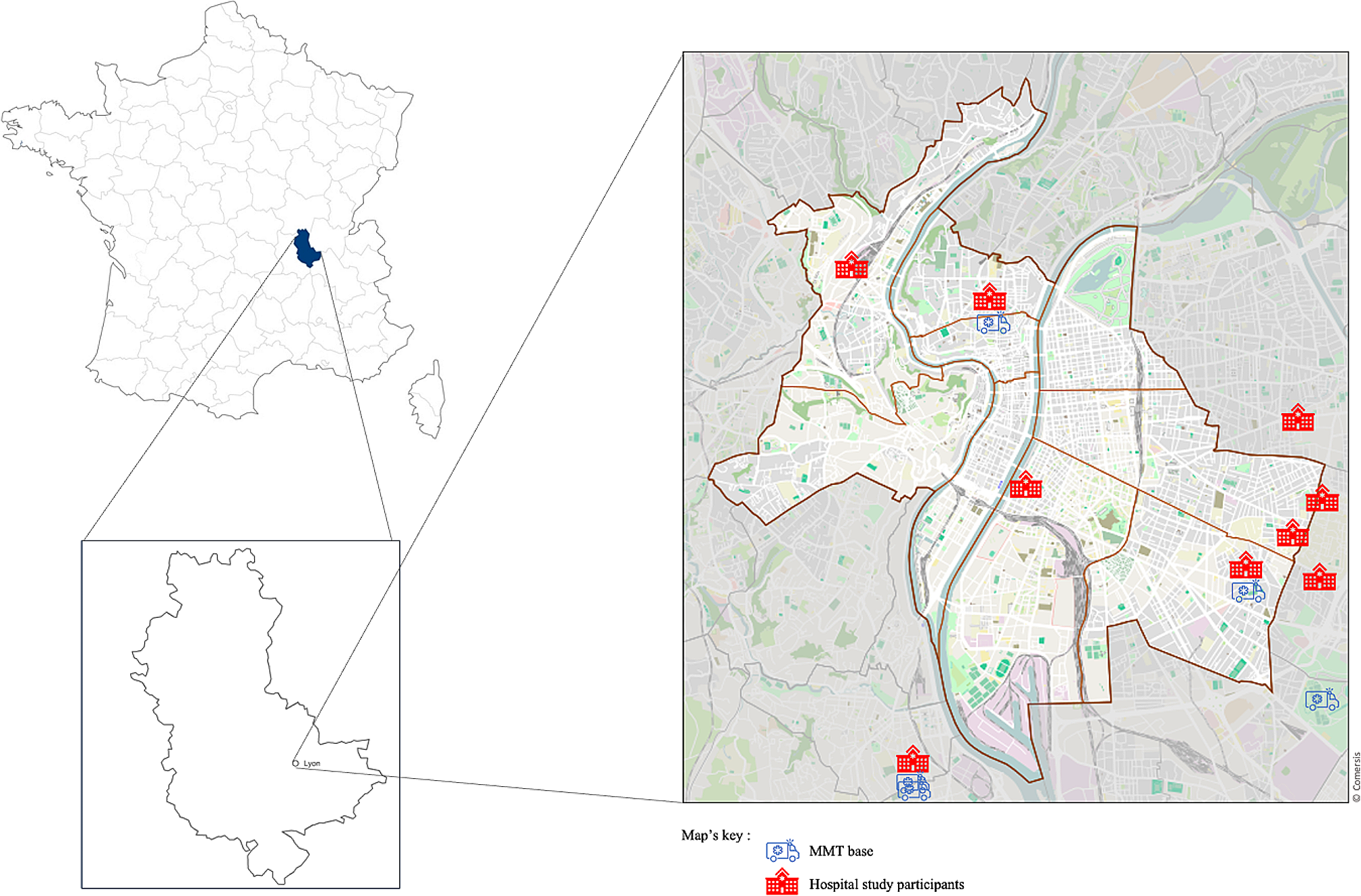

This study was conducted at the Hefei Second People's Hospital, Hefei, China, between 1 January 2020, and 31 December 2020. Hefei Second People's Hospital, a national CPR training centre in China, is a large referral hospital and a tertiary A-level hospital that has 2583 inpatient beds and over 2 million annual emergency and outpatient visits. Our hospital health service manages out-of-hospital health emergencies in Hefei city and rescues more than 350 patients who experience OCHA each year. Our CPR team consists of two systems. The first system, which performed STD-CPR or CO-CPR out of the hospital, consists of six emergency rescue stations. Each team in the station consists of two junior emergency physicians, one paramedic, and one ambulance driver. The second system, which performed advanced life support, consists of one senior emergency physician, six junior emergency physicians, a head nurse, and several registered nurses from the emergency intensive care unit (EICU). All members were trained to perform two CPR methods according to the American Heart Association guidelines. Abdominal compression–decompression training was performed under the supervision of the manufacturer’s monitoring staff.
The trial was retrospectively registered in the Chinese Clinical Trial Registry (registered number: ChiCTR2100049581). The study was approved by the Ethics Committee of the Second People’s Hospital of Hefei (The Affiliated Hefei Hospital of Anhui Medical University, approval number 2020-Science-025), and the requirement for informed consent was waived. This trial is a single-centre, prospective, randomized trial in which chest compression CPR after OHCA will be compared with a method that combines chest compression and abdominal compression–decompression CPR.
PatientsThe inclusion criterion was OHCA in patients at least 18 years of age. The exclusion criteria were patients aged 80 years or older or patients with any contraindications for the abdominal compression–decompression technique, including pregnancy, history of recent thoracic or abdominal trauma/surgery, known terminal or end-stage disease, or severe neurologic impairment.
Randomization and blindingRandomization was performed by a statistics professional from our hospital. A random number table was generated using SPSS 18.0 software. Numbers from the table were assigned on a unified basis by the professional. A blinded assessor evaluated the patient’s neurological prognosis. The outcome assessors and trial statisticians were also blinded. The clinical team responsible for the participants (physicians, nurses, and others) and involved with direct patient care were not blinded to the allocation group due to the inherent difficulty in blinding the intervention.
Data collection and follow-upData were recorded following Utstein resuscitation registry templates, including baseline characteristics, witnesses, bystander CPR, first monitored rhythm, epinephrine use, aetiology, event location and comorbidities.
All patients who were included in this study had a primary cardiac arrest, but cardiac arrests that occur outside the hospital are not always witnessed immediately. Therefore, the exact time from cardiac arrest to the initiation of CPR may not have been accurately determined in all patients in our study; therefore, the no-flow time was waived. Patients were followed-up by three methods: outpatient visits, inpatient visits, or telephone calls. The patients were followed up by a blinded assessor every 1 to 3 months after discharge from the hospital, and the endpoint of the follow-up was the date of death or July 30, 2021.
Outcome measuresThe primary outcome measure was the return of spontaneous circulation (ROSC). Secondary outcome measures were survival to hospital admission, survival to hospital discharge, and neurological outcomes at hospital discharge.
Neurological outcomes were assessed by the Cerebral Performance Category (CPC) scale. The CPC scale categorizes neurological outcomes as follows: CPC 1, good performance; CPC 2, moderate disability; CPC 3, severe disability; CPC 4, comatose or persistent vegetative status; and CPC 5, brain death or patient death [13]. The prognosis in the two groups was compared by classifying the CPC 1 or 2 patients as having a good neurological outcome and those patients with a CPC ≥ 3 as having a poor neurological outcome.
Sample sizeWe referred to another similar study at the time of the sample size estimation for this study [12]. The study had 80% power to find a significant result with a threshold two-sided p value of 0.05 if the expected proportion of ROSC was approximately 20%. The sample size was re-estimated as 122 patients for each group, and the final sample size was increased to approximately 150 patients due to a dropout rate of 20% (for details, see the supplementary materials).
Instrument and interventionAbdominal CPR compression–decompression instrumentThe instrument, produced by Beijing Germari Medical Equipment Co., Ltd., consisted of three components: a display panel, pressure application handles, and a negative pressure device. A compression plate on the bottom of the negative pressure device had to be placed on the epigastrium. After turning on the device, negative pressure was generated, which caused a tight bond between these compression plates and the patient's abdomen. When we performed compression, the compression force was approximately 186 mmHg when the indicator light was on. While decompression was performed, the decompression force was approximately 112 mmHg.
The operating parameters were as follows: (1) The abdominal compression force was limited to 50 kg, and the decompression force was limited to 30 kg; the force levels were controlled from a light-emitting diode (LED) display panel on top of the instrument. (2) The compression–decompression frequency was marked by an audio signal with a frequency of 100 times/min. Images of the device are shown in Fig. 1.
Fig. 1
The abdominal compression–decompression device and its use. A, B The abdominal compression–decompression device. C The training of emergency physicians. D The performance of CO-CPR
InterventionsIn principle, CPR attempts were performed according to current American Heart Association guidelines [1]. Patients were randomized to receive either CO-CPR or STD-CPR treatment. For CO-CPR, chest compression was performed in alternation with abdominal compression–decompression, i.e., when the chest was compressed, the abdomen simultaneously would be decompressed, and vice versa. Abdominal compression was performed at a rate of 100 times/min and a depth of 5–10 cm. The rate of abdominal compression–decompression cycles to chest compression cycles, marked by the audio signal and delivered in alternation, was set to 1:1. The effectiveness, safety, and stability of the abdominal compression–decompression device used in this study have been verified in human studies [11, 12].
All methods were performed following relevant regulations and guidelines. Defibrillation was administered as needed. All patients were ventilated with a bag-valve mask during resuscitation during the out-of-hospital period. After referral to the hospital, all patients received orotracheal intubation and respiration with the aid of a rebreathing bag. Targeted temperature management with a target temperature of 33 °C was performed in each patient. The two CPR methods were applied until either ROSC or resuscitation was deemed futile by the treating emergency physicians.
Statistical analysisPASS 15 software was used to calculate the sample size (NCSS, LLC., Kaysville, Utah, USA). Statistical analysis was performed using SPSS version 18.0 (SPSS Inc., Chicago, IL, USA) and GraphPad Prism 8.0 (GraphPad Software, La Jolla, California, USA). The continuous variables are presented as the mean ± standard deviation (SD) and were analysed by the independent samples t test, while primary and secondary outcome analyses were conducted using Fisher’s exact or Pearson chi-square (\(^\)) tests for comparison. Kaplan–Meier analysis with the log-rank test was plotted to compare the survival curves of the two groups at the end of the follow-up. A p value less than 0.05 was considered statistically significant.
留言 (0)