
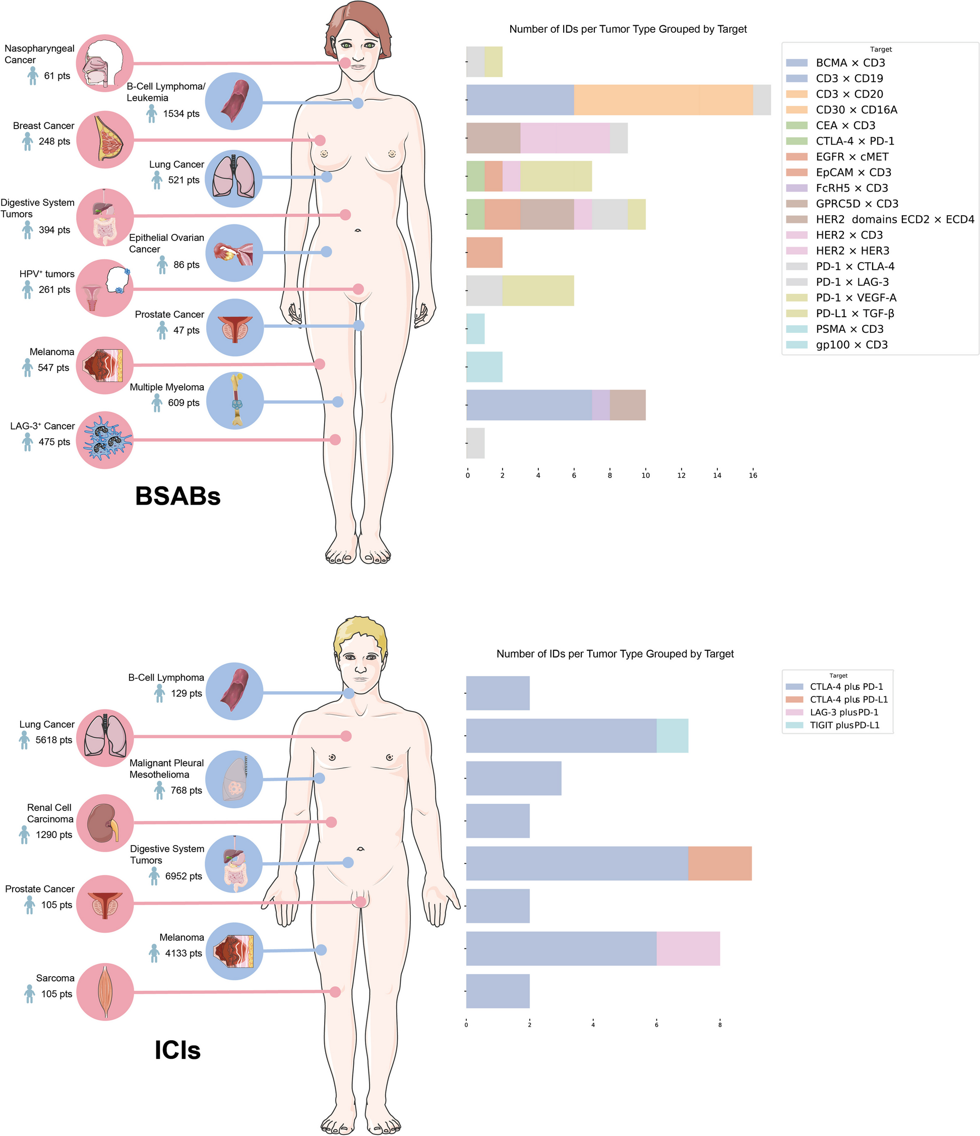
記住我


TIPE3 mRNA expression were analyzed including 171 normal individuals and 179 patients with PC. Compared with normal tissues, TIPE3 mRNA was significantly raised in PC tissues. IHC results of 188 PC tissue specimens from the retrospective cohort also demonstrated that TIPE3 expression was increased in PC tissues. And immuno-staining found that TIPE3 mainly localized in both cytoplasm and membrane of PC cells (Fig. 1(T1) A-B).
Fig. 1
T1: The expression and clinical significance of TIPE3 in PC. A Representative IHC staining of TIPE3. B IHC sum scores were applied to assess TIPE3 in PC specimens. C) IHC staining of TIPE3 in PC tissues with or without lymph node metastasis. D IHC sum scores were applied to assess TIPE3 in PC specimens with or without lymph node metastasis. E TIPE3 expression in negative lymph node and metastatic lymph node. F Survival analysis according to TIPE3 expression in 188 PC patients. G Prognostic Nomogram of 188 PC patients. H Calibration curves of the OS nomogram. (I-K) DCA for the OS nomogram at 1-year I 2-year J and 3-year K. ***, P < 0.001. T2. The clinical significance of TIPE3 A IHC sum scores were applied to determine TIPE3 in PC. B IHC sum scores were applied to assess TIPE3 expression in PC specimens. C Survival analysis using TIPE3 in 66 PC patients. *, P < 0.05; ***, P < 0.001. T3. TIPE3 promoted malignant biological behaviors of PC cells. A Detection of TIPE3 mRNA expression using qRT-PCR. B TIPE3 expression were detected after TIPE3 silencing or overexpression using qRT-PCR. C CCK8 analysis was conducted after TIPE3 silencing in AsPC-1 and PANC-1 cells. D CCK8 analysis was conducted after TIPE3 overexpression in PC cells. E Trans-well assays were performed after TIPE3 silencing or overexpression in AsPC-1 cells. F Trans-well assays were performed after TIPE3 silencing or overexpression in PANC-1 cells. *, P < 0.05; **, P < 0.01; ***, P < 0.001. T4. TIPE3 promoted tumor progression and metastasis in mice. A Tumor growth curve of orthotopic xenograft mouse. B Representative pictures of primary tumors in pancreas. B Tumor volume in NC, shTIPE3 and TIPE3 group, respectively. D Tumor weight in NC, shTIPE3 and TIPE3 group, respectively. E Survival curves of different groups. F The representative of bioluminescent images in metastatic mouse model. G Number of liver metastatic foci were recorded. H Number of peritoneal metastatic tumors were recorded. *, P < 0.05; **, P < 0.01; ***, P < 0.001
The baseline characteristic of this cohort was shown in Table S1. The increased TIPE3 in tumor tissues was correlated with lymph node metastasis (P < 0.001) and TNM stage (P < 0.001) (Fig. 1 (T1) C-D, Table S1; Table S5). Moreover, we also detected TIPE3 expression in 30 negative lymph nodes and 30 metastatic lymph nodes, and results showed that positive TIPE3 rate was significantly higher in metastatic lymph nodes (Fig. 1 (T1) E).
High TIPE3 expression is associated with poor survival of PC patientsTo determine the role of TIPE3 in overall survival (OS) of PC patients, univariate analysis with K-M method was conducted. In this cohort, patients with advanced N stage (P < 0.001) and poor differentiation (P < 0.001) had lower OS. Advanced TNM stage (P = 0.067) also tend to indicate lower OS, although this tendency was not of statistical significance, which may results from limited number of stage III-IV patients (Table S2). Importantly, high TIPE3 expression (P < 0.001) also led to unfavorable prognosis.
Multivariate analysis was further performed and results found that high level of TIPE3 (P = 0.013) was an independent unfavorable prognostic factor (Table S2, Fig. 1(T1) F).
Construction of the nomogram based on TIPE3 expressionAccording to the aforementioned results, tumor differentiation, 8th edition AJCC N stage, and TIPE3 expression was established to predict the overall survival rate (Fig. 1(T1) G). Results showed the C-index was up to 0.679 (95% CI: 0.630–0.728). The calibration curve for the OS probability at 1, 2, or 3-year showed favorable calibration of the nomogram (Fig. 1(T1) H). In addition, DCA of the nomogram indicated that the model had a favorable net clinical benefit for predicting survival rates (Fig. 1 (T1) I-K).
Clinical significance of TIPE3 expression in the prospective cohortOur previous results found that TIPE3 was closely correlated with the prognosis of PC using a retrospective cohort and nomogram analysis. We further investigated the expression and clinical significance of TIPE3 expression in PC using a prospective cohort that consisting of 66 PC patients. The expression of TIPE3 was also raised in tumor tissues, and elevated TIPE3 expression was associated with lymph node metastasis as well (P = 0.030) (Table S3, Fig. 1 (T2) A-B; Table S6). Then univariate and multivariate analysis were conducted, revealed that poor tumor differentiation (P = 0.002) and high TIPE3 level (P = 0.018) were closely associated with worse survival. Notably, high TIPE3 expression was also identified as an independent prognostic factor for PC patients in the prospective cohort (Table S4, Fig. 1(T2) C).
TIPE3 accelerates tumor progression in vitroOur previous results found that TIPE3 might be involved in the progression of PC. We observed that all the eight PC cells (AsPC-1, MIA PaCa-2, CFPAC-1, PANC-1, BxPC-3, Capan-1, Patu-8988 and SW-1990) presented moderate to high TIPE3 expression (Fig. 1 (T3) A). Specifically, we chose two cell lines (AsPC-1 and PANC-1) with highest TIPE3 expression to perform gene-knockdown experiments via lentivirus transfection of TIPE3-shRNAs. The knockdown and overexpression efficiency was testified (Fig. 1 (T3) B). The effect of TIPE3 on malignant behaviors of tumor cells was evaluated via CCK-8 assay and Transwell assay. Results showed that TIPE3 silencing attenuated the proliferation, migration and invasion capacities of PC cells. In addition, to testify the related conclusions, TIPE3 was overexpressed in PC cells, CCK-8 and Transwell assays were repeated. The proliferation, migration and invasion capacities were enhanced after TIPE3 overexpression (Fig. 1 (T3) C-F).
TIPE3 promotes tumor progression in vivoOrthotopic xenograft mouse model was established using stable TIPE3 silenced (LV-shTIPE3) or TIPE3 overexpressed (LV-TIPE3) AsPC-1 cells. Tumors from LV-shTIPE3 group showed slower growth, smaller size and lighter weight (Fig. 1 (T4) A-D). Mice from the shTIPE3 group also presented increased survival compared to controls (86.8 vs 88.3 days). While tumors from LV-TIPE3 group presented increased size and weight, and TIPE3 overexpression led to decreased survival rate (86.8 vs 83.4 days) (Fig. 1 (T4) E).
To evaluate the function of TIPE3 on the metastasis of PC, a metastatic mouse model was constructed. We found that mice in the LV-shTIPE3 group presented reduced number of distant metastasis compared with LV-shNC group. The size of metastatic tumors were also smaller in the LV-shTIPE3 group. While in the LV-TIPE3 group, the number and size of metastatic tumor were markedly increased compared to control (Fig. 1 (T4) F-H). All these results demonstrated that TIPE3 promoted tumor progression and metastasis in vivo.
TIPE3 promoted RAC1 expression in PCRAC1 could participate in tumor progression, especially the metastasis of PC. Research also found that other members of TIPE family functions through interacting with RAC1. So we hypothesized that TIPE3 may promoted PC progression via targeting RAC1. Therefore, we conducted the following experiments. First, RAC1 expression was observed in PC specimens using IHC, and results showed that RAC1 expression was also raised in PC tissues compared to the normal tissues (Fig. 2 (T1) A-B). Importantly, RAC1 expression was closely correlated with TIPE3 expression in PC tissues (Fig. 2 (T1) C). Moreover, decreased RAC1 expression was shown in TIPE3 silencing PC cells, whereas elevated RAC1 expression was shown in TIPE3 overexpressed PC cells (Fig. 2 (T1) 4D-E). Furthermore, RAC1 expression was also closely associated with TIPE3 expression in xenograft tumor tissues. In addition, the expression of RhoA and MMP9, a RAC1 downstream target, were also detected, and results showed both the expression of RhoA and MMP9 expression were related with TIPE3 expression in tumor tissues (Fig. 2 (T1) F-G). Taken together, these results indicated that TIPE3 promoted RAC1 expression in PC.
Fig. 2
T1: TIPE3 increased RAC1 in PC. A IHC staining of RAC1. B IHC sum scores were applied to determine RAC1 expression. C IHC sum scores of RAC1 in PC tissues with low or high TIPE3 expression. D Western-blot was conducted to detected RAC1 expression after TIPE3 silencing or overexpression. E Quantified results of western-blot for RAC1 expression in PC cells. F IHC staining of TIPE3, RAC1, RhoA and MMP9 in orthotopic xenograft tumors. G IHC sum scores of TIPE3, RAC1, RhoA and MMP9 expression in orthotopic xenograft tumors. *, P < 0.05; **, P < 0.01; ***, P < 0.001. T2. TIPE3 accelerated the malignant behaviors of PC cells in a RAC1-dependent manner. A NSC23766 (50 μM) was used in CCK8 analysis. B CCK8 assays was measurement after RAC1 silencing in PC cells. C Trans-well assays after treatment of NSC23766 (50 μM). D Trans-well assays were measurement after RAC1 silencing in PC cells. E CCK8 assays were measurement in TIPE3 silencing PC cells that pretreated with NSC23766 (50 μM) or transfection with RAC1 siRNA. F CCK8 assays were conducted in TIPE3 overexpressed PC cells that pretreated with NSC23766 (50 μM) or transfection with RAC1 siRNA. G Trans-well assays were conducted in TIPE3 silenced PC cells that pretreated with NSC23766 (50 μM). H Trans-well assays were conducted in TIPE3 silenced PC cells that transfected with RAC1 siRNA. I Trans-well assays were conducted in TIPE3 overexpressed PC cells that pretreated with NSC23766 (50 μM). J Trans-well migration and invasion assays were conducted in TIPE3 overexpressed PC cells that transfected with RAC1 siRNA
TIPE3 promotes tumor progression in a RAC1-dependent mannerTo explore whether TIPE3 promoted tumor progression via activating RAC1, NSC23766 and RAC1 silencing were used in the subsequent experiments. Both treatments dramatically suppressed the malignant behaviors of PC cells (Fig. 2 (T2) A-D). Notably, TIPE3 knockdown decreased the proliferation, migration and invasion of PC cells, while NSC23766 treatment and RAC1 silencing eliminated this effect. Consistently, NSC23766 treatment and RAC1 silencing also blocked the impact of TIPE3 overexpression on the malignant behaviors in PC cells (Fig. 2 (T2) E-J). These data indicated that TIPE3 promoted PC progression in a RAC1-dependent manner.
Pancreatic cancer (PC) has a high degree of malignancy and poor prognosis. Identifying effective biomarker is essential for the precise stratification and the development of targeted therapies, which is crucial for improving the prognosis of these patients [14, 15]. For the first time, we demonstrated that TIPE3 served as an independent prognostic biomarker in PC, which would contribute to stratifying patients with high risk and poor prognosis. Mechanically, TIPE3 promoted the progression of PC via targeting RAC1. These finds identified a potential biomarker for prognostic prediction of PC, and preliminarily revealed the functions and related mechanism of TIPE3 in PC.
As the last discovered TIPE family members, TIPE3 was demonstrated to be a novel regulatory molecule in a number of tumors recently, but the function of TIPE3 in PC remains unknown, especially the correlation between TIPE3 expression and patients’ prognosis is unclear [16]. In the present study, we constructed both retrospective cohort and prospective cohort to explore the clinical value of TIPE3 in PC tissues. This study shows that the level of TIPE3 in PC tissue is increased, which was closely associated with lymph node metastasis and TNM stage. Metastatic lymph nodes showed significantly higher positive TIPE3 expression rate, indicating that tumor cells with positive TIPE3 expression had increased metastatic capacity. Of note, high TIPE3 expression served as an independent unfavorable prognostic factor for PC patients.
At present, most of the clinical decision that made for PC patients largely depend on the TNM system. Data from related randomized controlled trials or prospective studies are also limited [4, 5, 14]. This study established a nomogram for predicting the OS for PC patients based on TIPE3 expression. Moreover, to testify our conclusion, we constructed another prospective cohort with 66 patients. These results revealed that TIPE3 act as an ideal marker for predicting patients’ prognosis, which is an essential supplement to the oncogenic role of TIPE3 and provided novel theoretical basis for the progress of PC. Therefore, we strongly recommend a routine TIPE3 staining for PC tissues. We then further investigated the role of TIPE3 both in vivo and in vitro. It is worth noting that we constructed three in vivo models to prove that TIPE3 promotes tumor progression and metastasis in PC.
RAC1 is a key regulator during tumor progression. It has been reported that RAC1 hyperactivation and up-regulation is closely correlated with enhanced growth and metastasis in numerous tumors, including PC. RAC1 has become a standard for tumor stratification and a promising therapeutic target due to its crucial role in tumor progression [17, 18]. The present study also primarily demonstrated that TIPE3 promoted tumor progression via up-regulating RAC1 expression in PC.
Previous research demonstrated that another TIPE family member, TIPE2, was also involved in regulating the activity of RAC1. The N-terminal lysine and arginine residues are essential for the interaction between TIPE2 and RAC1 [19, 20]. All the four members of TIPE family were of high homology, but the expression and roles of different TIPE family members varies a lot. The N-terminus of TIPE family members might be crucial for their functions [8]. Therefore, clarifying the key domain that responsible for the functions of TIPE3 in PC is necessary in further study. In addition, a larger prospective cohort is needed to evaluate the sensitivity and specificity of TIPE3 expression in predicting the prognosis of PC patients.
留言 (0)