
記住我
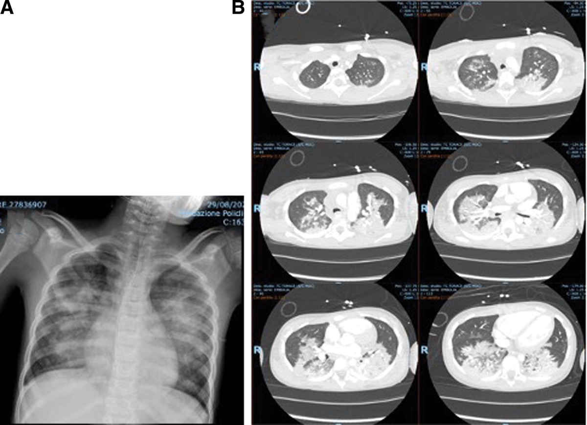

A 1-year-old girl was admitted to our hospital in November 2011 with a 10-day history of fever, worsening cough and hypoxemia. Her medical history was notable for a left multicystic kidney and esophageal atresia, which was surgically repaired without complications. On admission, physical examination revealed a temperature of 38 °C, heart rate of 160 beats per minute, respiratory rate of 70 per minute, and oxygen saturation of 97% in ambient air. The white blood cell count was 15,810 cells/mm3 (with differential of 74% neutrophils, 14.5% lymphocytes, 9.4% monocytes and 2.1% eosinophils), and serum C-reactive protein level was 2.81 mg/dL. Rapid tests for respiratory syncytial virus and human metapneumovirus infection were both negative. Chest radiograph on admission showed diffuse bilateral peribronchial cuffing and mild hyperinflation in the right lower lung field (Fig. 1). She was diagnosed with viral asthmatic bronchitis and was treated with beta-agonist bronchodilators and ampicillin/sulbactam intravenously for suspected secondary bacterial infection. On day 4 of admission, ampicillin/sulbactam was discontinued following negative blood culture results, and oral erythromycin was introduced to suppress bronchial inflammation. On day 6, her symptoms worsened with severe wheezing, dyspnea and hypoxemia (oxygen saturation of 90% in ambient air). She was started on supplemental oxygen and intravenous methylprednisolone (1 mg/kg/dose thrice daily). She improved temporarily but worsened on day 13. Her white blood cell count was 22,660 cells/mm3, and C-reactive protein was 6.28 mg/dL. Methylprednisolone was discontinued, and intravenous ampicillin/sulbactam was restarted. On day 14, chest radiograph showed multiple bilateral nodules (Fig. 1). She continued to have high fevers and respiratory distress despite changing antibiotics to cefotaxime on day 14 and then to panipenem/betamipron on day 16. Computed tomography (CT) scan of the chest performed on day 17 revealed multiple, bilateral, well-defined nodules and consolidation in the right lower lobe (Fig. 2). Blood cultures taken on day 13 remained negative, and several sputum cultures taken after admission detected only normal respiratory flora. Further laboratory tests revealed the diagnosis.
 FIGURE 1.:
 Chest radiograph images on Day1, Day14 and Day32.
 FIGURE 2.:
 CT images on Day17, 1 month and 6 months.
For Denouement see P. 783.
DENOUEMENT(Pediatr Infect Dis J 2022;41:783–785)Continued from P. 782.
On day 17, a urine antigen test (BinaxNOW Legionella, Alere, Waltham, Maria) returned positive. In addition, both polymerase chain reaction (MIP gene; SRL, Tokyo, Japan) and culture of sputum were positive for Legionella, and serum anti-Legionella antibody by indirect fluorescent antibody test (SRL, Japan) showed a titer of 1:25. The antibiotic was changed from panipenem/betamipron to ciprofloxacin. On day 18, her temperature decreased, and her respiratory symptoms improved without reexacerbation. She was treated with ciprofloxacin for 7 days and thereafter with oral tosufloxacin for 7 days. At 1 month after completion of ciprofloxacin, chest CT continued to show consolidation in the right lower lobe and ground glass opacity (Fig. 2). At 6 months, almost all aberrant findings were improved.
Legionella pneumonia is caused by the Gram-negative aerobic bacteria Legionella pneumophila and is considered a rare pathogen in immunocompetent children. Since 2001 in Japan, the average reported number of cases in children under 15 years of age was only one case per year despite a reporting requirement for all diagnosed cases. Most reported pediatric cases of Legionella pneumonia were in newborns, with only 4 cases in children 1–4 years of age in the last 10 years.1 Similarly in the United States, most cases occurred in people ≥50 years of age, and the incidence in children under 10 years old was low (1/1255 case in 2017, 3/835 cases in 2016).2 Additionally, almost all newborn cases of Legionella pneumonia have reportedly occurred in preterm babies or in those with an underlying disease, leading to an immunocompromised state.3,4 Recently, there have been some reports describing community-acquired Legionella pneumonia in children.5,6 One of these reports was neonate and the other one was an adolescence who had anorexia nervosa, both might have underlying immunosuppressed condition. This patient was admitted with a respiratory infection that gradually deteriorated until treatment with a quinolone on day 17 of hospitalization after which the child began to improve.
We considered 4 risk factors for Legionella pneumonia in children: (1) newborn, (2) immunodeficiency (particularly, cellular immunity), (3) asthma and/or chronic pulmonary disease, and (4) steroid use.7–10 Moreover, some recent reports have also mentioned the risk of adrenocorticotropic hormone therapy.11,12 Among these risk factors, this case experienced steroid use only and the dosage administered was not high (1 mg/kg, thrice daily), although the duration was relatively long (7 days) for viral bronchitis.
Here, we thought there were 2 possible explanations for the transmission route of L. pneumophila, community-acquired or hospital-acquired due to possible airborne contact in the hospital environment. Regarding the first possibility, the reported incubation period of L. pneumophila is relatively long (2–14 days), thus suggesting that community-acquired L. pneumophila should be considered as the route of transmission among inpatients if the clinical symptoms appear within the first 2 weeks of hospitalization. Regarding the possibility that this patient became infected after hospitalization, we investigated environmental contamination and evaluated other patients in the pediatric ward. Samples for L. pneumophila culture were collected from the water supply (tap water, humidifiers and bath water) in the pediatric ward. There was no evidence of contamination of the environment or a silent outbreak in the pediatric ward.
This is the first report of a 1-year-old girl with Legionella pneumonia, which exacerbated after receiving short-duration steroid treatment in hospital. In addition, the CT findings were examined sequentially along with symptoms.
In this case, we first suspected Legionella pneumonia because of the poor response to broad-spectrum antibiotic treatment and confirmed our diagnosis by performing sputum polymerase chain reaction and serum antibody testing. Because Legionella pneumonia may have been underdiagnosed in children, urine antigen test is useful because of its convenience. As for Legionella serogroup-1, the sensitivity and specificity were reported around 85% and 99%, though needed to keep caution about the other serogroup and other Legionella species.13,14
There are no reports in the past literature describing disease course and chest CT findings in immunocompetent children, especially in the infancy, with Legionella pneumonia. Throughout the course of CT imaging in this case, the following findings were considered to be characteristic. First, there were bilateral multiple lesions, which showed various mixed appearances (well-defined consolidation directly under the pleura accompanied by air bronchogram intermingled with nodular ground glass opacities). Second, there was no pleural effusion. These two features seemed to be similar to those observed in adults.6,15–17
In summary, we have presented a 1-year-old patient with Legionella pneumonia, confirmed by sputum culture, polymerase chain reaction and serology, which serves as a reminder that this pathogen should be considered in patients with refractory pneumonia.
ETHICS COMPLIANCEAuthors have taken ethical considerations for patient and her parents. Informed consent was obtained from the parents.
REFERENCES 1. National Institute of Infectious Diseases J: Legionellosis, January 2008-December 2012. IASR. 2013;34:155–157. 2. CDC: Legionella (Legionnaires’ disease and pontiac fever) Available at: https://www.cdc.gov/legionella/about/index.html. 3. Greenberg D, Chiou CC, Famigilleti R, et al. Problem pathogens: paediatric legionellosis–implications for improved diagnosis. Lancet Infect Dis. 2006;6:529–535. 4. Allgaier J, Lagu T, Haessler S, et al. Risk factors, management, and outcomes of legionella pneumonia in a large, nationally representative sample. Chest. 2021;159:1782–1792. 5. Leruste A, Rambaud J, Picard C, et al. Successful pediatric ECMO in a rare case of septic shock due to a community-acquired Legionella infection. Med Mal Infect. 2017;47:68–70. 6. Sammori M, Funakoshi H, Fukai Y, et al. First case of Legionella pneumonia in a female patient with anorexia nervosa. J Paediatr Child Health. 2021;57:950–952. 7. Abernathy-Carver KJ, Fan LL, Boguniewicz M, et al. Legionella and Pneumocystis pneumonias in asthmatic children on high doses of systemic steroids. Pediatr Pulmonol. 1994;18:135–138. 8. Myers C, Corbelli R, Schrenzel J, Gervaix A. Multiple pulmonary abscesses caused by Legionella pneumophila infection in an infant with croup. Pediatr Infect Dis J 2006;25:753–754. 9. Heine S, Fuchs A, von Müller L, et al. Legionellosis must be kept in mind in case of pneumonia with lung abscesses in children receiving therapeutic steroids. Infection. 2011;39:481–484. 10. Huong Ple T, Hien PT, Lan NT, et al. First report on prevalence and risk factors of severe atypical pneumonia in vietnamese children aged 1-15 years. BMC Public Health. 2014;14:1304. 11. Musallam N, Bamberger E, Srugo I, et al. Legionella pneumophila and pneumocystis jirovecii coinfection in an infant treated with adrenocorticotropic hormone for infantile spasm: case report and literature review. J Child Neurol. 2014;29:240–242. 12. Shachor-Meyouhas Y, Ravid S, Hanna S, et al. Legionella pneumophila pneumonia in two infants treated with adrenocorticotropic hormone. J Pediatr. 2017;186:186–188.e1. 13. Edelstein PH, Jørgensen CS, Wolf LA. Performance of the ImmuView and BinaxNOW assays for the detection of urine and cerebrospinal fluid Streptococcus pneumoniae and Legionella pneumophila serogroup 1 antigen in patients with Legionnaires’ disease or pneumococcal pneumonia and meningitis. PLoS One. 2020;15:e0238479. 14. Wong AYW, Johnsson ATA, Ininbergs K, et al. Comparison of four streptococcus pneumoniae urinary antigen tests using automated readers. Microorganisms. 2021;9:827. 15. Kim KW, Goo JM, Lee HJ, et al. Chest computed tomographic findings and clinical features of legionella pneumonia. J Comput Assist Tomogr. 2007;31:950–955. 16. Sakai F, Tokuda H, Goto H, et al. Computed tomographic features of Legionella pneumophila pneumonia in 38 cases. J Comput Assist Tomogr. 2007;31:125–131. 17. Miyashita N, Higa F, Aoki Y, et al. Clinical presentation of Legionella pneumonia: evaluation of clinical scoring systems and therapeutic efficacy. J Infect Chemother. 2017;23:727–732.
留言 (0)