
記住我


The present study consisted of 72 typically developing participants between the ages of 6 and 18 (mean ± std age = 12.11 ± 3.77 years old, including 30 individuals identified by caregivers as female) that were randomly selected from the Neurodevelopment of Reasoning Ability (NORA) dataset (Wendelken et al. 2011, 2016, 2017; Ferrer et al. 2013). All participants were right-handed; for additional demographic and socioeconomic information see Supplementary Table 1. In total, 61 of these participants were also included in prior research on sulcal depth (Voorhies et al. 2021). All participants were screened for neurological impairments, psychiatric illness, history of learning disability, and developmental delay. All participants and their parents gave their informed assent/consent to participate in the study, which was approved by the Committee for the Protection of Human Participants at the University of California, Berkeley.
Data acquisitionImaging dataMRI data were collected on a Siemens 3 T Trio system at the University of California Berkeley Brain Imaging Center. High-resolution T1-weighted MPRAGE anatomical scans (TR = 2300 ms, TE = 2.98 ms, 1 × 1 × 1 mm voxels) were acquired for cortical morphometric analyses.
Behavioral dataAll 72 participants completed a matrix reasoning task (WISC-IV), which is a widely used measure of abstract, nonverbal reasoning (Ferrer et al. 2013; Wendelken et al. 2016, 2017). Two additional control measures were included when available: processing speed (N = 71) and verbal working memory (N = 64). Reasoning performance was measured as a total raw score from the WISC-IV matrix reasoning task (Wechsler 1949; mean ± std = 25.65 ± 6.01). Matrix reasoning is an untimed subtest of the WISC-IV in which participants are shown colored matrices with one missing quadrant. The participant is asked to “complete” the matrix by selecting the appropriate quadrant from an array of options. Previous factor analysis in this dataset (Ferrer et al. 2013) showed that the matrix reasoning score loaded strongly onto a reasoning factor that included three other standard reasoning assessments consisting of the Block Design subtest of the Wechsler Abbreviated Scale of Intelligence (WASI; Wechsler 1999), as well as the Analysis Synthesis and Concept Formation subtests of the Woodcock-Johnson Tests of Achievement (Woodcock et al. 2001).
Processing speed was computed from raw scores on the Cross Out task from the Woodcock-Johnson Psychoeducational Battery-Revised (WJ-R; (Brown et al. 2012)). In this task, the participant is presented with a geometric figure on the left followed by 19 similar figures. The participant places a line through each figure that is identical to the figure on the left of the row. Performance is indexed by the number of rows (out of 30 total rows) completed in 3 min (mean ± std = 22.1 ± 6.75). Cross Out scores are frequently used to estimate processing speed in developmental populations (McBride-Chang and Kail 2002; Kail and Ferrer 2007).
Verbal working memory was measured via raw scores of the Digit Span task from the 4th edition of the Wechsler Intelligence Scale for Children (WISC-IV; Wechsler 1949). The Digits Forward condition of the Digit Span task taxes working memory maintenance, whereas the Backward condition taxes both working memory maintenance and manipulation. In Digits Forward, the experimenter reads aloud a sequence of single-digit numbers, and the participant is asked to immediately repeat the numbers in the same order; in Digits Backward, they are asked to immediately repeat the numbers in the reverse order. The length of the string of numbers increases after every two trials. The Forwards task has eight levels, progressing from 2 to 9 digits. The Backwards task has seven levels, from 2 to 8 digits. Participants are given a score of 1 for a correct answer or a 0 for an incorrect answer. Testing on a given task continues until a participant responds incorrectly to both trials at a given level, after which the experimenter recorded a score out of 16 for Digits Forward (16 total trials; mean ± std = 9.03 ± 2.24) and a score out of 14 for Digits Backward (14 total trials; mean ± std = 5.84 ± 2.12).
Morphological analysesCortical surface reconstructionAll T1-weighted images were visually inspected for scanner artifacts. FreeSurfer’s automated segmentation tools (Dale et al. 1999; Fischl and Dale 2000; FreeSurfer 6.0.0) were used to generate cortical surface reconstructions. Each anatomical T1-weighted image was segmented to separate gray from white matter, and the resulting boundary was used to reconstruct the cortical surface for each participant (Dale et al. 1999; Wandell et al. 2000). Each reconstruction was visually inspected for segmentation errors, and these were manually corrected when necessary.
Cortical surface reconstructions facilitate the identification of shallow tertiary sulci compared to post-mortem tissue for two main reasons. First, T1 MRI protocols are not ideal for imaging vasculature; thus, the vessels that typically obscure the tertiary sulcal patterning in post-mortem brains are not imaged on standard resolution T1 MRI scans. Indeed, indentations produced by these smaller vessels that obscure the tertiary sulcal patterning are visible in freely available datasets acquired at high field (7 T) and micron resolution (100–250 μm; Lüsebrink et al. 2017; Edlow et al. 2019). Thus, the present resolution of our T1s (1 mm isotropic) is sufficient to detect the shallow indentations of tertiary sulci but is not confounded by smaller indentations produced by the vasculature. Second, cortical surface reconstructions are made from the boundary between gray and white matter; unlike the outer surface, this inner surface is not obstructed by blood vessels (Weiner et al. 2018; Weiner 2019).
Defining the presence and prominence of the para-intermediate frontal sulcusWe first manually defined the pimfs within each individual hemisphere in tksurfer (Miller et al. 2021b). Manual lines were drawn on the inflated cortical surface to define sulci based on the most recent schematics of pimfs and sulcal patterning in LPFC by Petrides (Petrides 2019), as well as by the pial and smoothwm surfaces of each individual (Miller et al. 2021b). In some cases, the precise start or end point of a sulcus can be difficult to determine on a surface (Borne et al. 2020). Thus, using the inflated, pial, and smoothwm surfaces of each individual to inform our labeling allowed us to form a consensus across surfaces and clearly determine each sulcal boundary. For each hemisphere, the location of the pimfs was confirmed by three trained independent raters (E.H.W., W.I.V., J.K.Y.) and finalized by a neuroanatomist (K.S.W.). Although this project focused on a single sulcus, it took the manual identification of all LPFC sulci (2448 sulcal definitions across all 72 participants) to ensure the most accurate definitions of the pimfs components (for descriptions of these LPFC sulci see: Petrides 2019; Miller et al. 2021a, b; Voorhies et al. 2021; Yao et al. 2022).
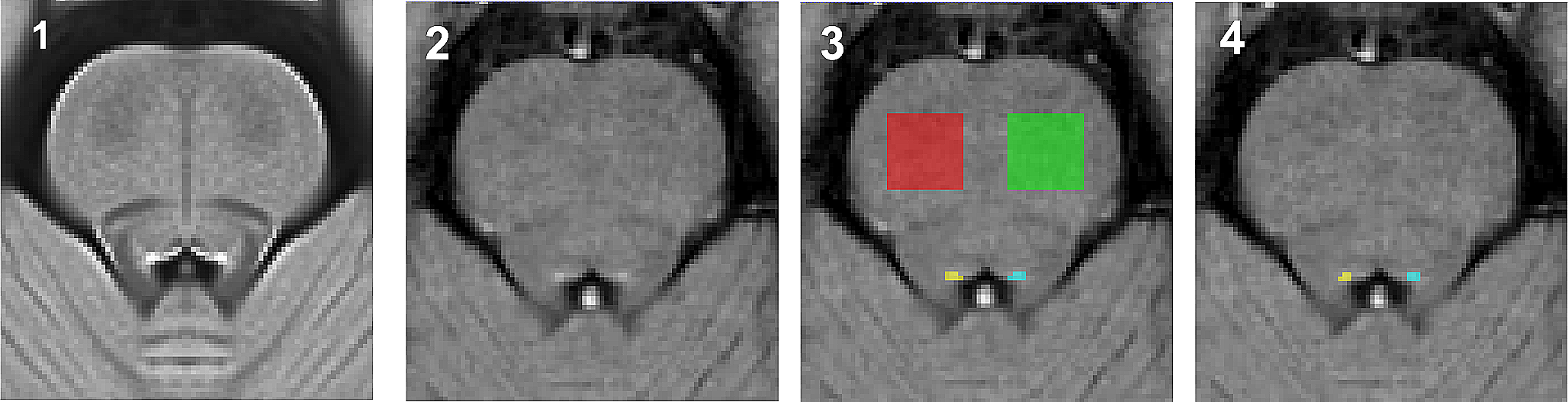
Individuals typically have three to five tertiary sulci within the middle frontal gyrus (MFG) of the lateral prefrontal cortex (Miller et al. 2021a, b; Voorhies et al. 2021; Yao et al. 2022). The posterior MFG contains three of these sulci, which are present in all participants: the anterior (pmfs-a), intermediate (pmfs-i), and posterior (pmfs-p) components of the posterior middle frontal sulcus (pmfs; Miller et al. 2021a, b; Voorhies et al. 2021; Yao et al. 2022). In contrast, the tertiary sulcus within the anterior MFG, the para-intermediate frontal sulcus (pimfs), is variably present. A given hemisphere can have zero, one, or two pimfs components (Fig. 1A; Supplementary Fig. 1; Voorhies et al. 2021; Yao et al. 2022).
Fig. 1
The para-intermediate frontal sulcus: A tertiary sulcus in lateral prefrontal cortex with pronounced individual differences. A Pial (top) and inflated (bottom) left hemispheres (sulci: dark gray; gyri: light gray; cortical surfaces are not to scale) depicting the four types of the para-intermediate frontal sulcus (pimfs): (i) both components present, (ii) neither present, (iii) dorsal component present, (iv) ventral component present. The prominent sulci bounding the pimfs are also shown: the horizontal (imfs-h) and ventral (imfs-v) intermediate frontal sulci and inferior frontal sulcus (ifs). These four sulci are colored according to the legend. B Stacked bar plot depicting the incidence of the pimfs components in both hemispheres across the sample (N = 72 participants). The incidence of the pimfs is highly variable. In each hemisphere, it is more common for participants to have two components than a single component or no component (***ps < 0.0001); the distribution of incidence does not differ between hemispheres (p = 0.30). When only one component was present in a given hemisphere, it was equally likely to be a dorsal or ventral component (ps > 0.30)
Drawing from criteria outlined by Petrides (2013, 2019), the dorsal and ventral components of the para-intermediate frontal sulcus (pimfs-d and pimfs-v) were generally defined using the following twofold criterion: (i) the sulci ventrolateral to the horizontal and ventral components of the intermediate frontal sulcus, respectively, and (ii) superior and/or anterior to the mid-anterior portion of the inferior frontal sulcus. Note that in this schematic (Petrides 2019), there is presently an unidentified sulcus located on the MFG between the pmfs-a and pimfs-d, which appears as a posterior branch of the imfs-h (below the star (*) symbol in the schematic). In the present work, we included this un-identified sulcus as the posterior extent of the imfs-h in our definitions of the imfs-h. In our sulcal definitions, our principled criteria always identified the pimfs-d below the imfs-h and the pimfs-v always below the imfs-v. Thus, with this criterion, the posterior component of the imfs-h was not confusable with our definition of the pimfs-d. Future work can seek to clarify the incidence and distinctiveness of this branch from the imfs-h. The location of each indentation was cross-checked using the inflated, pial, and smoothwm surfaces. We first confirmed the accuracy of this criterion by applying it to the individual participants with two identifiable pimfs. Next, we extended this criterion to label the cases in which an individual only had one component. We then compared incidence rates between components and hemispheres with a Chi-squared and Fischer exact test, respectively.
We quantified the prominence of the pimfs as its surface area (in mm2). The surface area values for each pimfs label were extracted using the mris_anatomical_stats function that is included in FreeSurfer (Fischl and Dale 2000). For those with two pimfs components, the surface area was extracted as a sum of both components together (via a merged label with mris_mergelabel function (Dale et al. 1999)) and for each individual component separately. We also considered normed values. To normalize pimfs surface area by the surface area of the PFC, we automatically defined the PFC in both hemispheres of each participant with the mris_annot2label –lobesStrict function and then extracted surface area values with the mris_anatomical_stats function (Fischl and Dale 2000).
Behavioral analysesRelating the presence of the pimfs to reasoning performanceTo assess whether the presence of the pimfs in each hemisphere is related to reasoning performance, we first conducted an analysis of covariance (ANCOVA) with number of components in the left and right hemispheres (two or less than two) as factors and assessment age as a covariate. There was not a robust relationship between age and the number of pimfs components (left: p = 0.059, right: p = 0.31; Supplementary Fig. 2A). Sex was not associated with either matrix reasoning (p = 0.65) or the number of components (left: p = 0.27, right: p = 0.80), and including sex as a factor in the ANCOVA did not affect the model results, or result in any effects with sex (ps > 0.44). Therefore, sex was dropped from the final model. Next, to determine whether the presence of a specific pimfs component was related to reasoning performance, we ran a second ANCOVA with left and right hemisphere presence of the pimfs-v and pimfs-d (yes, no) as factors and age as a covariate. Although age differed as a function of the presence/absence of one of the four pimfs components in one hemisphere (left pimfs-d: p = 0.021; all other ps > 0.20; Supplementary Fig. 2B), this collinearity did not, according to the conventional variance inflation factor (VIF) threshold of five (James et al. 2014), affect the model results (VIF < 2). Further, there were no sex differences in the presence/absence of pimfs components (ps > 0.37), and including sex as a factor in the second ANCOVA did not affect the model results, or result in any effects with sex (ps > 0.75). Therefore, sex was dropped from the final model.
Control behavioral analysesTo ascertain whether the relationship between left pimfs-v presence and cognition is specific to reasoning performance, or generalizable to other general measures of cognitive processing (Kail and Ferrer 2007), we tested this sulcal-behavior relationship with two other widely used measures of cognitive functioning: processing speed and working memory maintenance and manipulation. Specifically, we ran three ANCOVAs with left pimfs-v presence (yes, no) as a factor and assessment age as a covariate.
Matching analysisTo confirm that differences in the sample size and age distribution did not drive the effect of left pimfs-v presence on reasoning scores, we conducted variable-ratio matching on age (ratio = 3:1, min = 1, max = 5) with the MatchIt package in R (https://cran.r-project.org/web/packages/MatchIt/MatchIt.pdf). The optimal ratio parameter was determined based on the calculation provided by (Ming and Rosenbaum 2000). To accommodate variable-ratio matching, the distance between each member of each group was computed by a logit function:
$$Estimate\, _ =Pr\,(_ = 1| X) = \frac^_\beta }}}$$
$$Distance\,(_}, _}) =_ - _$$
where X is participant age in groups without (i) and with (j) a pimfslh_ventral. Matches were determined by greedy nearest-neighbor interpolation such that each participant in the smaller group received at least one, and up to five, unique matches from the larger group.
A weighted linear regression was then run in the matched sample with left pimfs-v presence and age as predictors of reasoning to confirm the robustness of our initial finding with the whole sample. We then employed a two-pronged analysis to assess and verify the unique variance explained by left pimfs-v presence, while accounting for age-related effects on reasoning. First, we ran a Chi-squared test to compare the previously described weighted-regression model to a weighted-regression model with age only. Second, as described and implemented in prior work (Voorhies et al. 2021; Yao et al. 2022), we fit these two weighted-regression models with leave-one-out cross-validation (looCV), which is suitable for our sample size. Since these are nested models (the largest model contains all elements in the smaller models), the best fit was determined as the model with the lowest cross-validated RMSEcv and the highest R2cv value.
Relating the size of the pimfs to reasoning performanceTo test whether the prominence (surface area) of the pimfs was related to reasoning performance, we implemented a multiple linear regression with surface area of pimfs (combined if two were present) in left and right hemispheres as predictors, while controlling for assessment age. Sex was not included, as it was not related to surface area in either hemisphere (left: p = 0.16, right: p = 0.78), and including sex as a factor in the regression did not affect the model results and did not uncover any effects involving sex (ps > 0.53). Despite there being a significant correlation between age and left pimfs SA (r = 0.24, p = 0.042; Supplementary Fig. 2C) and a trending correlation between age and right pimfs SA (r = 0.20, p = 0.087; Supplementary Fig. 2C), this collinearity did not affect the model results (VIFs < 5); thus, age and left and right pimfs SA were included in the model. As in the previous analysis, we first compared the pimfs SA model to age alone with a Chi-squared test and then further validated with looCV and repeated K-fold (fivefold, 10 repeats) cross-validation methods. Finally, to assess whether prefrontal surface area affected the model, we also ran an exploratory linear regression with normed surface area of the pimfs (by hemispheric PFC surface area) in left and right hemispheres with the covariate assessment age as predictors. See the Supplementary Materials for an in-depth description of these results.
Statistical testsAll statistical tests were implemented in R v4.1.2 (https://www.r-project.org/). Incidence Chi-squared tests were carried out with the chisq.test function from the R stats package. Fisher’s exact tests were carried out with the fisher.test function from the R stats package. All ANCOVAs were implemented using the lm and Anova functions from the R stats and cars packages. Effect sizes for the ANCOVA effects are reported with the generalized eta-squared (η2G) metric. Linear models were run using the lm function from the R stats package. Leave-one-out and K-fold cross-validation analyses were carried out with the train.control and train functions from the R caret package. The effect of each pimfs model was compared to the effect of age alone with the anova function from the R stats package.
留言 (0)