
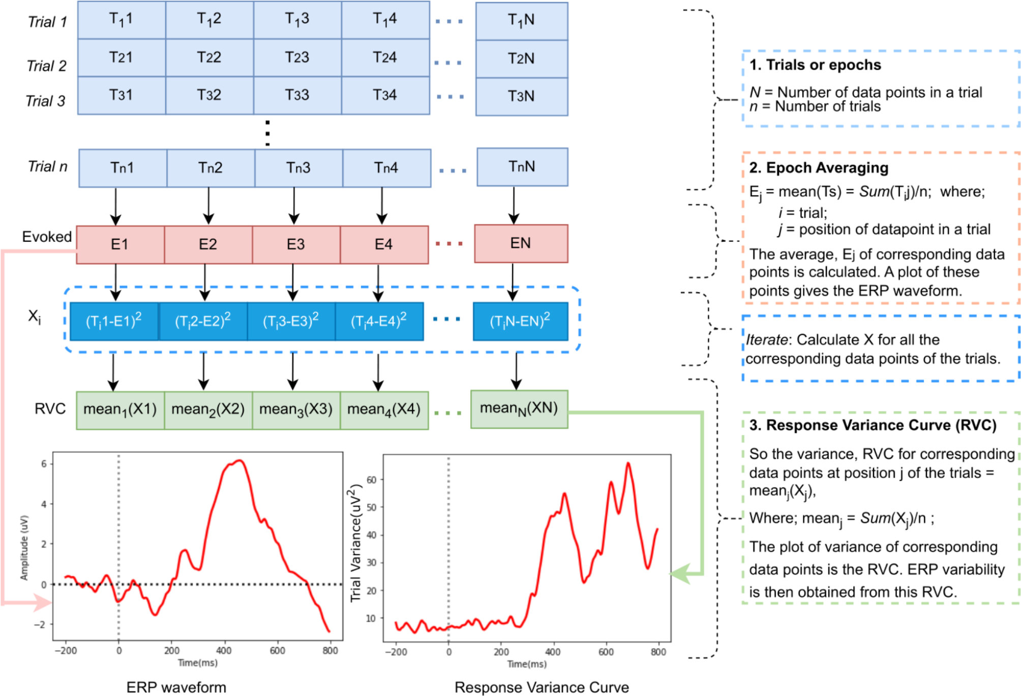
記住我
We identified five novel variants in the PSEN1 gene (Family A: p.Val103_Ser104delinsGly, Family B: p.Ala275Thr, Family C: p.Lys395Ile, Family D: p.Pro264Se, Family E: p.Ile414Thr). No additional variants were detected in PSEN2, APP, or other dementia causative genes (e.g., MAPT, GRN, and TARDBP). The main clinical features of the PSEN1 variant carriers are summarized in Table 1. The mean age at onset in newly identified families was 43.5 years (range 36–54), which is consistent with early-onset AD due to PSEN1.
Family A: p.Val103_Ser104delinsGlyThe proband (patient III1, Fig. 2a) came from a family with a history of early-onset dementia (<65 years) in three generations. Cognitive complaints emerged at age 39 years, characterized by memory loss, word-finding difficulties, and geographic disorientation. The proband was first assessed one year after symptom onset: MMSE score was 19, CDR® was 1, and Functional Activities Questionnaire (Pfeffer) score was 11. Brain MRI showed global atrophy, and FDG-PET revealed hypometabolism in the posterior cingulate cortex, precuneus, and temporoparietal cortices. One year later, the proband started to have myoclonus and the MMSE score was 13. Two years later, the proband was unable to identify family members, got lost inside the house, and could no longer recognize himself in the mirror; MMSE score was eight. APOE haplotype was E3/E3. The patient passed away at the age of 44 years due to complications of dementia (pneumonia), 5 years after disease onset. Seizures were described in the last months of his disease. The proband had one sibling diagnosed with early-onset AD (patient III2, Fig. 2a), as well as two first-degree cousins (patient III5 and III6, Fig. 2a).
Fig. 2
Identification of DIAD variants in densely affected Alzheimer’s disease (AD) pedigrees. Individuals with MCI and dementia have been classified as symptomatic and are represented with shaded rhombus. All generations under the family average age at onset were excluded, and gender has been masked to maintain anonymity. Diagonal lines represent deceased individuals. All symptomatic participants in the study were labeled with symptomatic age at onset. If the age at onset was unknown, the data were labeled as not available (Unk). Arrows indicate the index case. (+) indicate those individuals with DNA, all of whom are mutation/variant carriers. (−) indicate those individuals with DNA, all of whom are NOT mutation/variant carriers. For asymptomatic mutation carriers under the family age, the results of genetic testing were excluded to prevent potential disclosure of mutation status
Sequencing of the proband DNA revealed a PSEN1 variant that would result in the deletion of the amino acids valine at codon 103 and serine at codon 104 and the insertion of a glycine in place, without changing the translation-reading frame (in-frame) (p.Val103_Ser104delinsGly). The proband’s symptomatic sibling and cousins carry the same variant and the asymptomatic aunt (patient II5, Fig. 2a) and sibling (patient III3, Fig. 2a) did not carry the variant, suggesting that the variant segregated with disease in this family.
To explore whether these variants of unknown significance in PSEN1 impact Aβ levels in a manner consistent with known pathogenic mutations in PSEN1, we used a N2A-PS1/PS2 KO cell line in which endogenous presenilin genes are deleted [22, 25]. PSEN1 p.Val103_Ser104delinsGly produced a significant increase in the extracellular Aβ42/40 ratio compared with PSEN1 WT, which was consistent with known pathogenic mutations (Fig. 3).
Fig. 3
Cell-based model to assess the impact of variants of unknown significance in PSEN1 on Aβ levels. A Diagram of the location of variants of unknown significance in PSEN1. B Mouse N2A-PS1/PS2 KO cells were transiently transfected with plasmids containing APP WT and PSEN1 WT, known pathogenic mutation (ΔE9), or a variant of unknown significance. After 48 h, media was collected and analyzed for Aβ42 and Aβ40 by ELISA. B. Ratio of Aβ42/40 expressed relative to PSEN1 WT. Aβ42 (white box) and Aβ40 (gray box) levels expressed relative to PSEN1 WT. Graphs represent mean ± SEM. Significance indicated by Dunnett’s t-test (*, p < .05). C. Cells lysates were analyzed by SDS-PAGE and immunoblotting as described in the “Methods” section. Immunoblots were probed with 6E10 (full-length APP). The immunoblot is representative of 2 independent experiments
Finally, to determine pathogenicity, we used the algorithm as described by Hsu et al. [24]. PSEN1 p.Val103_Ser104delinsGly segregates with the disease. This variant was absent from the large population-based gnomAD genome and exome databases (Table 2), with no prior documentation in the medical literature. A missense variant in the same position (p.Val103Gly) [26] was previously reported and identified as a variant of unknown significance. The absence of a rare variant in large, population-based cohorts suggests that the variant may be pathological in nature and thus evolutionarily selected against. This residue is highly conserved between PSEN1 and PSEN2. In a cell model, PSEN1 p.Val103_Ser104delinsGly, produced Aβ profiles consistent with known pathogenic mutations. Thus, the PSEN1 p.Val103_Ser104delinsGly variant meets the proposed criteria for designation as pathogenic.
Table 2 Variants of unknown significance evaluated by the pathogenicity algorithmFamily B: p.Ala275ThrThe index case (patient II1, Fig. 2b) in this family developed depressive symptoms and loss of episodic memory at 45 years old, followed by aberrant motor symptoms and obsessive-compulsive tendencies, manifesting with increased cleaning behaviors. By 47 years, language problems ensued, and at age 49 (age at which was first assessed) profound expressive aphasia was noticed. There was no history or signs of parkinsonism, myoclonus, psychotic symptoms, or seizures. Brain MRI revealed hippocampi and temporoparietal atrophy. Brain SPECT revealed right posterior parietal hypoperfusion. The proband was diagnosed with early-onset AD. The proband’s parent (patient I1, Fig. 2b) had AD with onset at age 56 years and death at 66 years.
Sequencing of the proband DNA (Fig. 2) revealed a single base pair substitution (GCC to ACC) at codon 275 in exon 8 of PSEN1, resulting in an alanine-to-threonine change (p.Ala275Thr). PSEN1 p.Ala275Thr was absent in from both gnomAD genome and exome databases. This residue was highly conserved between PSEN1 and PSEN2. PSEN1 p.Ala275Thr has not been reported in ClinVar, but two variants in the same position (p.A275V, p.A275S) were previously reported and identified as pathogenic [27, 28], supporting the damaging effect of amino acid changes in position 275. Cells expressing PSEN1 pAla275Thr produced a significant increase in Aβ42 and Aβ40 levels without altering the ratio (Fig. 3). These findings suggest that PSEN1 p.Ala275Thr is a likely pathogenic variant.
Family C: p.Lys395IleThe proband (patient III2, Fig. 2c) was identified in a Brazilian family with three generations of early-onset AD and mean age-at-symptomatic onset of 51.3 years. The proband’s symptoms started at the age of 51 years, with episodic memory problems and later development of anomia and geographic disorientation. The proband had no major neuropsychiatric symptoms aside from apathy. Neurological examination revealed a postural tremor of both hands, which had been present since 30s (suggestive of essential tremor) and had mild rigidity in his upper right limb. His first MMSE score (at age 54 years) was 16, with progressive worsening. Four years after the first assessment (seven years after the onset), MMSE score was six. First MRI was interpreted as “normal.” EEG was normal. APOE haplotype was E3/E3. Blood tests did not reveal an alternative cause for dementia.
The proband’s parent (patient II1, Fig. 2c) was diagnosed with AD and parkinsonism, with age-at-symptomatic onset of 55 years, and the grandparent (patient I1, Fig. 2b) had similar symptoms with onset at 60 years. The proband’s parent died at age 53 due to a workplace accident. The proband’s sibling (patient III3, Fig. 2a) was diagnosed with early-onset AD with symptomatic onset at age 55 years.
Sequencing of the proband revealed a single base substitution (AAA to ATA) at codon 395 in exon 11 of PSEN1, resulting in a replacement of the lysine amino acid at codon 395 by isoleucine (PSEN1 p.Lys395Ile). The variant was present in several family members, segregating with symptomatic disease. The proband’s sibling carried the same variant (patient III3, Fig. 2c). The asymptomatic older sibling (patient III1, Fig. 2a) did not carry the variant (at the time of testing the older sibling was eight years older than the mean family age-at-symptomatic onset). PSEN1 p.Lys395Ile was absent from population-based databases (gnomAD genome and exome) and not reported in ClinVar (as of Dec 2021). Lysine at position 395 is highly conserved across species, suggesting that its replacement by isoleucine may be deleterious. In a cell model, PSEN1 p.Lys395Ile led to a significant increased extracellular Aβ42/40 ratio. Applying the algorithm for assessing pathogenicity, we propose that PSEN1 p.Lys395Ile represents a pathogenic variant.
Family D: p.Pro264SerThe proband (patient III2, Fig. 2d) was identified in a family with three generations of early-onset AD and with a mean age-at-symptomatic onset of 48.5 years. The proband started forgetting messages at work when she was 45 years old. Memory problems worsened over the coming 1–2 years, eventually requiring cessation of work. The proband had episodes of geographic disorientation and became more disorganized. During the first clinical assessment (at age 48 years), the MoCA score was 22 (points lost for impaired clock draw and inability to recall five words after a delay). Neuropsychological testing confirmed impaired verbal and visual recall, with relative preservation of semantic and phonemic fluency.
Sequencing of the proband (patient III2, Fig. 2d) revealed a single base pair substitution (CCT to TCT) at codon 264 in exon 8 of PSEN1, resulting in a proline-to-serine change (PSEN1 p.Pro264Ser). This variant was not identified in ClinVar (as of Dec 2021) and was absent from gnomAD genome and exome databases (Table 1). This residue is highly conserved between PSEN1 and PSEN2. Another variant in the same position (p.Pro264Leu) [29, 30] was previously reported and identified as pathogenic, supporting the damaging effect of amino acid change in position 264. Cells expressing PSEN1 p.Pro264Ser produced a significant increase in the extracellular Aβ42/40 ratio (Fig. 3). Thus, applying the algorithm for assessing pathogenicity, we propose that the PSEN1 p.Pro264Ser represents a likely pathogenic variant.
Family E: p.Ile414ThrThe proband (patient III1, Fig. 2e) developed irritability and apathy at 51 years old, followed by progressive memory loss. At baseline assessment, the proband scored 23/30 on the MMSE. Global CDR® was 0.5 (very mild dementia). Reassessment one-and-a-half years later revealed rapid progression in the clinical course, scoring 16/30 on the MMSE. Global CDR® was 2 (moderate-severity dementia). Ideomotor apraxia was present on examination. No myoclonic jerks or abnormal movements were observed. APOE haplotype was E3/E4. The proband’s parent developed a “progressive memory decline” at age 50, and several family members (Fig. 2) showed similar clinical presentation. This family originated in a remote community in rural Mexico, with a few family members dispersed across Mexico City. To date, 6 of 12 family members within the age at onset range have reported cognitive impairment, three of whom are deceased. Segregation of the variant was not possible in this family. The mean age-at-symptomatic onset in this family is 59.0 years (range 50–74).
Sequencing of the proband (patient III1, Fig. 2e) revealed a single base pair substitution (ATT to ACT) at codon 414 in exon 11 of PSEN1, resulting in an isoleucine-to-threonine change (PSEN1 p.Ile414Thr). PSEN1 p.Ile414Thr was absent in 250 Mexican mestizo exomes and from both gnomAD genome and exome, and has not been reported in ClinVar (as of December 2021). This residue is highly conserved between PSEN1 and PSEN2. Cells expressing PSEN1 p.Ile414Thr produced Aβ42 and Aβ40 levels similar to PSEN1 WT and did not alter Aβ in a manner consistent with a known pathogenic mutation. While the bioinformatic predictions are consistent with potential pathogenicity, bioinformatic findings alone are not sufficient to define pathogenicity. Based on the current evidence (insufficient segregation data in the affected family and in vitro results indicating that PSEN1 p.Ile414Thr does not alter Aβ), we propose that PSEN1 p.Ile414Thr represents an AD risk factor.
Finally, for all the novel variants, N2A PS1/PS2 KO cells were co-transfected with APP and PSEN1 variants to further analyzed for APP expression. Expression was confirmed with immunoblotting, suggesting the alteration in Aβ42/40 levels in PSEN1 variants was independent of APP expression (Fig. 3C).
留言 (0)