Human samples
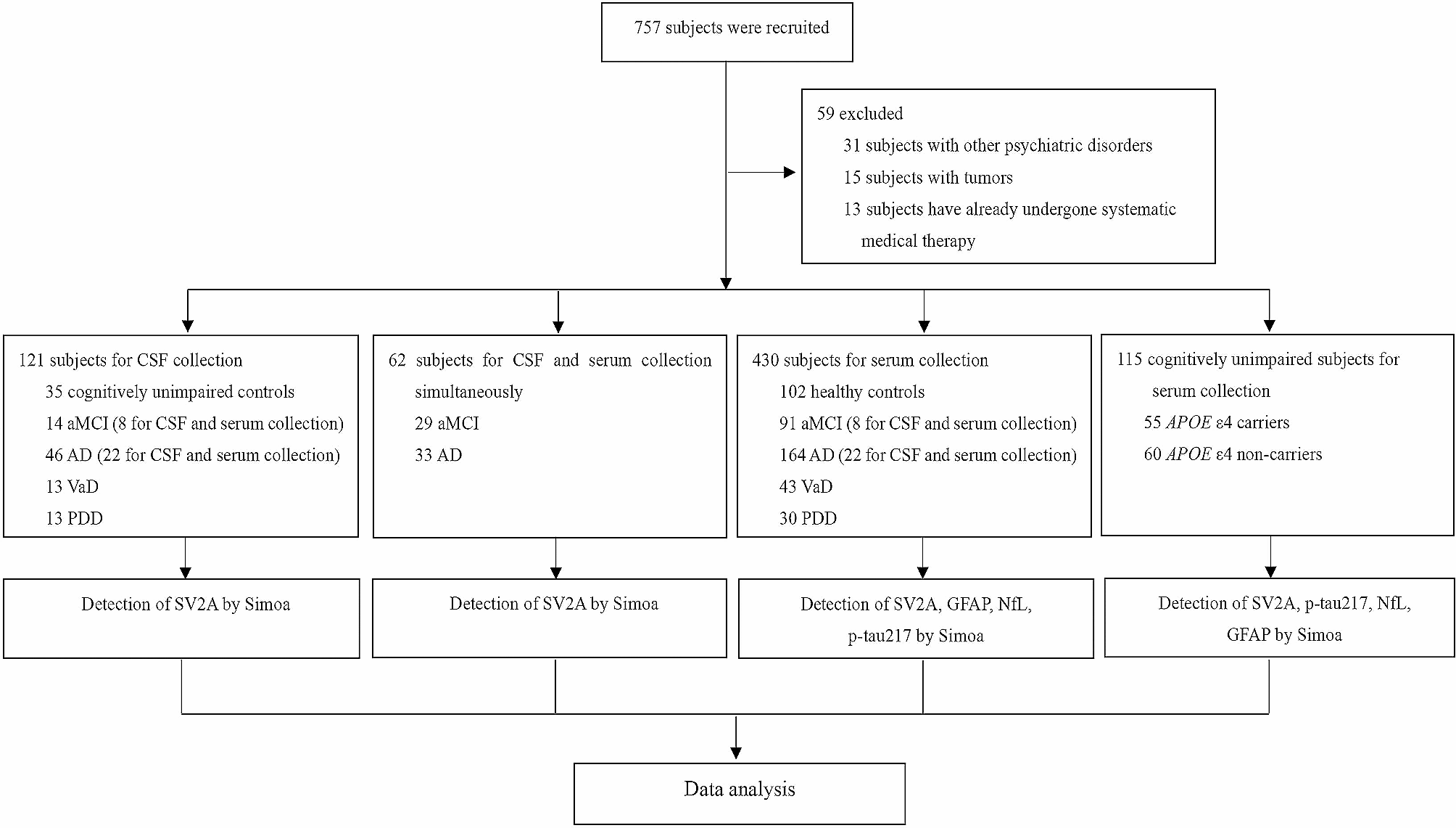
A total of 90 subjects were enrolled for cerebrospinal fluid (CSF) detection: 30 patients with normal cognition including peripheral nerve disease (n = 8), myelopathy (n = 8), dural arteriovenous fistula (n = 5), cerebrovascular malformation (n = 4), and body weakness (n = 5); 30 patients with mild cognitive impairment (MCI) of Alzheimer’s type; and 30 patients with Alzheimer’s dementia. All the patients were diagnosed by doctors of the Neurology Department of Xuanwu Hospital according to the criteria of NINCDS-ADRDA. Approximately 500 μl of CSF was collected and stored at −80°C for further analyses. Then, 100 subjects were enrolled for serum collection: 30 patients with MCI, 30 patients with Alzheimer’s dementia, and 40 age-matched healthy controls. The same diagnostic criteria were applied. Approximately 1 ml of serum was obtained and stored at −80°C. The design of this study was approved by the ethics committee of Xuanwu Hospital of Capital Medical University and conducted in accordance with the guidelines of the Declaration of Helsinki.
Animal models
2-, 5-, and 8-month-old male APPswe/PS1dE9 (APP/PS1) mice (n = 6 for each age group) and male C57BL/6 mice with the corresponding ages (n = 6 for each group) were purchased from Nanjing Biomedical Research Institute of Nanjing University (Nanjing, China). After a week’s balance, the mice were euthanized. Serum samples and brain tissues were collected for further analyses. In a separate experiment, 5-month-old male APP/PS1 mice (n = 12) and 5-month-old male C57BL/6 mice (n = 6) were used for ML264 administration. APP/PS1 mice were further divided randomly into two groups, namely, the AD control group (n = 6) and the ML264-treatment group (n = 6). The mice were injected intraperitoneally with 5 mg/kg ML264 (MedChemExpress) or vehicle (PBS) every other day for 15 weeks. After 15 weeks of treatment, the mice were subjected to behavioral tests. At the end of the test, all the animals were sacrificed, and the brain tissues were collected for further analyses. All the mice were housed in a specific pathogen-free room under a 12-h/12-h light/dark cycle and given free access to diet. All the animal procedures were performed in accordance with the criteria outlined in the Guide for the Care and Use of Laboratory Animals (National Institutes of Health, Bethesda, MD) and approved by the Animal Care and Use Committee of Xuanwu Hospital of Capital Medical University.
Morris water maze test
The Morris water maze is a white circular pool (120 cm in diameter) filled with water and dyed with milk to appear white. The pool was divided into four quadrants of equal areas and labeled clockwise as quadrants A, B, C, and D. A platform was placed at the center of one of the quadrants of the pool and submerged 2 cm below the water. In this test, the platform was set in quadrant B. On the day before the experiment was initiated, the mice were acclimated to swimming for 60 s in the absence of the platform. Then, they were given four times of training with an inter-trial interval of 1 h each day for 4 consecutive days, and their escape latencies were recorded. If a mouse did not reach the platform within 60 s, it would be guided to the platform by the experimenter. On day 5, a probe test was performed. In this test, the platform was removed from the pool with a cutoff time of 60 s, and the time they first crossed the hidden platform and the swimming path was recorded.
Lentivirus
The full-length KLF5 cDNA (Genbank ID: NM_009769) or APPswe cDNA (Genbank ID: NM_201414) was amplified through polymerase chain reaction (PCR), subsequently cloned into the pLV-CMV vector to construct KLF5-overexpressing lentivirus (KLF5-over) and APPswe-overexpressing lentivirus (APP-over), and confirmed by DNA sequencing. Short-hairpin RNA (shRNA) targeting KLF5 (shRNA-KLF5) and BACE1 (shRNA-BACE1) were purchased from Syngentech. The target sequences are shown in Supplementary Table S1. A lentivirus was produced in accordance with the manufacturer’s protocol. The titer of the purified virus was determined through flow cytometry.
Cell culture and cell treatment
HEK293T, SH-SY5Y, and HT22 cells were purchased from ATCC. HEK293T and HT22 cells were cultured in Dulbecco’s minimum Eagle’s medium (Biological Industries) containing 10% (v/v) fetal bovine serum (Biological Industries) and 1% (v/v) penicillin/streptomycin (Invitrogen, CA, USA), while SH-SY5Y cells were maintained in a minimum essential medium (Biological Industries) supplemented with 10% (v/v) fetal bovine serum (Biological Industries) and 1% (v/v) penicillin/streptomycin at 37°C in a humidified atmosphere containing 5% CO2. For lentivirus transfection, HT22 cells were seeded in 12-well culture plates at a cell density of 1 × 10⁵/well 1 day before transfection. Then, lentiviral vectors were transfected into cells by using polybrene. After 72–96 h, the cells were harvested for further analyses. For PMA treatment, PMA (MedChemExpress) was dissolved in DMSO, and SH-SY5Y cells were seeded in 10-cm culture plates at a cell density of 3 × 10⁷/plate 1 day before the treatment. Then, PMA was added to the medium with a final concentration of 100 nM. After 2 h, the cells were harvested for ChIP assay.
Luciferase assay
For plasmid constructions, the genomic fragments that contained all of the putative KLF5-binding sites on the BACE1 promoters (~2.0 kb upstream from the transcription start site) and the mutation constructs that mutated the putative KLF5-binding site on the promoter at −1556 to 1547 (site 1), −1467 to −1458 (site 2), −1264 to −1255 (site 3), −988 to −979 (site 4), and −599 to −590 (site 5) were constructed through base synthesis. All the products were subcloned into the pGL4.1-promoter vector (Promega, WI, USA) and confirmed by DNA sequencing. For luciferase assay, HEK 293T cells were co-transfected with pcDNA3.1-KLF5 (300 ng) or pcDNA3.1 (300 ng) vector and one of the constructs and pRL-CMV Renilla luciferase by using jetOPTIMUS transfection reagent (PolyPlus). After 48 h, the cells were lysed for luciferase activity. Firefly luciferase activity was normalized to Renilla luciferase activity.
RNA extraction and quantitative real-time PCR
Total RNA was extracted from brain tissue samples or cultured cells by using Trizol reagent (Invitrogen) in accordance with the manufacturer’s protocol. A reverse transcription reaction was performed with 1.0 μg of total RNA and 5× All-In-One RT MasterMix (ABM). TB GreenTM Premix Ex TaqTM II (TaKaRa) was utilized for quantitative PCR in a final volume of 20 μl on a LightCycler 480 system (Roche) under the following conditions: 95°C for 30 s followed by 40 cycles of amplification (95°C for 5 s and 60°C for 30 s). Melting curve analysis from 60 to 90°C and continuous fluorescence were performed following amplification. The average threshold cycle (Ct) of fluorescence units was applied to analyze the mRNA levels, which were normalized to a housekeeping gene (β-actin or GAPDH). The relative level of each gene was calculated using the 2-ΔΔCt method. The primers are shown in Supplementary Table S2.
Protein extraction and western blot
The cells or brain tissue samples were homogenized in RIPA buffer to extract total proteins. Protein concentrations were examined using the BCA protein assay (Thermo Fisher Scientific, CA, USA). Equal amounts of total protein were separated on 8% or 15% sodium dodecyl sulfate-polyacrylamide gels and then transferred onto polyvinylidene difluoride membranes (EMD Millipore, Darmstadt, Germany) by using the Trans-Blot TurboTM blotting system (Bio-Rad). The following primary antibodies were used: rabbit anti-KLF5 (1:1000, Proteintech, 21017-1-AP, Wuhan, China), rabbit anti-BACE1 (1:1000, Abcam, ab183612, Cambridge, UK), and rabbit anti-APP-CTF (1:1000, Proteintech, 27320-1-AP). An enhanced chemiluminescence Western HRP substrate kit (EMD Millipore) was used to detect specific proteins. The target bands were checked in accordance with the “Observed Molecular Weight” of the manufacturer’s instructions (KLF5: 51-55KD, BACE1: 68-70KD) and all target bands were consistent with the reported studies [15, 16].
Chromatin immunoprecipitation
A chromatin immunoprecipitation (ChIP) assay was performed using a PierceTM agarose ChIP kit (Thermo Fisher Scientific) in accordance with the manufacturer’s protocol. SH-SY5Y cells and hippocampal tissues of 2-, 5-, and 8-month-old APP/PS1 mice were prepared for the assay. Briefly, the SH-SY5Y cells were treated with PMA (100 nM) for 2 h to induce KLF5, and hippocampal tissues (100–120 mg) were homogenized with a mortar and pestle for single-cell suspension preparation. Then, the cells were cross-linked in 10 ml of 1% formaldehyde (Thermo Fisher), and DNA was fragmented to 200–1000 bp in length through enzyme digestion. Each 150 μl of chromatin solution was immunoprecipitated using 10 μg of monoclonal anti-KLF5 (Santa Cruz, CA, USA) or IgG control overnight at 4°C. Input and immunoprecipitated samples were digested with proteinase K to reverse the cross-linking at 65°C for 90 min. DNA was purified and further analyzed through qPCR by using the primers against the BACE1 promoter (Supplementary Table S2).
Immunofluorescence and immunofluorescence laser confocal microscopy assay
Frozen sections of formalin-fixed brain tissues with 5-μm thickness were prepared. They were permeabilized with 0.5% Triton X-100, and antigens were retrieved. Then, the sections were blocked with 3% goat serum and labeled with mouse anti-BACE1 antibody (1:50 dilution; Santa Cruz) and rabbit anti-KLF5 antibody (1:200 dilution; Proteintech) overnight at 4°C. They were subsequently labeled with Texas red-conjugated goat anti-mouse and FITC-conjugated goat anti-rabbit secondary antibodies (Santa Cruz). Nuclei were stained with 4′,6-diamidino-2-phenylindole (Santa Cruz). After being mounted, the samples were analyzed through fluorescence microscopy or laser confocal microscopy.
Aβ plaque histology
Amyloid plaques were stained with Thioflavin-S. Frozen sections of formalin-fixed brain tissues with 5-μm thickness were prepared, rinsed with TBS thrice, stained with 0.3% Thioflavin-S (Invitrogen) in 50% ethanol for 8 min, washed with 50% ethanol thrice, and rinsed with TBS for 5 min. Then, they were covered with a glass cover by using an antifade mounting medium and observed under a fluorescence microscope.
β-Secretase activity assay
The activities of BACE1 in cells and mouse brains were determined using a β-secretase activity assay kit (Abcam). Briefly, cells and brain tissues were homogenized with an extraction buffer and then incubated with a reaction buffer and a β-secretase substrate at 37°C for 1 h. The BACE1 activity was measured using a multifunctional plate reader (Beckman Coulter) with Ex/Em = 335/495 nm.
ELISA
Aβ1-40 and Aβ1-42 levels in the cell culture medium and brain extract and KLF5 levels in the serum were measured using the corresponding sandwich ELISA kits in accordance with the manufacturer’s instructions (Aβ1-40 and Aβ1-42, Mlbio, and KLF5, Abebio, China).
Statistical analysis
Data were expressed as mean ± SEM and statistically analyzed using two-tailed Student’s test, Mann–Whitney test, one-way ANOVA with Tukey’s test, or two-way ANOVA in GraphPad Prism 6 (GraphPad, La Jolla, CA). Differences among groups were considered to be statistically significant at P < 0.05.

留言 (0)