
記住我


An 18-year-old boy with Behçet’s disease (HLA-A26 positive) who had previously undergone aortic valve replacement with an 18-mm ATS Open Pivot AP360 mechanical aortic valve prosthesis (ATS Medical, Inc, Minneapolis, MN) at the age of 12 years, presented sudden-onset general fatigue and was emergently transferred to the regional hospital. Since initial aortic valve replacement, 10 mg a day of prednisolone sodium succinate, 12 mg once weekly of methotrexate, 750 mg every 4–6 weeks of infliximab, and 35 mg every 2 weeks of alendronate sodium hydrate had been administered.
Chest X-ray showed displacement of the implanted mechanical valve (Fig. 1A, C). From a retrospective point of view, slight displacement of the valve prosthesis had been already confirmed 1 week ago at the scheduled follow-up (Fig. 1B). An echocardiogram revealed mobile valve prosthesis and severe aortic regurgitation. Just before leaving for our hospital for surgical treatment, a completely detached valve prosthesis was floating in the ascending aorta (Fig. 1C).
Fig. 1
Chest X-ray findings 3 months ago (A), 1 week ago (B), at emergent admission to the regional hospital (C), at leaving for our hospital (D), and at arriving at our hospital (E). Black arrows indicating mechanical valve
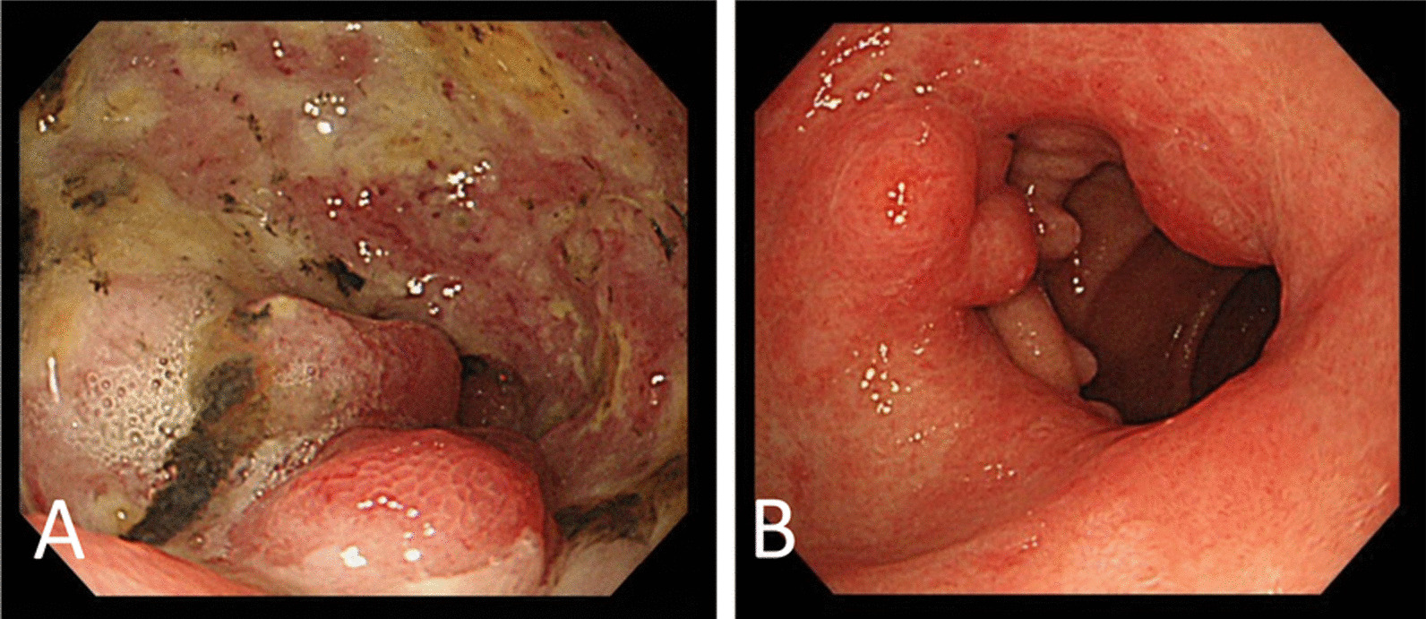
On arrival, the valve prosthesis was stuck to the transverse arch (Fig. 1D). Emergent removal of the previous mechanical valve from the aortic arch and redo aortic valve replacement were performed under total circulatory arrest. After a removal of fibrous tissues around annulus on where previous valve prosthesis was placed, 14 pairs of pledgeted 2–0 polyester terephthalate braided, non-everting mattress suture from deep subannular left ventricular endocardium to the aortic wall just above the true annulus were placed to prevent recurrent detachment of implanted valve prosthesis. After that, a newly fashioned 24-mm ATS Open Pivot AP360 valve could be implanted in the supra-annular position without any annular enlargement procedures. The patient was back to the intensive care unit with extracorporeal membrane oxygenation support. Histopathology of the surrounding tissue around a previous valve prosthesis showed no bacterial infection sign, then only inflammatory change was noted (Fig. 2A, B).
Fig. 2
A Photomicrograph (×2, scale bar = 500 μm) by elastica van Gieson staining showed fibrous tissue with granulation and degeneration. B Photomicrograph (×200, scale bar = 50 μm) by hematoxylin and eosin staining showed granulation tissue and degenerated collagen fibers, with microvasculature, fibroblast, and inflammatory cells
Extracorporeal membrane oxygenation was successfully decannulated 5 days later. Intra-arterial balloon pump and hemodialysis could be weaned off on post-operative day 8. Intra-cranial computed tomography was performed on post-operative day 29, which revealed the hypoxic ischemic change in bilateral basal ganglia and multiple old microinfarctions. Left ventricular dysfunction has remained since then, but catecholamine support could be discontinued 2 months later. Now 3 months have been passed since the emergent surgery. He was still supported by phosphodiesterase III inhibitor, but no longer supported by mechanical ventilator. Left ventricular end-diastolic diameter and ejection fraction were 43 mm and 25%, respectively.
Sixty mg a day of soluble predonine had been initiated since post-operative day 13, which then was gradually tapered to 30 mg a day for 3 months.
留言 (0)