The multiple linear regression model generated in this study to estimate AL, along with the application of this estimated AL in two different datasets, confirmed observations made previously that SE and K can be used to predict AL [15, 21]. Furthermore, we have shown that ALest can be applied for correcting eye transverse magnification error in FAZA measurements of OCTA image. Post-cycloplegic data from the training dataset were used to generate the relationship between AL, SE, and K. Here, the use of post-cycloplegic training data may increase the accuracy of the regression formulae as cycloplegia mitigates the varying effects of tonic accommodation [22,23,24,25].
The ALest formula in the present study did not include age as a variable, as population studies show that age may not have a statistically significant impact on axial length [26, 27]. Contrary to this is a study by Atchison et al. [28], which found older emmetropic eyes have longer axial lengths than younger emmetropic eyes. However, the correlation was poor (R2 = 0.04) and there were no significant trends for males or females alone. Atchison et al. [28] noted that this weak trend was primarily due to increases in lens thickness, with minimal changes in vitreous depth. Changes in lens thickness may contribute to changes in refractive error [26, 29], and thus will be reflected in the formula for ALest in the present study. Nevertheless, the performance of ALest in different age groups may be explored in the future.
Validation of ALest against ALact using Bland-Altmann analysis in the Raine Study Gen2-20 year follow-up cohort resulted in a lower mean difference and LoA for post-cycloplegic SE compared to non-cycloplegic SE datasets. Application of the ALest formula on the post-cycloplegic data of the Raine Study Gen2-20 year follow-up cohort would appropriately produce the smallest mean difference due to the mitigation of the effects of accommodation on SE. The larger mean difference between ALest and ALact observed in the non-cycloplegic SE data of the Raine Study Gen2-20 year follow-up group compared to that of the OCTA cohort may be explained by the relationship between age and cycloplegia. In a younger age group, it is known that the effect of accommodation is stronger, hence cycloplegia will result in a greater reduction in SE due to reduction in myopia. The mean ages of the Raine Study Gen2-20 year follow-up cohort and the OCTA cohort were 20 and 36 years, respectively. Hence, accommodation would have a greater impact on the non-cycloplegic SE values in the Raine Study Gen2-20 year follow-up cohort compared to that of the OCTA cohort, resulting in a greater mean difference.
In the present study, both ALest and ALact was applied in the Littmann-Bennett formula to correct for magnification error in FAZA. It is important to note that while the Littmann-Bennett formula is treated as the gold standard in magnification error calculation, a recent study by Lal et al. [30] questions the use of the Littmann-Bennett formula in correcting OCTA image magnification error and concluded that SE should be considered in conjunction with AL when correcting OCTA image magnification factor in patients with anterior eye changes, such as in contact lens use or post refractive surgery. ALest and ALact can be reliably applied and compared in FAZA magnification error correction using the Littmann-Bennet formula in the present study, as all participants had healthy anterior segments. Furthermore, Lal et al. [30] corrected for magnification error of vessel density, which is less sensitive and more difficult to correct for than FAZA and benchmarked their findings against a study by Obadas et al. [31] for clinical significance. However, these two studies used different imaging and analysis protocols. Lal et al. used the Zeiss AngioPlex, Cirrus HD-OCT 5000, Carl Zeiss Meditec Inc., which defines vessel density as the total vessel length by the image size within the central 1 mm macula zone in a 3 × 3 mm acquisition area whilst Obadas et al. [31] used the Revue XR Avanti system, Optovue, which defines vessel density as the area of all white pixels within an OCTA image divided by the total image size of the whole 6 × 6 mm image. Due to the differences in methodologies, the conclusions by Lal et al. [30] cannot be applied to our study.
The mean difference of the ALest values with those of ALact values have been reported in previous studies. Estimations of AL by Grosvenor and Scott [32] using linear regression models with corneal radius and SE yielded a mean difference of 0.38 mm (95% LoA: − 0.60, 1.36) mm, which is higher than the mean difference found in the present study. This may be due to the inaccuracies that arise from the type of linear regression model used by Grosvenor and Scott [32], which analysed the relationship between the AL/K ratio and SE values. Kim et al. [21] estimated AL using a formula derived from the Gullstrand model of the eye, and reported a mean difference of 0.18 mm (95% LoA: − 0.75, 1.10) mm. This larger mean difference reported may be due to the inaccuracies introduced into the AL estimation formula due to the assumptions adopted with the use of a Gullstrand model eye. In a study by Morgan et al. [13], a linear regression model based on post-cycloplegic SE and K of 144 participants was also used to estimate AL values. This model showed a mean difference of 0.13 mm (95% LoA: − 0.73, 0.99) mm when applied to a separate validation cohort of 1046 participants aged between 6 to 22 years. This mean difference is higher but comparable to that reported in this study and may be so due to the generation of a linear regression from a small cohort.
Others have utilised semi-automated computer processing to facilitate AL estimation. Tang et al. [33] compared machine learning with traditional multiple regression formulae in developing a method for estimating AL. Regression models were based on age, gender, K, SE and white-to-white diameter. Tang et al. [33] found that machine-learning methods outperformed traditional multiple regressions model for estimating AL, with the strongest machine-learning model AL prediction model having a R2 of 0.86, which is considerably robust, however Tang et al. [33] did not report a mean difference or 95% LoA. A deep-learning algorithm applied to colour fundus photographs was used by Dong et al. [34] to estimate AL, and resulted in R2 = 0.59 and mean difference of 0.16 mm (95% LoA: − 0.60, 1.27) mm. This is a higher mean difference and wider 95% LoA than observed in the present study. AL estimation using deep neural network visual interpretation of fundus images by Jeong et al. [35] yielded R2 = 0.67 while the mean difference and 95% LoA were not reported. The R2 value of AL estimation in the present study is higher than in the estimation methods of Dong et al. [34] and Jeong et al. [35]. Hence, even though these studies are promising for the use of semi-automated estimation of AL, further research in this area is needed to improve on these methods.
Whilst semi-automated computer processing may aid in AL estimation, there is value in traditional estimation techniques such as those described in this study owing to its ease of use. This does not discount the accuracy of the ALest. In comparisons of the magnification error correction performance of ALest with the AL estimation methods presented in the aforementioned studies [13, 21, 32,33,34,35], it is important to note that even marginal improvements in the accuracy of estimation methods may provide significant advances in magnification error correction for FAZA, and possibly for other fundus parameters like vessel density.
The ALest demonstrated in the present study has robust agreement with a high ICC and narrow LoAs when compared against ALact in correcting transverse magnification error. Since the work published by Sampson et al. [3], multiple studies have acknowledged the inaccuracy in fundus analysis without correcting for magnification error and have proceeded to include AL-based correction of fundus parameter analyses [20, 36, 37]. This is particularly true for FAZA measurements, especially in high myopes [38, 39]. In addition to FAZA correction, magnification error correction has been used in studies investigating macular vessel density, impacting on the accuracy of these measurements [40].
Here, FAZA values corrected with ALact were not significantly different from uncorrected FAZA values or FAZA corrected with ALest (P > 0.05, Table 3). Sampson et al. [3] also did not find a significant difference between corrected and uncorrected FAZA measurements with sample size of 67 eyes. However, the authors demonstrated that not correcting for magnification factor contributed to an error of FAZA measurements of more than 5% for 74% of their participants. This is similar to the error found in the OCTA participants of the present study when comparing uncorrected FAZA with FAZA corrected with ALest and ALact. FAZA correction with AL also did not reach significance in a study be Linderman et al. [8] with a sample of the 232 eyes. However, Linderman et al. [8] pointed out that not correcting FAZA for the magnification factor resulted in errors to FAZA of up to 31% (0.07 mm2). This is comparable to the percentage changes in FAZA in OCTA participants when comparing uncorrected FAZA with FAZA corrected with ALest and ALact.
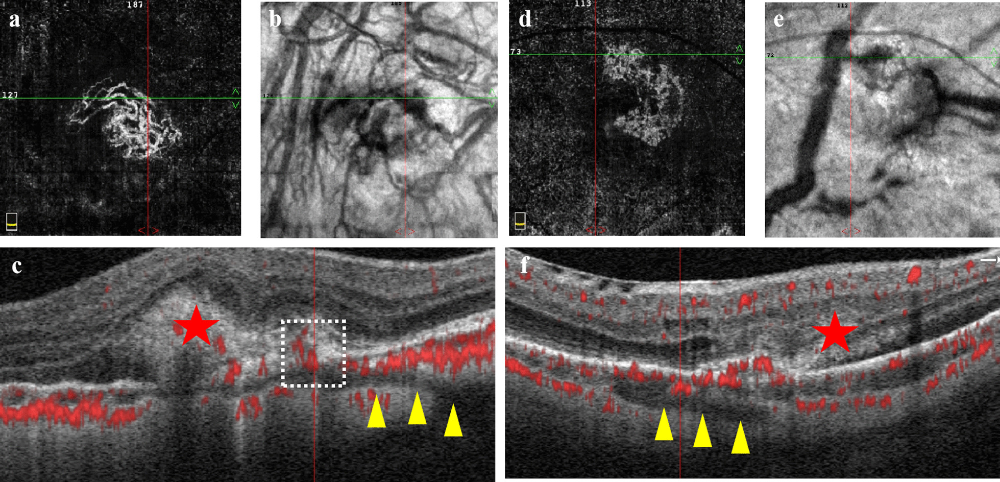
In line with these studies, whilst FAZA correction for magnification error did not reach statistical significance, there was an important increase in the accuracy of FAZA measurements after correction for eye magnification factor. As seen in Fig. 2c, d, the deviation from the true FAZA value is higher for uncorrected FAZA than FAZA corrected with ALest. Therefore, FAZA corrected by the ALest is more accurate than uncorrected FAZA. This is confirmed when adjusting FAZA for magnification error with ALest resulted in a twofold reduction in the width of the 95% LoA, and a lower frequency of exceeding the CR in the present study, compared to uncorrected FAZA. This increase in accuracy is important for standardising FAZA measurements to enable comparisons of FAZA across studies. It may also provide more sensitive monitoring of FAZA in participants over time. Understanding this importance, in cases where ALact is not available for FAZA correction, it is advisable to correct for magnification error in FAZA measurements using ALest rather than accepting uncorrected measurements.
The use of ALest in magnification error correction may be important in assessing the presence of disease or for monitoring disease progression in cases where actual AL measurements are unavailable. Currently, magnification factor correction is being performed more commonly when monitoring fundus parameters in diseased states such as diabetic retinopathy and diabetic macular oedema [41]. Traditionally, magnification factor correction is performed with ALact measured with an ocular biometer. These devices have become increasingly popular in clinical practice and are necessary in managing myopia, in conjunction with routine corneal parameters. However, there may be instances where retrospective correction of magnification error in OCTA images is needed to be performed in the absence of axial length measurements; our study allows for this if K and SE are known.
In clinics without access to these biometry devices, the present study suggests that ALest may have a role in correcting magnification factor for monitoring fundus parameters in diseases that impact the fundus. The use of ALest may also provide for a more cost-effective means of performing magnification factor correction than using ALact, as specialised biometry devices are not required. Future studies should assess the performance of ALest in magnification error correction in a range of diseased states including diabetic retinopathy, age related macular degeneration and glaucoma. Additionally, conditions that impact the anterior segment of the eye, such as keratoconus, cataracts, and refractive surgery, may result in changes to refractive error and K results independent of AL [30], and thus the performance of ALest in magnification error correction in these conditions should also be studied.
Future studies may also wish to assess the performance of ALest in myopic or hyperopic cohorts specifically, as the training, validation and test cohorts of the present study include participants with a wide range of refractive errors. In addition, FAZA magnification error correction with ALest resulted in an overestimation of FAZA for shorter AL and an underestimation of FAZA for longer AL, when compared to magnification error correction of FAZA with ALact (Fig. 2c; Additional file 4: Fig. S4). This may indicate the need for further refinement of the ALest formula in myopic or hyperopic conditions. However, clinicians should be reassured that many of these differences in FAZA corrected with ALest and ALact are subtle, with 98.9% of measurements falling within the CR. Rates of myopia may vary between ethnicities in paediatric populations [42, 43]. However, the impact of ethnicity on rates of myopia in adults is less clear [44]. Nevertheless, the Raine Study Gen2-20 year follow-up cohort is predominately Caucasian, and hence the performance of ALest in cohorts of different ethnic backgrounds will be an opportunity for further study.
The instruments used to determine K and autorefraction in the training and validation cohorts were different from those of the test cohort. Since measurements from different machines may vary for a given parameter, future studies may test the performance of ALest across different instrumentations and measurement protocols. Additionally, future studies may choose to use larger ranges of AL, K and SE to assess if differences between uncorrected FAZA and FAZA corrected with ALest or ALact reach statistical significance and is of clinical relevance. It is important to note that though FAZA has a strong dependence on magnification error correction, the effect of image correction also impacts vessel density. Vessel density magnification error correction is also influenced by the FAZA included in the region of analysis. To truly determine the effectiveness of the proposed formula for ALest, future studies should expand the use of ALest in assessing the accuracy in vessel density magnification error correction.


留言 (0)