
記住我


 Significance Statement
Hyperphosphatemia is a major complication in the later stages of CKD, causing vascular calcification. We have identified 54 kidney-enriched genes, 19 of which are expressed in renal primary proximal tubule cells. One of the proximal tubule–specific genes, TMEM174, interacted with NPT2A, and its knockdown attenuated the reduction of NPT2A protein by fibroblast growth factor 23 (FGF23) and parathyroid hormone (PTH) treatments in proximal tubule cells. TMEM174 knockout mice had significantly increased levels of serum phosphate, FGF23, and PTH, resulting in vascular calcification.
AbstractBackground The proximal tubules play a critical role in phosphate (Pi) homeostasis by reabsorbing Pi via sodium-dependent Pi cotransporters. NPT2A is a major proximal-specific Pi cotransporter, whose expression is regulated by circulating hormones, such as parathyroid hormone (PTH) and fibroblast growth factor 23 (FGF23). In this study, we aimed to find a novel regulator in Pi homeostasis.
Methods Using RNA-seq and RT-qPCR analysis, we identified proximal tubule cell–enriched genes. We next used RNAi screening of the identified proximal tubular cell–enriched genes to identify a novel proximal tubule–specific gene that contributes to FGF23- and PTH-mediated inhibition of Pi uptake and NPT2 reduction. We created mice lacking this novel regulator of Pi homeostasis to examine whether the novel regulator contributes to Pi homeostasis in vivo.
Results We identified 54 kidney-enriched genes, 19 of which are expressed in renal primary proximal tubule cells. One of the proximal tubule–specific genes, TMEM174, interacted with NPT2A, and its knockdown blocked the reduction of NPT2A protein by FGF23 and PTH treatments in human and opossum proximal tubule cells. TMEM174 KO mice had significantly increased levels of serum Pi, FGF23, and PTH, resulting in vascular calcification.
Conclusions TMEM174 is a novel regulator of Pi homeostasis that interacts with NPT2A.
Inorganic phosphate (Pi) is crucial for several important cellular processes, including energy metabolism, bone formation, signal transduction, or as a constituent of phospholipids and nucleic acids.1,2 During a steady state, phosphate levels are balanced by Pi excretion in the urine and feces. Pi in the plasma or extracellular fluid undergoes one of three fates: transport into cells, deposition in bones or soft tissue, or elimination, predominantly by the kidneys. Maintenance of normal serum phosphate levels is primarily achieved through a tightly regulated process of Pi reabsorption from the glomerular filtrate. Within the nephron, approximately 85% of Pi reabsorption occurs within the proximal tubule.2,3 The remainder of the nephron plays a minor role in Pi regulation, and the transporters involved have yet to be identified.1,3 Within the proximal tubule, Pi transport from the ultrafiltrate across the proximal tubule epithelium is an energy-dependent process that requires sodium.
The three renal sodium-dependent Pi cotransporters, NPT2A (SLC34A1), NPT2C (SLC34A3), and PiT-2 (SLC20A2), are all positioned in the apical brush border membrane (BBM) of renal proximal tubule cells and use energy derived from the transport of sodium down their gradients to move Pi from the luminal filtrate into the cell.1⇓–3 Renal control of Pi reabsorption is mainly regulated by two phosphaturic hormones: (1) parathyroid hormone (PTH) and (2) fibroblast growth factor 23 (FGF23). PTH causes decreased renal reabsorption of Pi by decreasing the abundance of NPT2A, NPT2C, and PiT-2 in the renal proximal tubule BBM.4,5 However, we previously reported that the mechanisms underlying PTH regulation of the Pi cotransporters are very different.6 FGF23 is an endocrine hormone produced in osteoblasts in response to increases in serum Pi.7⇓–9 To exert its physiologic effects on the proximal tubule, FGF23 requires the presence of a cofactor, Klotho (KL), which is produced in the renal tubules and activates FGF receptors (FGFRs) in the renal proximal tubles.10,11 FGF23 reduces the expression and activity of NPT2A through the activation of MAPK in the renal proximal tubules.7 Pi is a well-established risk factor for all-cause and cardiovascular mortality in patients with CKD12⇓⇓–15 and in the general population.16,17 Because the kidney has a key role in Pi regulation, Pi homeostasis is disturbed in patients with CKD. Therefore, patients with ESKD have extremely high levels of PTH and FGF23, yet they have higher levels of serum Pi and lower levels of calcitriol (1,25[OH]2D).18⇓⇓⇓⇓–23 These observations suggest CKD is a state of FGF23/PTH resistance. Serum Pi is a major inducer of the production and secretion of PTH and FGF23 in the parathyroid gland and bone, respectively.1,24 Extremely high levels of serum Pi, PTH, and FGF23 are all central factors in CKD-mediated cardiovascular complications, such as vascular calcification. A transmembrane (TMEM) protein is a type of protein that spans biologic membranes. Many of these proteins extend through the lipid bilayer of the plasma membrane, but others are located on the membrane of organelles. Over 250 TMEMs have been identified, but the biologic functions of many of them remain unknown. TMEM174 is a kidney-specific type-III TMEM protein and is expressed in various renal carcinomas. The transcription of the TMEM174 gene is regulated by AP-1 and CREB transcription factors.25⇓–27
In this study, we found that a proximal tubular–specific TMEM protein, TMEM174, is involved in Pi homeostasis. Mice lacking TMEM174 develop severe hyperphosphatemia and vascular calcification.
MethodsAnimalsTo generate TMEM174 knockout (KO) mice, we inserted a NEO cassette onto the exon 1 and exon 2 of TMEM174 (Supplemental Figure 2A). In brief, a DNA fragment encompassing a part of exons 1 and 2 of TMEM174 was amplified by PCR using the BAC clone RP23 as a template. A left homologous arm encompassing the DNA sequence upstream of exon 1 was PCR amplified and cloned at the ClaI site, upstream of the Neo cassette of the pBS-neo vector. Subsequently, a right homologous arm, containing the DNA sequence on part of exon 2 and downstream of exon 2, was PCR amplified and cloned at the NotI site, downstream of the Neo cassette. The targeting vector was linearized and introduced by electroporation into murine B6/129 hybrid EC7.1 embryonic stem cells. At the Bioengineering Core Facility at the University of Colorado Denver, karyotypically normal embryonic stem cell clones were microinjected into C57BL/6 blastocysts to produce chimeric founders.28,29 TMEM174 KO mice were backcrossed ten times with C57Bl6 or DBA/2J mice. Blood was drawn after a 4-hour fasting period. For PCR genotyping, genomic DNA was amplified with a forward primer (5′-ACCTTTATTCTTCTAAACAA-3′), reverse primer (5′-TGGTGTAGTACTGAGGGGGACTTGG-3′), and Neo primer (5′-ACCTGGTTGTCATGGAGGAG-3′). For the Pi adaptation study, animals were fed a low-Pi diet (TD.85010, 0.1% Pi/0.6% calcium [Ca]) or a high-Pi diet (TD.85349, 1.2% Pi/0.6% Ca) for 7 days.30 Animal experiments were approved by the Institutional Animal Care and Research Advisory Committee at the University of Colorado Denver. Concentrations of plasma PTH and FGF23 were analyzed with PTH and FGF23 ELISA kits (Immutopics, San Clemente, CA). 1,25(OH)2D levels were measured with an EIA kit (Fisher Scientific). Serum creatinine was analyzed by liquid chromatography with tandem mass spectrometry.28 Serum Pi, Ca, magnesium, sodium, potassium, and chloride levels were analyzed with a dry-chemistry autoanalyzer at the Comparative Pathology Core Facility at the University of Colorado Denver.
Cell CulturePrimary renal proximal tubular cells (PCS-400-010) were purchased from ATCC and were grown and maintained at 37°C, in an atmosphere of 5% carbon dioxide, in a growth media (PCS- 400-030) in the absence/presence of 10–100 nM FGF23 (2604-FG; R&D System) and 100 ng/ml of a secretory form of KL, which was synthesized and purified from HEK293-expressing pcDNA3.1-KL-His cells at the Cell Technologies Core Facility at the University of Colorado Denver. Opossum proximal tubular (OK-P) cells were grown and maintained in the absence/presence of 10–100 nM PTH (Bachem H-5460), as previously described.5,6 Cells were treated with 20 nM small interfering RNAs (siRNAs; ON-Target Plus siRNA library, LP_73887; Horizon Discovery) or OK-P TMEM174 siRNA (rCrUrCrArUrGrArUrArArUrGrUrUrGrCrArUrUrUrArUrUGA and rCrArArGrUrArCrCrArArGrGrCrArUrCrUrCrUrCrArCrUTT; Integrated DNA Technology) with RNAiMax reagent overnight. The primary renal proximal tubular cells are crude and show heterogeneous expression of NPT2A. The primary renal proximal tubular cells expressing NPT2A were isolated by single selection.5,29,31 In brief, primary renal proximal tubular cells were split into 0.6 cells per well in 96 wells. The levels of NPT2A mRNA in the single cells were determined by quantitative RT-PCR (RT-qPCR). We used a clone that highly expresses NPT2A.
RNA-SequencingTotal RNAs were isolated from the kidneys of 12-week-old male C57B6/J mice (n=3) using a Direct-zol kit. mRNA-sequencing (mRNA-seq) library construction and sequencing were performed at Novogene, in accordance with the manufacturer’s instructions, using a HiSeq4000 system (Illumina), as previously described.32
Western Blot AnalysisBBM vesicles were prepared from mouse kidney by the Ca2+ precipitation method, as described previously.5,6 BBM vesicle protein samples were subjected to SDS-PAGE. The separated proteins were transferred by electrophoresis to polyvinylidene difluoride membranes (Immobilon-P; Millipore). The membranes were treated with affinity-purified anti-NPT2A antibody6 (1:1000; or a custom polyclonal antibody against CLGVLSQHNATRL [Biomatik]), anti-NPT2C antibody6 (1:1000), PiT2 antibody (1:1000; H130; Santa Cruz Biotechnology), anti-TMEM174 antibody (1:1000; CSB-PA023755LA01HU for the immunoblot of mouse and human TMEM174 [Flare Biotech]; NBP1-82023 for the immunoblot and immunohistochemistry/immunofluorescence analysis of human TMEM174 [Novus Biologicals], or a custom polyclonal antibody for the immunoblot analysis of opossum TMEM174 [Biomatik]), anti-NHERF1 (1:5000; A302-974A; Bethyl Laboratories), Ca-sensing receptor (1:1000; 73303; Cell Signaling Technology), FGFR1 (1:1000; 9740; Cell Signaling Technology), PTHR1 (1:500; 50100; BiCell Scientific), EGF receptor (1:1000; 18986-1-AP; Proteintech), anti-FLAG M2 (1:1000; F1804; Sigma-Aldrich), and anti-actin (1:2000; 66009; Proteintech). Goat anti-rabbit or anti-mouse IgG–horseradish peroxidase conjugate was used as the secondary antibody, and signals were detected using Supersignal ECL reagents (Thermo Fisher). The uncropped pictures are shown in Supplemental Figure 2.
ProtterAmino acid sequences of human TMEM174 were obtained from the National Center for Biotechnology Information. Transmembrane topology of TMEM174 was predicted and visualized using Protter version 1.0.33
CoimmunoprecipitationOK-P cells were transfected with plasmids containing FLAG-NPT2A or FLAG-NHERF1 (28291; Addgene). Cell and kidney protein lysates were prepared using coimmunoprecipitation buffer (150 mM sodium chloride, 1% nonidet P-40, 50 mM Tris, pH 8.0). Lysate (500 μg) was incubated with either 40 μl of anti-DYKDDDDK G1 Affinity Resin (GenScript) or 50 μl of anti-NPT2A–conjugated protein A magnetic beads (73778S; Cell Signaling Technology) overnight at 4°C. Resins were washed five times with coimmunoprecipitation buffer.29 Immunoprecipitated proteins were analyzed using immunoblot analysis with anti-TMEM174 and VeriBlot for IP Detection Reagent (ab131366; Abcam).
RNA AnalysisqRT-PCR assays were performed using an Applied Biosystems StepOne Plus qPCR instrument. Quantitative expression values were calculated by the absolute standard curve method, using plasmid template (Open Biosystems) containing each target gene cDNA. The primer sequences are shown in Supplemental Table 1.
Immunohistochemical AnalysisImmunostaining analysis of human kidney sections (Zyagen) was performed as described previously.28,34 For immunostaining, the section was incubated with affinity-purified anti-TMEM174 antibodies (1:1000, recognizes human TMEM174 but not mouse TMEM174; NBP1-82023) overnight at 4°C. Sections were then treated with rabbit peroxidase (Dako) for 1 hour at room temperature. Immunoreactivity was detected by treatment with ImmPACT DAB peroxidase substrate (Vector Laboratories, Burlingame, CA). For immunofluorescence staining, the section was incubated with affinity-purified anti-TMEM174 antibodies (1:1000; NBP1-82023), Lotus tetragonolobus lectin (an renal apical membrane marker; FL-1321-2; Vector Laboratories) and anti-villin mAbs (sc-58897; Santa Cruz Biotechnology) overnight at 4°C. Immunoreactivity was detected by FITC, Alexa Fluor 488–conjugated anti-mouse IgG (A-11001; Thermo Fisher Scientific) or Alexa Fluor 568–conjugated anti-rabbit IgG (A-11011; Thermo Fisher Scientific). Prepared samples were observed with an Olympus FluoView FV1000 confocal microscope. Images were obtained at a resolution of 1024 × 1024 pixels. Calcified lesions in the aortic sinus were analyzed using Alizarin red staining, as previously described.28,35 We were not able to perform immunohistochemistry of TMEM174 in mouse kidneys. Flare Biotech TMEM174 antibody (CSB- PA023755LA01HU) detects mouse TMEM174 protein by immunoblot analysis but not immunohistochemistry, suggesting the antibody may recognize only denatured TMEM174 protein.
Pi Uptake of Renal Proximal Tubule CellsThe medium was replaced and incubated with uptake solution containing 0.1 mM KH232PO4 (1 μCi), as previously described.28,36,37 Routine uptake was performed at room temperature for 6 minutes and then stopped by washing the cells three times with cold stop solution. Cells were solubilized with 0.1 N sodium hydroxide, and the signals of 32P were counted using liquid scintillation.
Statistical AnalysesData were collected from more than two independent experiments and are reported as the mean±SEM. Statistical analysis was performed using a two-tailed unpaired t test for two group comparisons. Either one-way ANOVA with a Student–Newman post hoc test or two-way ANOVA with a Tukey post hoc test was used for multiple group and treatment comparisons. Significance was accepted at P<0.05.
ResultsIdentification of a Kidney-Specific Protein that Regulates Pi HomeostasisBecause (1) hyperphosphatemia has a much higher prevalence in patients with CKD compared with patients with other chronic diseases and (2) NPT2A, which is responsible for the majority (approximately 80%) of Pi reabsorption, is specifically expressed in the apical BBM of renal proximal tubule cells, we hypothesized that other kidney-specific proteins contribute to Pi homeostasis.1,10 To find a novel kidney-specific protein, we performed RNA-seq analysis with 20 mouse tissues (GSE189618 and Supplemental Table 1) and identified 54 kidney-enriched genes, including NPT2A (SLC34A1) and KL. We next examined which of the 54 kidney genes are expressed in human primary renal proximal tubule cells, which are a major site of Pi reabsorption. qPCR analysis of mouse tissue RNAs found that renal proximal tubule cells express 19 out of the 54 kidney-enriched genes at high levels (Supplemental Table 1). To identify the gene that affects NPT2A expression in proximal tubule cells, we treated human proximal tubule cells with a customized siRNA library. The proximal tubule cells were transfected with 30 nM siRNA, using RNAiMax reagents for 24 hours, and then treated with 10 nM FGF23, 100 ng/ml secretory KL, and 2 μg/ml heparin for 1 hour. All of the siRNAs reduced the target gene by >60% (Figure 1A). We found that TMEM174 siRNA significantly blocked the reduction of Pi uptake and NPT2A protein (t1/2 of approximately 0.9 hours, approximately 70 kD38) mediated by FGF23-KL in human proximal tubule cells (Figure 1, B–D, Supplemental Figure 3A). Levels of TMEM174 were not altered by FGF23 treatment (Figure 1D, Supplemental Figure 3B). Consistently, TMEM174 knockdown increased protein levels of NPT2A (approximately 110 kD)37,39 in OK-P cells and blocked PTH-induced NPT2A reduction (t1/2 of approximately 1.9 hour), whereas PTH treatment increased protein levels of TMEM174 (Figure 1E, Supplemental Figure 3, C and D). We next determined whether TMEM174 regulates PTH and FGF23 signaling and endocytosis: TMEM174 siRNA treatment did not alter FGF23- or PTH-mediated protein kinase C and extracellular signal–regulated kinase activation in human proximal tubule cells or OK-P cells (Supplemental Figure 3, E and F). In addition, EGF-mediated EGF receptor endocytosis was intact in OK-P cells treated with TMEM174 siRNA (Supplemental Figure 3G). These data suggest that PTH signaling, FGF23 signaling, clathrin- mediated endocytosis, and lysosomes are not affected by TMEM174. A short-term treatment with TMEM174 siRNA did not affect mRNA levels of NPT2A, NPT2C, or PiT2 (Supplemental Figure 3, H–K) in OK-P cells. Using RT-qPCR analysis with 20 different mouse tissues, we confirmed that TMEM174 is specifically expressed in the kidney (Figure 2A). We next determined whether TMEM174 is localized in the apical membrane of proximal tubules. Immunohistochemistry revealed that TMEM174 is abundantly expressed in human renal proximal tubules (Figure 2B). TMEM174 and NPT2A are colocalized with Lotus tetragonolobus lectin–labeled α-linked L-fucose and villin, which are the apical membrane markers of proximal tubules (Figure 2C). Immunofluorescence confocal microscopic analysis showed that TMEM174 and NPT2A are colocalized in the plasma membrane of human renal proximal tubule cells (Figure 2D). TMEM174 is a type-III TMEM protein (Figure 2E). Immunoprecipitation analysis showed TMEM174 binds to NPT2A, but not to the PDZ NaPi2 anchoring protein NHERF1,40 in OK-P cells and mouse kidneys (Figure 2, F and G).
 Figure 1.
 A novel kidney-specific protein regulates Pi homeostasis. siRNA library screening identified that the TMEM174 gene regulates Pi uptake in cells expressing NPT2A. (A) siRNA knockdown efficiency, (B) total Pi uptake, and (C) levels of NPT2A protein. Human renal tubular cells expressing NPT2A were treated with 20 nM siRNA and then with FGF23 (10 nM) in the presence of 100 ng/ml secretory KL and 2 μg/ml heparin for 1 hour. (D) TMEM174 and NPT2A protein expression in human proximal tubular cells treated with 20 nM TMEM174 siRNA and then treated with FGF23 (10 nM) for 1–4 hours in the presence of 100 ng/ml secretory KL, 2 μg/ml heparin, and 0.1% BSA. Flarebio and Novus TMEM174 antibodies were used for human proximal tubular cells. The asterisk (*) represents nonspecific protein signal. (E) TMEM174 and NPT2A protein expression in OK-P cells treated with 20 nM TMEM174 siRNA and then with PTH (10 nM) for 1–4 hours in the presence of 0.1% BSA. Biomatik TMEM174 antibody was used for OK-P cells. Statistical analysis was performed using a one-way ANOVA. The experiments were repeated three times. ***P<0.001, **P<0.01 (versus Scr+FGF23), #P<0.05 (versus Scr-FGF23).
 Figure 2.
 TMEM174 is specifically expressed in the apical membrane of renal proximal tubules and is a NPT2A binding protein. (A) RT-qPCR analysis of TMEM174. (B) Immunohistochemistry and (C) immunofluorescence show specific localization of TMEM174 in the apical membrane of human renal proximal tubules. FITC-labeled Lotus tetragonolobus lectin (LTL), mouse anti-Villin mAb, and Novus rabbit anti-TMEM174 polyclonal antibody were used for immunohistochemistry and immunofluorescence. (D) Topology of TMEM174. (E) Immunofluorescence analysis shows colocalization of TMEM174 and NPT2A in the plasma membrane of human proximal tubule cells. The experiments were repeated twice. (F and G) Immunoprecipitation (IP) analysis shows physical interaction of TMEM174 and NPT2A. (F) FLAG-NPT2A or FLAG-NHERF1 was transfected into OK-P cells for 24 hours. Biomatik TMEM174 antibody was used for the IP study. (G) Total protein lysates of kidneys from wild-type (WT) and TMEM174 KO mice were immunoprecipitated with anti-NPT2A. TMEM174 protein was detected with Flarebio anti-TMEM174 antibody. The asterisk (*) represents nonspecific bands.
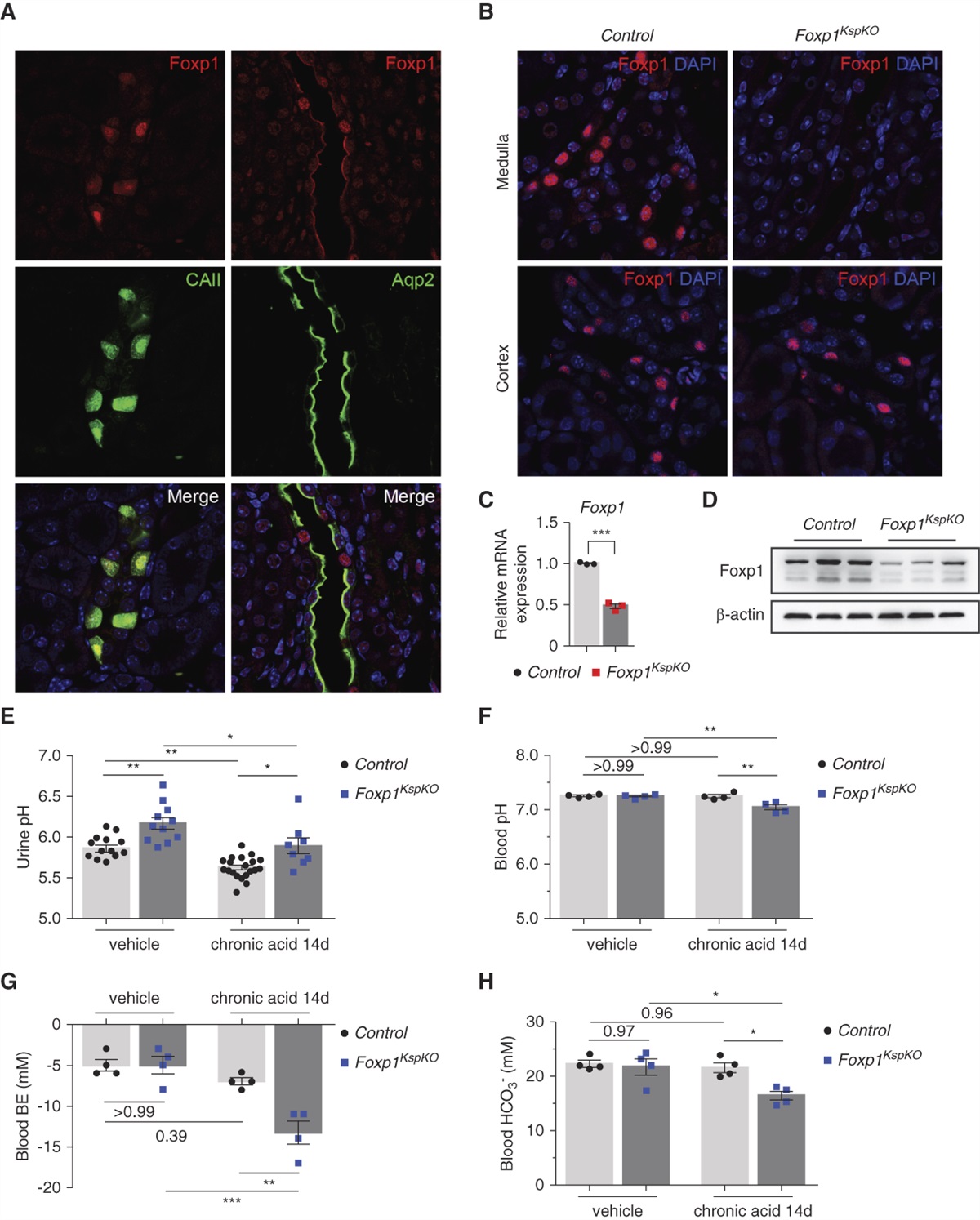
TMEM174 KO Mice Have Severe Hyperphosphatemia and Vascular CalcificationTo study the physiologic role of TMEM174 in the regulation of Pi homeostasis and CKD-mediated cardiovascular complications, we generated TMEM174 KO mice using the procedures we previously used for other mouse strains (Supplemental Figure 1). TMEM174 KO mice had a normal growth rate (Supplemental Figure 4A). Levels of serum creatinine, Ca, magnesium, sodium, potassium, and chloride and renal morphology were normal in TMEM174 KO mice (Supplemental Figure 4, B–H). However, the effect of TMEM174 deficiency on the parameters involved in renal Pi homeostasis was striking. As shown in Figure 2, TMEM174 KO mice had two-fold higher serum phosphate levels than wild-type mice (Figure 3A). Levels of FGF23 and PTH were also drastically higher in the serum of TMEM174 mice (Figure 3, B and C). Levels of 1,25(OH)2D were not altered by TMEM174 deficiency (Figure 3D). Levels of NPT2A protein were significantly higher in the renal BBM of TMEM174 KO mice (Figure 3, E and F, Supplemental Figure 5A). Although NPT2C levels were reduced, levels of PiT2 and NHERF1 were not changed by TMEM174 deficiency (Figure 3E, Supplemental Figure 5). Levels of kidney PiT2 were not changed, whereas NPT2A and NPT2C mRNA levels were reduced by TMEM174 deficiency (Supplemental Figure 5). Total Pi uptake of renal BBM was increased in TMEM174 KO mice, whereas sodium-independent Pi uptake was not changed between wild-type and TMEM174 KO mice (Figure 3, G and H). Urinary Pi, but not Ca, excretion was significantly reduced in TMEM174 KO mice (Figure 3, I and J). Levels of CYP27B1 and CYP24A1 were significantly higher in TMEM174 KO mice (Supplemental Figure 5). These observations could explain the normal levels of 1,25(OH)2D in TMEM174 KO mice. Levels of renal Ca-sensing receptor and intestinal NPT2B protein were not altered by TMEM174 deficiency. FGFR1 protein expression was increased, whereas PTHR1 was reduced, in the kidneys of TMEM174 KO mice (Supplemental Figure 5). More importantly, TMEM174 KO mice developed severe vascular calcification (Figure 3, K–M). To determine whether (1) dietary Pi manipulations affect TMEM174 expression and (2) if TMEM174 is involved in the regulation of sodium-dependent Pi transporters through dietary Pi, wild-type and TMEM174 KO mice were fed a low-Pi (0.1%) diet or a high-Pi (1.2%) diet for 7 days. Feeding with a low-Pi diet reduced levels of serum Pi in both wild-type and TMEM174 KO mice. Under both low-Pi and high-Pi conditions, however, levels of serum Pi were significantly higher in TMEM174 KO mice than wild-type mice (Figure 4A). Neither dietary Pi nor TMEM174 genotype affected levels of serum Ca (Figure 4B). A high-Pi diet significantly reduced levels of TMEM174 protein in the renal BBM of wild-type mice (Figure 4, C and D). Upon feeding of a high-Pi diet, levels of NPT2A and NPT2C were drastically reduced in the BBM of wild-type mice. Levels of NPT2A protein were significantly higher in the BBM of TMEM174 KO mice than wild-type mice under a high-Pi diet, whereas levels of NPT2C protein were significantly lower in TMEM174 KO mice under both high- and low-Pi diets (Figure 4, C, E, and F). Neither dietary Pi level nor TMEM174 genotype affected levels of PiT2 and NHERF1 protein (Figure 4, C, G, and H). Dietary Pi levels did not affect levels of TMEM174 and NPT2A, whereas a high-Pi diet reduced levels of NPT2C and PiT2 mRNA (Figure 4, I–L). TMEM174 mice had significantly lower levels of NPT2A mRNA under a high-Pi diet. Although levels of TMEM174 and NPT2A were not affected by dietary phosphate, NPT2C and PiT2 mRNA levels were significantly reduced by a high-Pi diet. Upon high-Pi feeding, levels of NPT2A mRNA were lower in TMEM174 KO mice than wild-type mice. In addition, TMEM174 deficiency reduced levels of NPT2C mRNA under both high-Pi and low-Pi conditions (Figure 4, I–L).
 Figure 3.
 Disruption of Pi homeostasis by TMEM174 deficiency. Serum (A) Pi, (B) FGF23, (C) PTH, and (D) 1,25(OH)2D in 8-week-old TMEM174 KO male mice fed a chow diet. (E) TMEM174, renal Pi cotransporters, and NHEFR1 in the renal BBM of TMEM174 KO mice. Flarebio TMEM174 antibody was used to detect mouse TMEM174. (F) Immunofluorescence analysis of NPT2A in the kidneys of TMEM174 KO mice.25⇓–27 (G) Total Pi uptake and (H) sodium-independent Pi uptake in the renal BBM of TMEM174 KO mice. Urinary (I) Pi and (J) Ca excretion of TMEM174 KO mice. Urine was collected in metabolic cages for 24 hours. (K) Alizarin red staining, (L) calcified lesions, and (M) aortic Ca content in the aortic sinus of TMEM174 KO mice. Statistical analysis was performed using a two-tailed t test. *P<0.05, **P<0.01, ***P<0.001. WT, wild type.
 Figure 4.
 Effect of dietary Pi on renal TMEM174 and sodium-dependent Pi cotransporter expression in wild-type (WT) and TMEM174 KO mice. Levels of serum (A) Pi and (B) Ca in mice fed either a low-Pi (0.1%) or a high-Pi (1.2%) diet for 7 days. (C) Immunoblot analysis of TMEM174 (Flarebio), NPT2A, NPT2C, PiT2, and NHERF1 in the renal BBM of wild-type and TMEM174 KO mice (n=3) fed either low-Pi or high-Pi diet. (D–H) The densitometry analysis of the immunoblot analysis shown in (B): (D) TMEM174, (E) NPT2A, (F) NPT2C, (G) PiT2, and (H) NHERF1. Levels of (I) TMEM174, (J) NPT2A, (K) NPT2C, and (L) PiT2 in the kidneys of TMEM174 KO mice fed either a low-Pi or high-Pi diet. Statistical analysis was performed using a two-way ANOVA test with the Turkey multiple comparisons post hoc test. *P<0.05, **P<0.01, ***P<0.001.
DiscussionTMEM174 was previously identified as a kidney-enriched TMEM protein.26,27,41 However, the functions of the protein have not been elucidated. In this study, we used mRNA-seq to confirm that TMEM174 protein is a kidney-specific TMEM protein. In addition, TMEM174 is selectively localized in the apical membrane of renal proximal tubule epithelial cells. TMEM174 regulates Pi homeostasis by decreasing Pi uptake and retaining NPT2A in the BBM. In TMEM174 KO mice, levels of serum Pi were selectively increased by increasing expression of NPT2A. Due to hyperphosphatemia, TMEM174 KO mice showed other features of CKD-dependent complications, such as drastic increases in FGF23 and PTH and vascular calcification. A previous study found the localization of TMEM174 in the endoplasmic reticulum.26 The difference between the previous study and our study could be due to the use of different cell lines. The previous study used HEK cells, which are not proximal tubular cells, do not develop microvilli, and do not express endogenous TMEM174. Because plasma membrane proteins are glycosylated in the endoplasmic reticulum, some of the TMEM174 proteins could be found in the endoplasmic reticulum.
Although the precise mechanism by which TMEM174 deficiency increases levels of NPT2A protein is still unclear, this study demonstrated the TMEM174 protein binds to NPT2A and increases NPT2A protein in cultured cells and mouse kidneys. Because TMEM174 binds to NPT2A but not NHERF1, it is possible that TMEM174 competes with NHERF1 to destabilize NPT2A protein. We had other interesting observations: (1) levels of NPT2A mRNA were significantly reduced in the kidneys of TMEM174 KO mice, (2) both NPT2C protein and mRNA expression were reduced in the kidneys of TMEM174 KO mice, (3) TMEM174 knockdown blocked FGF23 and PTH-mediated NPT2A degradation in proximal tubule cells, and (4) TMEM174 KO mice had normal levels of other electrolytes (sodium, potassium, magnesium, Ca2+, and chloride ions). However, TMEM174 siRNA treatment did not affect NPT2A or NPT2C mRNA expression in OK-P cells. In addition, levels of NPT2C protein and mRNA were tightly associated with levels of serum Pi. These results suggest the reduction of renal NPT2C through in vivo TMEM174 deficiency were indirect and probably due to hyperphosphatemia. These observations suggest the effect of TMEM174 is specific to NPT2A and that TMEM174 increases the degradation of NPT2A. Further studies will be required to elucidate (1) a molecular mechanism by which TMEM174 selectively increases the degradation of NPT2A protein, (2) whether TMEM174 alterations directly contribute to CKD-dependent vascular calcification, and (3) whether TMEM174 could be a pharmacologic target for CKD-dependent complications.
DisclosuresJ. Blaine reports receiving research funding from Calliditas Therapeutics (as site principal investigator for a trial of budesonide in IgA nephropathy) and Novartis (as site principal investigator for a trial of an agent to treat IgA nephropathy); serving as medical director of a Fresenius dialysis unit; and having ownership interest in Pfizer Inc. (owns 150 shares with spouse). All remaining authors have nothing to disclose.
Author ContributionsJ. Blaine, A.L. Keenan, and M. Miyazaki reviewed and edited the manuscript; J. Blaine and M. Miyazaki provided supervision and were responsible for funding acquisition; A.L. Keenan and M. Miyazaki were responsible for investigation; A.L. Keenan, M. Miyazaki, and S. Miyazaki-Anza were responsible for data curation and formal analysis; M. Miyazaki wrote the original draft and was responsible for project administration, validation, and visualization; and M. Miyazaki and S. Miyazaki-Anza conceptualized the study and were responsible for methodology.
Data Sharing StatementThe raw data are deposited in the National Institutes of Health Gene Expression Omnibus (accession GSE189618).
Copyright © 2022 by the American Society of Nephrology
留言 (0)