
記住我


As an emerging class of immune-oncology therapy, therapeutic cancer vaccines have demonstrated robust tumor-specific immunogenicity and antitumor activity in patients with melanoma, glioblastoma and other cancers in the past decade1,2,3,4,5. Different from the previously developed cancer vaccines targeting tumor-associated antigens that overexpress in tumor cells, the recent cancer vaccines trigger specific immune responses against neoantigens that are produced by genetic mutations in tumor cells, thus potentially avoiding off-target effects6,7. However, the immunogenicity of tumor neoantigens alone is often disappointing, and the use of immune adjuvants and/or delivery vehicles is an effective approach to improve the immunogenicity8,9. Immune adjuvants can stimulate the antigen-presenting cells (APCs) to provide the necessary costimulatory signals for successful antigen presentation, while delivery vehicles can enhance the uptake and processing efficiency of neoantigens by APCs10,11.
Currently, some promising nanocarriers with intrinsic immune adjuvant properties, such as polymeric and lipid nanoparticles with stimulator of interferon genes (STING) pathway activation ability, have been developed12,13. These nanocarriers can ensure that immune activation and antigen delivery occur in the same APCs, which is necessary for effective antigen presentation. Inspired by the body’s natural immune defenses against bacterial invasion, our group has developed two different types of nanocarrier based on bacterial membrane materials for cancer vaccine delivery14,15. Owing to the large amounts of pathogen-associated molecular patterns (PAMPs), the bacterial membrane materials can act as excellent nanocarriers with intrinsic immune adjuvant properties16,17,18. To make these nanocarriers more suitable for use in cancer vaccines, two different loading methods for known and unknown tumor neoantigens have been applied for different application purposes, respectively (Table 1).
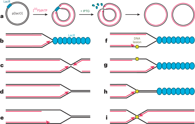
Table 1 Properties and applications of two types of nanocarrier based on bacterial membrane materials for cancer vaccinesIn the first study, the bacteria-derived outer-membrane vesicles (OMVs) were extracted through ultracentrifugation, which are the natural nanosized vesicles secreted by Gram-negative bacteria (Fig. 1a)19,20. To simplify the display of tumor neoantigens on OMVs, we employed a Plug-and-Display system to decorate OMVs through genetic engineering (Fig. 1a)14. The Plug-and-Display system comprises the tag/catcher protein pairs, and the protein catchers were fused with the surface protein ClyA on OMVs21,22. Then, the OMVs decorated with different protein catchers can simultaneously display multiple tumor neoantigens labeled with the corresponding tags, thereby rapidly constructing the tumor nanovaccines (Fig. 1a). These OMV-based nanovaccines have been used to inhibit tumor growth and metastasis, which are more suitable for customized design when the tumor neoantigens have been identified and/or can be identified. According to the identified antigen information, the individualized matched neoantigens were synthesized and displayed on the OMV-based nanocarriers to create antigen-suited nanovaccines for patients with cancer.
Fig. 1: Preparation of OMV- and HM-NPs for cancer vaccines.
a, Preparation of OMV-based nanocarriers. The catchers (SpC and SnC) in the Plug-and-Display system are fused with the ClyA protein (ClyA-catchers) on OMVs’ surface, through E. coli genetic engineering. Then, the engineered bacteria-derived OMVs are extracted through ultracentrifugation. The OMVs decorated with two different catchers can simultaneously display multiple tumor neoantigens (Ag) labeled with corresponding tags (SpT and/or SnT), thereby rapidly constructing the antitumor nanovaccines. b, Preparation of HM-NPs. Tumors are surgically removed from tumor-bearing mice to obtain TM. E. coli are treated with lysozyme to remove the cell wall to generate protoplasts, and the cytomembrane is extracted with an extraction buffer to prepare E. coli cytomembrane (EM). HM vesicles are generated by mixing EM and TM, followed by extruding through an extruder. PLGA nanoparticles (NPs) as polymeric cores are then added to generate HM-NPs. Figure adapted from ref. 14 under a Creative Commons licence CC BY 4.0 (a) and ref. 15, AAAS (b).
In the second study, we developed hybrid membrane-based nanocarriers (HM-NPs), containing the bacterial cytoplasmic membrane from Escherichia coli (EM) and tumor cell membrane (TM) from the resected autologous tumor tissues (Fig. 1b)15. Briefly, the poly(lactic-co-glycolic acid) (PLGA) nanoparticles were constructed as a core, and the EM and TM were simultaneously coated onto the PLGA nanoparticles to form the final HM-NPs (Fig. 1b). The EM acts as the immune adjuvant to activate the innate immune response, and the autologous tumor neoantigens in the TM are the key messengers for transmitting the tumor-specific identification to the adaptive immune system23,24,25,26. These HM-NPs possess unique advantages as personalized cancer vaccines when the tumor neoantigens are not readily available and are more suitable for prevention of postoperative recurrence.
Here we provide protocols for the construction of these two types of vaccine based on bacterial membrane materials and the matched tumor antigens from different sources, and the potential applications and limitations of the nanovaccines are discussed. The characterization methods are validated and presented in detail, and the antigen delivery effect and immune stimulation mechanisms were validated both in vitro and in vivo.
Development of the protocolDevelopment of OMV-based nanocarriers and vaccinesOwing to their excellent immunostimulatory ability, OMVs are attractive candidates as vaccine carriers in the field of prophylactic vaccines against pathogenic microorganisms20. For example, the OMV-based vaccines against Group B meningococcus, namely MeNZB, have effectively decreased the incidence and mortality of meningitis in New Zealand27. In addition to preventing the invasion of their parental microorganisms, OMVs can be employed as a display platform of heterologous antigens to form vaccines against other pathogens through genetic engineering, chemical bonding and physical mixing28. To exploit OMVs as nanocarriers for cancer vaccines, the loading method of tumor neoantigens is the key issue. First, we adopted fusion expression to display a pattern antigenic epitope of ovalbumin (OVA), OVA257–264 (SIINFEKL) on the OMVs’ surface, in which OVA257–264 was fused with the C terminal of the surface protein ClyA (ClyA-OVA) on OMVs29. The strong antigen-specific immune response and antitumor effect induced by ClyA-OVA OMVs indicated that OMVs are outstanding vaccine nanocarriers for displaying tumor neoantigens to elicit antitumor immunity.
However, the tumor neoantigens generated from the gene mutations have high heterogeneity and variability30, making it impractical and time-consuming to produce OMV-based nanovaccines for every patient via fusion expression with neoantigens. The antigen display of tumor vaccine carriers should be rapid and flexible to meet the clinical requirement. Therefore, we employed the Plug-and-Display system to decorate OMVs, including a SpyTag (SpT)/SpyCatcher (SpC) pair and a SnoopTag (SnT)/SnoopCatcher (SnC) pair21,22. The SpC and SnC catchers were fused with the ClyA protein (ClyA-catchers, CC) on OMVs’ surface and can spontaneously bind to the neoantigens labeled with SpT and SnT tags through isopeptide bond formation, respectively. These bioengineered CC OMVs were able to simultaneously and rapidly display multiple tumor neoantigens, thereby being competent for rapid preparation of the customized cancer vaccines when tumor antigens can be predicted and identified.
Development of HM-NPs and vaccinesSurgical resection is the primary treatment option for most solid tumors, and the recurrence and metastasis after surgery are still unmet clinical challenges31,32. Vaccination after surgery to induce the antigen-specific immune responses to eliminate residual tumor cells may have wide application33. TMs contain a high proportion of antigenic motifs and have similar antigen patterns as are displayed on the surface of tumor cells in vivo34,35. However, autologous tumor antigens can be recognized as ‘self’, and they are more likely to induce antigen-specific tolerance rather than antitumor immunity36. The body’s immune defenses against bacterial invasion are rather sensitive; utilizing bacterial constituents that act as adjuvants to enhance immunogenicity is a promising strategy to overcome this limitation.
However, lipopolysaccharide (LPS) and other cell wall components of bacteria as the first line of contact with immune cells could cause an undesirable immunopathological state23. The bacterial cytoplasmic membrane, which is spatially separated from the organism’s cell wall, may be used as a potential adjuvant to reduce the magnitude of ‘danger signals’17,18. Membrane fusion is a technology that can confer a hybrid membrane (HM) with properties inherited from two source cell membranes24,25,26. Thus, we developed the immunotherapy strategy using bacterial cytoplasmic membranes from E. coli and autologous tumor membranes from resected tumor tissue to confer a HM with the properties inherited from two source cell membranes15. These HM-NPs were able to codeliver antigen and adjuvant to APCs.
Overview of the proceduresFirst, we describe the preparation of the two types of nanocarrier based on bacterial membrane materials and the related nanovaccine constructions (Procedure 1, Steps 1–18 for OMV-based nanocarriers; Procedure 2, Steps 1–38 for HM-NPs). Next, the methods and processes for characterization of physicochemical properties and biological components are described in detail (Procedure 1, Steps 19–30 for OMV-based nanocarriers; Procedure 2, Step 39 for HM-NPs). Then, the antigen delivery efficiency and the immune response analysis are performed (Procedure 1, Steps 31–48 for OMV-based nanocarriers; Procedure 2, Step 40 for HM-NPs). Finally, we discuss the strategies to evaluate the in vivo antitumor effect in different mouse models using multiple antigens (Procedure 1, Step 49 for OMV-based nanocarriers; Procedure 2, Step 41 for HM-NPs). More detailed information on the two procedures is provided below.
Procedure 1: OMV-based nanocarriers and vaccinesThrough genetic engineering, we splice the gene fragments fusing SpC or SnC and ClyA (ClyA-SpC and ClyA-SnC) respectively, and insert them into the co-expression plasmid pETDuet-1. Then, the co-expression plasmid is transformed into the engineered bacteria E. coli Rosetta (DE3). Finally, CC OMVs are extracted from the culture medium in massive quantities by bacterial fermentation and ultracentrifugation, followed by antigen display, vaccine preparation and functional characterization. In this protocol, the size and morphology of CC OMVs are characterized using dynamic light scattering (DLS) and transmission electron microscopy (TEM), respectively. The antigen display ability is evaluated by western blot. Finally, CC OMVs are used to display multiple tumor antigens including OVA257–264 (termed ‘OTI’, an epitope that can stimulate the production of MHC class I-restricted OVA-specific CD8+ T cells in mice), OVA223–339 (ISQAVHAAHAEINEAGR; ‘OTII’, an epitope that can stimulate the production of MHC class II-restricted OVA-specific CD4+ T cells in mice), an antigenic epitope of tyrosinase-related protein 2 (TRP2), TRP2180–188 (SVYDFFVWL) and an antigenic epitope of Adpgk (CGIPVHLELASMTNMELMSSIVHQQVFPT)37,38. The in vitro and in vivo immune response and antitumor effect are determined in the pulmonary metastatic melanoma model and subcutaneous colon cancer model.
Procedure 2: HM-NPs and vaccinesHM-NPs are synthesized by preparation of the HM of EM and TM and coating the HM onto the synthesized PLGA nanoparticles. In this protocol, the size and morphology of HM-NPs are characterized using DLS and TEM, respectively. Immunogold staining for specific markers to identify EM and TM on HM-NPs is also observed by TEM. The enhanced tumor antigen uptake and maturation of bone marrow-derived dendritic cells (BMDCs) by HM-NPs are evaluated by flow cytometry and enzyme-linked immunosorbent assay (ELISA). The mouse tumor models of 4T1, CT26 and B16-F10 cells are then used to investigate the antitumor immune effects of HM-NPs.
Applications of the methodPotential applications of OMV-based strategy for vaccine developmentIn this protocol, we used the OMV-based nanocarriers to rapidly display multiple antigens, including OTI, OTII, TRP2180–188 and Adpgk. Of course, this platform can be also employed to deliver any other antigenic peptide, including human and murine antigens. Because of the high compatibility, this platform is suitable for the preparation of customized cancer vaccines for each patient with cancer. In the future, if a tumor antigen library is established, we can quickly identify the antigen information of patients with tumors by gene sequencing, so as to make use of the rapid antigen display capability of this platform for the preparation of customized cancer vaccines. In addition, OMV-based nanocarriers can also be used for rapid screening and identification of tumor antigens owing to their excellent immune stimulation, antigen transport and rapid antigen display capabilities.
As mentioned earlier, previous studies have focused on using OMVs to build vaccines against microorganisms. Therefore, in addition to cancer vaccines, prophylactic vaccines against infectious diseases can also use our OMV-based nanocarriers, especially for pathogenic microorganisms with highly variable antigens, such as influenza virus.
Potential applications of HM-based strategy for vaccine developmentThe HM-NPs are integrated into a vaccine delivery nanoplatform with both bacterial cytoplasmic membranes and surgically derived TM to enhance both innate and adaptive immune responses. This HM-derived vaccine strategy is beneficial to multiple tumor models by codelivery of individualized tumor antigens and adjuvants into dendritic cells (DCs). In addition, these HM-NPs can be genetically or chemically modified to further expand their multifunctionality. The core of the nanocarriers can also load various cargos for integration of other treatment modalities to enhance the effects of immunotherapy.
Comparison with other methodsSeveral materials have been used to develop nanocarriers for tumor vaccine delivery, such as lipid, polymer, synthetic high-density lipoprotein and DNA origami39,40,41,42,43,44. Compared with these materials, the bacterial membrane material-based nanocarriers have a unique immune stimulation function and play the dual roles of immune adjuvants and antigen carriers. In addition, compared with the complexity of chemical synthesis, bacterial membrane material-based nanocarriers can be obtained in relatively large quantities through bacterial fermentation. This feature may ensure compatibility with industrial production in the future.
In the first Procedure, the Plug-and-Display system is employed to achieve the rapid display of peptide antigens on OMVs. The current clinical trials of tumor vaccines based on peptide antigens are mainly conducted by using a mixture of peptides and adjuvants1,3,4,5. Nanocarriers can improve stability and immune system targeting of the antigens and realize the codelivery of antigens and adjuvants9,10. Therefore, the tumor vaccines based on nanocarriers have received more and more attention9,10. As mentioned above, the bacteria-derived OMVs have the dual functions of both carriers and adjuvants, while the chemical synthetic nanocarriers are mainly delivery vehicles. Therefore, the nanovaccines based on OMVs do not require additional adjuvants. In addition, our study demonstrated that the OMV-based nanovaccines can activate an antigen-specific T-cell response, as distinct from the previously reported B-cell-mediated antibody responses in the OMV-based prophylactic vaccines against pathogenic microorganisms20,27.
More importantly, we employed the Plug-and-Display system to achieve the rapid display of multiple peptide antigens. Compared with the fusion expression, physical encapsulation or chemical conjugation, this flexible approach of antigen display makes our OMV-based nanocarriers more suitable for rapid preparation of customized cancer vaccines. The Plug-and-Display system has been used in the construction of vaccines against microorganisms based on virus-like particle45,46,47. We firstly demonstrate the feasibility of this system for constructions of tumor vaccines. Compared with virus-like particle, OMVs have the advantage of immune adjuvant function, despite the disadvantage of complex composition. Furthermore, the current practice for antigen encapsulation into the nanoparticles during the production process lacks flexibility to load multiple antigens under different scenarios41,43. The vehicle and antigen(s) in our nanovaccine can be separately synthesized and used to rapidly prepare the vaccine through a simple combination procedure before immunization. This modular design allows one to establish a neoantigen library in advance and rapidly select appropriate antigens for individual patients, which may reduce the production time and realize the bedside preparation of tumor vaccines for individual patients in the future14.
In the second Procedure, the cancer vaccines are constructed through hybrid fusion of the surgically removed TM and the bacterial cytoplasmic membranes. Traditional methods of autologous tumor cell-based vaccines use whole-cell tumor vaccines or tumor lysate vaccines to elicit immunity against the entire collection of antigens expressed by the tumor33,48,49. Although whole-cell tumor or tumor lysate vaccines open a promising area for cancer immunotherapy, therapeutic efficacy may be severely limited as the immunogenicity of antigens in these vaccine formulations may be diluted or inhibited by the unrelated proteins and nucleic acids. The current HM tumor vaccines are constructed through the fusion of the surgically removed TM and the bacterial cytoplasmic membranes, possessing unique advantages as personalized cancer vaccines with highly enriched antigens when the identified ones are not sufficient to trigger antitumor immunity or the neoantigens are not readily available.
Although TM are naturally enriched with a specific tumor antigen pool, these membrane antigens usually have low immunogenicity since tumorigenesis implies adaptation of tumor cells to the host immune system. Compared with the other tumor membrane-based nanocarriers, the introduction of the bacterial cytoplasmic membranes enhances the recognition of tumor membrane antigens by immune system. Meanwhile, bacterial cytoplasmic membranes also take advantage of the antibody-dependent, cell-mediated cytotoxicity of NK cells, which lyse malignant cells even before the stimulation of specific T cells in an antigen-independent manner. Bacteria-derived substances (inactivated or attenuated bacteria, lipids, proteins and/or nucleic acids) provide ‘danger signals’ that alert the immune system to the potential infection and invasion17. However, bacteria-derived formulations, especially LPS, can lead to severe side effects, such as cytokine storm and sepsis18. Using bacterial cytoplasmic membranes can reduce the magnitude of ‘danger signals’ since most of the bacterial LPS is removed. Meanwhile, the bacterial cytoplasmic membranes can be rapidly and manageably prepared from inexpensive cultures of bacteria. This vaccine formulation protocol provides an appropriate balance among accessibility, safety and effectiveness.
LimitationsSome limitations of OMV-based nanocarriers for cancer vaccines should be considered. First, the OMVs contain large amounts of proteins, polysaccharides, lipids and trace amounts of nucleic acids from bacteria, and the complicated components may cause difficulty for the quality control of vaccine production. In this Procedure, we use DLS to detect the particle size and TEM to observe the morphology for quality control of OMVs. The ideal OMVs should have a low polydispersity index (PDI, <0.2). Another quality control from the production aspect of OMV-based products may be the presence of some characteristic proteins in the OMVs, such as outer-membrane protein A/C/F, which can be used as the fingerprint for OMV identification20. Second, the amount of LPS in OMVs is the main component of endotoxin, and the elimination of LPS in OMVs by genetic engineering is a feasible option for further improvement50. Third, the antigens used in this protocol include model antigens and murine tumor antigens, and the adaptability of this platform to human tumor antigens needs further study. Fourth, ultracentrifugation is used to isolate and purify OMVs in this protocol, but this method may not be suitable for mass production. Appropriate purification methods for industrial practices are in urgent need, and chromatographic separation according to the size of OMVs is an optional solution. Finally, the Plug-and-Display system used to create CC OMVs may be further optimized. In addition to ClyA protein, there are some other surface proteins on OMVs that can be employed as scaffold for the Plug-and-Display system, such as hemoglobin protease and outer-membrane protein A/C/F20. In addition, updated versions of the Plug-and-Display system have been described, such as spycatcher003, with a stronger integration efficiency51. These novel scaffolds and Plug-and-Display systems should be further evaluated to determine whether they will be able to enhance the antigen display capability of the OMV-based nanocarriers.
One limitation of HM nanovaccines is that the immunosuppressive proteins of the TM have not been investigated. As tumor cells interact with the infiltration of immune cells and form the immune-suppressive environment, there is a high portion of immunosuppressive proteins. For example, the well-known immune checkpoint inhibitor, programmed cell death 1 ligand (PD-L1), is located in the TM52. Enhanced antitumor efficacy may be achieved by regulating these immunosuppressive factors. In addition, the contribution of other cell membranes to the antitumor immunity should be considered, since immune cells and stromal cells in the resected tumor tissue are not eliminated during HM nanovaccine preparation. Tumor cells could firstly be isolated and purified by the negative selection-based tumor cell isolation kit or culture plate adherent passaging, then the effectiveness of HM vaccines prepared from purified TM could be assessed in comparison. The fabricating methods could also be improved by the use of microfluidic methods instead of manual preparation and procedures. Last but not least, specific antigens on the TM recognized by the T cells are not yet identified. All these factors could either limit or benefit translation to the clinic.
In addition, the sterile production of the OMV-based nanocarriers and HM-NPs requires special attention. In the Procedure for the OMV-based nanocarriers, almost all the bacteria were removed by centrifugation at 5,000g for 10 min. In the following steps to separate OMVs from the supernatant, two filtration processes were carried out through 0.45 μm and 0.22 μm filters to ensure that the final OMVs did not contain bacteria. In the Procedure for the HM-NPs, the lysozyme and extraction buffer can lyse almost all the bacteria to obtain the cytoplasmic membranes of E. coli. In addition, in the following steps to prepare the HM-coated nanoparticles, the HM were extruded at least 13 times through the 400 and 200 nm cutoff sterile extruders, respectively. Therefore, the final HM-NPs do not contain bacteria. For future industrial-scale preparation, radiation sterilization may be suitable for large-scale production of the OMV-based nanocarriers and HM-NPs.
Experimental designSelecting tumor antigensIn our first Procedure, we adopt several model antigens (OTI and OTII) and murine tumor antigens (TRP2180–188 and Adpgk) to make tumor vaccines against different cancer cells. Other tumor antigens can be selected according to the cancer cell type. For example, the gp100 antigen can be used to establish a cancer vaccine against B16-F10 melanoma cells, and the gp70 antigen is suitable for the treatment of CT26 colon cancer cells53,54. In addition, the combination of mass spectrometry and exome sequencing can effectively predict and identify neoantigens in tumor cells, which lays the foundation for customized cancer vaccines38. These identified tumor antigens can also be used in the experiments according to our protocol. Once the tumor antigens are selected, the SpT or SnT tag needs to be added at the N terminal, and the tag-labeled antigen peptides are synthesized via the Fmoc solid-phase peptide synthesis method. When there is only one type of tumor antigen, either SpC–SpT or SnC–SnT pair can be used for rapid display, and there is no notable difference between them. If there are two types of tumor antigen, it is recommended to use different pairs to display, so as to increase the efficiency of display. If there are more than two types of tumor antigen, we recommend that each antigen can be displayed separately using either SpC–SpT or SnC–SnT pair to construct different nanovaccines, and eventually mix together for immunization.
Selecting murine cancer models and mouse strainsIn our two Procedures, several murine cancer models are used, including B16-F10 and OVA-expressing B16-F10 (B16-OVA) melanoma, MC38 and CT26 colon cancer and 4T1 breast cancer. According to the origin of the tumor cells, different mice were used to make tumor models, including BALB/c mice for CT26 and 4T1 and C57BL/6 mice for B16-F10, B16-OVA and MC38. This protocol is also suitable for use with other murine tumor models. If the tumor vaccine is to be built against human tumor cells, mice with a humanized immune system should be used.
Design of controlsIn the first Procedure, for OMV-based nanocarriers, the critical control group is a mixture of CN OMVs (from the bacteria expressing ClyA without the fused Catchers, ClyA-none) and tumor antigens, thus proving the importance of antigen display on CC OMVs for efficient immune stimulation. The control groups of separate antigens or separate empty CC OMVs are also necessary to illustrate the advantages of the system. If multiple antigens are displayed on the surface of CC OMVs to prepare tumor vaccines, control vaccines containing individual antigens should also be evaluated to dete
留言 (0)