
記住我


Cell irradiations were performed with a 241Am source (Eckert & Ziegler Isotope Products GmbH, Braunschweig, Germany). The source is sealed in an aluminum cover, and the circular active source window is 11.8 mm in diameter and covered by a 0.5 µm palladium coating. The activity was previously estimated to 405 ± 10 kBq [26].
A Passivated Implanted Planar Silicon (PIPS) detector A300 (Canberra, Mirion Technologies) in vacuum was used to measure the emission rate and energy spectrum of alpha particles from the 241Am source. All alpha particles reaching the detector are recorded as the detector thickness is greater than the alpha range for the alpha energies investigated in this work. Therefore, the detector efficiency is given by the geometrical efficiency. The source was positioned in the detector vacuum chamber at a 36 mm distance between the source window surface and detector surface.
Cell cultureThe prostate cancer cell line PC3 purchased from the American Type Culture Collection (ATCC®, Manassas, VA, USA) were grown in RPMI-1640 medium with L-glutamine (Biowest, Nuaillé, France) supplemented with 10% fetal bovine serum (Fisher Scientific, UK) and 1% penicillin–streptomycin (Fisher Scientific, UK). Cells were grown in T-25 flasks as a monolayer in a humified atmosphere in an incubator at 5% CO2 and 37 °C. Twenty thousand cells were seeded directly on glass coverslips placed at the bottom of wells on a 24-well plate one day prior to alpha irradiation, fixation, and fluorescent staining.
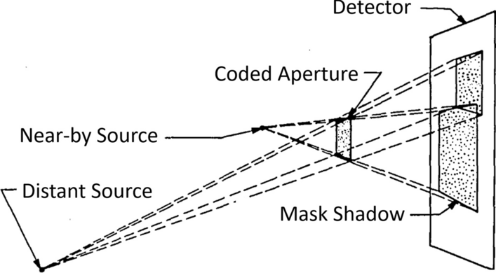
Alpha and X-ray irradiationWhen irradiating the PC3 cells with alpha particles, the cell culture media was removed to allow alpha particles to reach the cells (the alpha particle range in water is less than < 50 µm for the relevant particle energies) and the 241Am source was placed in a holder on top of the well plate with the source surface centered over the well opening. The distance between source window and the coverslips at the well bottom was 19 mm (Fig. 1). Cells were irradiated for 4, 8, and 12 min in triplicates. The 241Am source was immediately removed at the end of the irradiation, and fresh cell culture media was added to the well. To evaluate the damage caused by cells being without culture medium, cells were sham-irradiated for 12 min. The removal of cell media could potentially not only affect the background distribution of foci, but also affect the induction of γ-H2AX RIF and their repair, as compared to an irradiation setup in which the cells are continuously covered by cell media. This is a factor necessary to consider when evaluating the results, but it is beyond the aim of this study to further investigate this.
Fig. 1
Illustration of alpha irradiation of cells at bottom of well (not to scale). Arrow indicating the direction of the well axis, as referred to in the text. The 241Am source is placed in a holder centered over the well opening, irradiating adherent PC3 cells on the bottom. Irradiations are performed with cell media removed. The source-to-bottom distance is 19 mm, and the well is a cylinder of diameter 16.2 mm
To compare the appearance of γ-H2AX foci in control, alpha-, and X-ray-irradiated PC3 cells, cells plated on cover slips, as described above, were irradiated to 0.5 Gy on a XneX system (Xstrahl) with a 220 kV tube voltage at a dose rate of 2.1 Gy/minutes.
Immunofluorescence staining and imagingThirty minutes after irradiation, cells were fixated with 4% paraformaldehyde in PBS for 15 min, washed with PBS, permeabilized in 0.1% Triton X-100 solution (Sigma-Aldrich) for 5 min twice, washed with PBS in between and after. Cells were incubated with 1% bovine serum albumin (Sigma-Aldrich) for 30 min. Cells were then incubated with primary antibody (Anti-gamma H2A.X (phosphor S139) antibody [9F3] ab26350) (AbCam) for 1 h and subsequently rinsed with 0.05% Triton X-100 for 5 min twice with PBS rinse in between and after. Then, secondary antibody (Alexa Fluor® 647-conjugated AffiniPure F(ab')2 Fragment Donkey Anti-Mouse IgG) (Jackson ImmunoResearch) incubation lasted 30 min. Cells were rinsed twice in 0.05% Triton X-100 for 5 min. The cells were stained with DAPI (Thermo Scientific) for 15 min and then rinsed twice before mounting the coverslips on slides in antifade solution (Fluoroshield Abcam, ab104135).
Stained cells were imaged on a laser scanning confocal microscope (LSM 710 Confocal Microscope, Zeiss) with 63 × oil immersion objective. The pixel size in the horizontal plane (x and y) was 0.1318 µm and 0.3756 µm in the z-direction. To sample a large surface on the center of the coverslip area, tiled 16-bit images in a 3 × 3 grid were collected so that the resulting images had 3072 × 3072 pixels. Each grid had the length and width of 405 µm. At least 10 tiled grid images were collected for each coverslip. To generate a 3D mesh phantom, a smaller sample of cells on the non-irradiated controls were imaged as a z-stack. When imaging γ-H2AX foci, images were taken in the central 4*4 mm2 square of the cover slip.
Image foci segmentationImage processing was performed with MATLAB image processing tools (MATLAB R2020b). Cell nuclei were segmented from the DAPI signal color channel by first smoothing the image with a Gaussian filter, then converting the images to binaries by an adaptive threshold and performing a distance field and watershed transform to separate cell nuclei with touching borders. Cell nuclei were separated in a label matrix where pixels belonging to the same nucleus were given the same label/value. Nuclei area and major and minor axis length were sampled for each segmented label.
γ-H2AX foci within the boundaries of the segmented cell nuclei were similarly segmented from the γ-H2AX signal color channel and related to the respective nucleus. To reduce background, a median filtered image was subtracted from the original. Then, the images were smoothed by a Gaussian filter and converted to binaries and foci were individually segmented with a label matrix. Only foci with a minimal area of 9 pixels were segmented.
To exclude cell nuclei in the late stages of the cell cycle, often overexpressing γ-H2AX foci, the following inclusion criteria were used: Cell nuclei with a segmented DAPI surface of 35–209 µm2 (2000–12,000 pixels) were considered and nuclei with more than 20 foci were excluded.
As has been shown by Antonelli et al., the size of foci induced by alpha particle tracks can differ both from foci induced by X-rays and from background foci in non-irradiated cells [27]. To evaluate the induction of large foci, a second data set where only foci with an area above 30 pixels were included was generated, in similarity with the method used by Svetlicic et al. [28]. These results are from now on referred to as large foci.
GATE Monte Carlo simulationSimulations were performed using GATE [29] (v. 8.0) and Geant4 (v. 10.03). The low-energy electromagnetic physics list constructor emstandard_opt3 was used in all simulations.
Tools available in the GATE toolkit (Actors) were used to estimate the geometrical efficiency of the PIPS detector and to simulate the energy spectrum and directional distribution of alpha particles reaching the well bottom. Also, the number of alpha particle hits, total energy deposited, and absorbed dose in individual cell nuclei phantom at the bottom of the well was sampled with the GATE Dose Actor. A hit is defined as an alpha particle entering the scoring volume, i.e., the cell nucleus. The energy, LET, and energy imparted per alpha particle reaching a cell nuclei phantom volume were recorded with the GATE Energy Spectrum Actor.
In GATE, the emission from a source can be described by an imported emission spectrum. The normalized detected energy spectrum from the PIPS detector measurement was used to define the emission from the simulated 241Am source surface.
The PIPS detector geometrical efficiency was estimated through a simulation of the detector–source geometry to 3.4% (further described in Additional file 1: Fig. S1). Scaling the fluence at the detector surface to the source surface fluence in the PIPS measurements resulted in a surface rate of 2.4*105 alpha particles/second. This fluence was then implemented in the cell irradiation simulations.
Irradiation setup modelThe transport of alpha particles through the well to the bottom and the resulting energy loss in air were simulated by constructing a GATE model of the irradiation setup depicted in Fig. 1, consisting of the well, the source and a 20-µm-high water cylinder, at the well bottom. Visualization of the simulation geometry can be found in Additional file 1: Fig. S2.
The walls of the well were simulated as a 17-mm-high plastic hollow cylinder with an inner diameter of 16.2 mm and a wall thickness of 0.7 mm. The bottom was simulated as a 1-mm-thick plastic cylinder directly under the well walls. The 241Am source was simulated as previously described, placed above the well opening with a source-to-bottom distance of 19 mm. The surrounding volume between the well and the source was simulated as air.
Inside the 20-µm water layer, cell nuclei phantoms were placed 2.5 µm below the surface. All cell nuclei were simulated as volumes of water.
Cell irradiation simulations were performed, with total number of primary alpha particles emitted equal to the calculated fluence for 4, 8, and 12 min, as calculated from the source surface dose rate.
Constructing the elliptical cylinder phantomResults from the DAPI image segmentation were used as input in the creation of elliptical cylinder cell nuclei phantoms. A logistic distribution was fitted to the distribution of major axis of the segmented cell nuclei (Additional file 1: Fig. S4). Applying the inclusion criteria for DAPI area stated previously, meant excluding cell nuclei with a major axis length less than 4 µm or longer than 10 µm. The resulting probability distribution of the major axis lengths considered is pictured in Fig. 2a.
Fig. 2
Distribution of major axis lengths of PC3 cell nuclei in elliptical model (a). Measured from segmented DAPI-stained cell nuclei. Examples of cell phantom models (b). Elliptical cylinder phantoms (front row) generated from the axis lengths in (a). Examples of segmented 3D volumes (back row) from confocal imaging of DAPI-stained nuclei
Ten thousand elliptical cylinders were created to model as PC3 cell nuclei in the simulations (examples shown in Fig. 2b). The major axis lengths were generated by a random number generator, drawing numbers from the probability distribution. For all elliptical cylinders, the minor axes were 2/3 of its major axis length, and for all the height was 8 µm, which was the mean height of the segmented PC3 cells from the DAPI z-stack (described below). In the simulation, these phantom nuclei were randomly spread out to not overlap each other, in the central 4 × 4 mm2 area of the well bottom, to match the center area on the coverslip glass where the fluorescent imaging was performed. This way, the decreasing alpha particle fluence with increasing distance from the well center is considered controlled, as fluorescent microscopy imaging the whole surface of each coverslip would be too time-consuming.
Constructing mesh volume phantom3D models of PC3 cell nuclei were segmented from the reconstructed z-stack DAPI signal, examples in Fig. 2. A total of 105 cell nuclei were segmented. The binary voxel matrix was converted to a tessellated mesh surface (stl file format). The mesh volumes were read into the GATE simulation geometry individually as positioned in the original image volume. The image volume was repeated across the well bottom 95 times, thereby generating a total of 9975 cells nuclei phantoms, all within the central 4 × 4 mm2 area.
Statistical analysisTwo-sample Kolmogorov–Smirnov testThe control and sham-irradiated cells foci distributions were compared by the nonparametric two-sample Kolmogorov–Smirnov test to investigate if the time cells were without cell culture media-induced γ-H2AX foci. The test evaluates the difference between the cumulative density functions of the two data sets over the total range of both data sets. The null hypothesis assumes that the data sets are from the same continuous distribution. The null hypothesis was rejected at a 5% significance level.
Deconvoluting RIF and background fociThe detected γ-H2AX foci in irradiated cells are assumed to consist of radiation-induced foci (RIF) superimposed on an already existing foci background. To estimate the RIF distribution, the probability mass function (PMF) for RIF was deconvolved from the detected foci distribution, assuming that the detected foci in the sham-irradiated cells correctly described this background foci PMF.
For discrete values of \(y = 0,1,2,3 \ldots m\), the convolution \(p_\) of two PMFs \(p_\) and \(p_\) is the summation of a series of products of the two underlying PMFs, described as:
$$p_ \left( z \right) = \mathop \sum \limits_^ p_ \left( \right)p_ \left( y \right)$$
(1)
In this case, \(z\) is the number of foci, \(p_\) is the probability to detect \(z\) number of foci, modeled from the detected foci in irradiated cells, \(p_\) is the probability of the background foci, modeled from the resulting detected foci in sham-irradiated cells and \(p_\) is the unknown PMF of RIF. From this, \(p_\) for z = 0–15 was derived, and for each data set of detected γ-H2AX foci the RIF distribution was calculated (derivation further explained in Additional file 1).
Calculating simulated foci from simulated hitsLinear regression between mean number of simulated hits and mean detected RIF per cell nuclei was performed. Then, for each simulated hit, the hit was either kept or removed by a probability equal to the slope of the linear fit. This was performed for the large foci, with either elliptical or mesh phantom scoring the hits. After that, the distributions were assumed to represent simulated RIF.
留言 (0)