Preparation and characterization of hUC-EVs-ATO
We obtained human umbilical cord-derived MSCs (hUC-MSCs) from Beijing iCELL Biotechnology Co., Ltd. (Beijing, China). The characteristics of the hUC-MSCs are shown in the supplementary information, Additional file 1: Fig. S1. First, 1 × 10⁷ hUC-MSCs in 5 mL of culture medium were treated with 3 mg of ATO (Sigma-Aldrich, #311383) and then exposed to ultraviolet radiation (UBV, 300 J m−2) for 1 h. To obtain EVs primed with ATO, the culture supernatants were collected and centrifuged at 500 × g for 10 min to remove the cells and then centrifuged at 14,000×g for 2 min to remove the debris. Finally, the supernatants were further ultracentrifuged at 14,000×g for 1 h at 4 °C to pellet the EVs [36]. The pelleted EVs were washed and suspended in sterile phosphate-buffered saline (PBS) to form hUC-EVs-ATO for the following experiments. After lysing the EVs via NP-40 and applying an ultrasound crusher, the concentration of ATO in the EVs was measured by Q-Exactive mass spectrometry. Based on the recommendation of the International Society for Extracellular Vesicles [37, 38], the size distribution, concentration and surface markers (CD29, CD44, CD9 and CD81) of the generated hUC-EVs and hUC-EVs-ATO were characterized by nanoparticle tracking analysis (NTA), transmission electron microscopy (TEM) and flow cytometry.
Mouse aGVHD models
Male C57BL/6 mice and female BALB/c mice, 6–8 weeks old, were purchased from Charles River Laboratory (Beijing, China). The mice were kept in a specific pathogen-free environment, and all animal experiments were approved by the Ethical Committee of Peking University People’s Hospital.
The mouse aGVHD model was induced based on a previously described method [39]. Before transplantation, recipient BALB/c mice received water containing erythromycin (Solarbio, 250 mg/L) and gentamicin sulfate (Solarbio, 320 mg/L) for 7 days to prevent intestinal infection. BALB/c mice were irradiated with a myeloablative dose of 8 Gy prior to transplantation, after which T cell-depleted bone marrow (BM) cells (5 × 10⁶) supplemented with splenocytes (1 × 10⁷) harvested from donor C57BL/6 mice were infused into recipient BALB/c mice via the tail vein within 4 to 6 h. The aGVHD mice were randomly divided into four groups: One group was treated with PBS alone as a negative control, and the other groups were intraperitoneally injected with ATO (1 mg/kg), hUC-EVs and hUC-EVs-ATO for 5 consecutive days beginning on day 7 post-C57BL/6 cell transfusion. The number of EVs was approximately 1 × 10⁶/mouse, containing 4 nmol ATO. Moreover, we administered clodronate liposomes (Yeasen, Shanghai) by intravenous injection (5 mg/mL, 200 μL, every 4 days) to deplete the mouse macrophages starting on day 5 and then injected the abovementioned drugs after 48 h. The mice at the endpoint were killed on day 14 post-induction to perform the following experiments. All animal studies were conducted according to institutional guidelines.
Histological and clinical assessment
Target organs (skin, liver and gut) were extracted at the indicated time points (day 14 after transplantation). Tissue samples were fixed in 10% (v/v) formalin at 4 °C overnight and embedded in paraffin. Five-micrometer slices were used for hematoxylin and eosin (H&E) staining. As previously described [40], H&E staining of these target organs was used to evaluate the severity of aGVHD. All slides were observed using a NanoZoomer S360 digital slice scanner (C13220-01). Clinical assessment was implemented from 7 days after infusion of donor cells and was scored on the basis of skin integrity, fur texture, weight loss, posture and activity as previously described [41]. The scoring system denoted 0 as good and 2 as poor for the sum of each parameter, with a total score range of 0–10 points.
Immunofluorescence
Immunofluorescence staining was performed to detect M1 macrophages (F4/80+iNOS+ cells) and M2 macrophages (F4/80+CD206+ cells) in the liver and intestine. Primary antibodies included antibodies against F4/80 (CST, #700766), iNOS (Boster, BA0362) and CD206 (R&D, AF2535). Nuclei were stained with 4′,6-diamidino-2-phenyl-indole (DAPI, abs47047616). Confocal microscopy (TCS-SP8 STED 3X) was used to visualize the slides.
GVL model and bioluminescence imaging
To assess the GVL effect, 1 × 10⁵ A20 lymphoma cells (A20-luc, H-2d, National Collection of Authenticated Cell Cultures, China) expressing luciferase combined with T cell-depleted BM cells or T cell-depleted BM cells and splenocytes from donor C57/BL6 mice were infused into BALB/c recipients on the day of transplantation. From day 1 to day 5 after transplantation, hUC-EVs-ATO or PBS was given to the mice infused with BM cells, splenocytes and A20-luc. On days 7, 14 and 21 after transplantation, the mice were intraperitoneally injected with 200 μg firefly luciferin to evaluate the tumor burden with the Xenogen IVIS 100 Bioluminescent Imaging System (Caliper Life Sciences, Hopkinton, MA).
Generation and stimulation of macrophages
Bone marrow-derived macrophages (BMDMs) were collected from the femurs and tibia of 7-week-old C57BL/6 mice and cultured in RPMI-1640 medium containing 10% FBS (Gibco, 10099141), 50 ng/ml M-CSF (PeproTech, 315-02-10) and 1% penicillin–streptomycin (Gibco, PS2004HY) at 37 °C in 5% CO2 for 7 days. Microscopic observation and flow cytometry with PE/cyanine7-conjugated anti-F4/80 (BioLegend, 123114) and BV421-conjugated anti-CD11b (BioLegend, 101236) were used to identify the BMDMs. On the sixth day, 100 ng/mL LPS (Sigma-Aldrich, L4391) and 20 ng/mL IFN-γ (PeproTech, 500-P119-50) or 20 ng/ml IL-4 (PeproTech, 214-14) were added to induce BMDM-M1 or BMDM-M2 macrophages. Twenty-four hours later, the stimulated macrophages were treated with EV, ATO or EV-ATO for 24 h.
The RAW264.7 murine macrophage cell line was purchased from Zhong Qiao Xin Zhou Biotechnology (Shanghai, China) and cultured in RPMI-1640 medium containing 10% FBS and 1% penicillin–streptomycin. The induction of polarization and the intervention was identical to that described above.
Peritoneal macrophages were harvested from mice as reported previously [42]. Briefly, mice were injected intraperitoneally with 5 ml of ice-cold PBS (Solarbio, China). The mouse ascites was collected and centrifuged (350 g for 5 min at 4°) to obtain the cell pellets, which were then resuspended in incomplete medium. These cells were incubated in Petri plates for 3 h (5% CO2, 37 °C), unattached cells were discarded, and adherent cells were obtained. The identification of peritoneal macrophages was identical to that of BMDMs (Additional file 1: Fig. S2).
Quantitative real-time RT-PCR
Total RNA was obtained with TRIzol reagent (Thermo Fisher, USA, 15596026), and cDNA was synthesized from RNA using a reverse transcription kit (Takara, RR047A). Real-time PCR was performed using Power SYBR Green RT-PCR Reagent (Takara, RR820A) with specific primers based on the manufacturer’s instructions. GAPDH was used as an endogenous reference gene in each reaction, and all of the samples were assessed in triplicate. The PCR primer sequences are listed in the supplement (Additional file 2: Table S1).
Polarization conditions for T cells
CD4+ T cells were immunomagnetically isolated from the spleens of wild-type mice. For induction of Th1 differentiation, we cultured CD4+ T cells with anti-CD3/CD28 antibody (Dynabeads Mouse T activator, Life Technologies, Carlsbad, CA), IL-12 (10 ng/ml, R&D Systems) and anti-IL-4 (10 µg/ml, Bio X Cell) for 72 h. For induction of Th17 differentiation, we cultured CD4+ T cells with anti-CD3/CD28 antibody (Dynabeads Mouse T activator), IL-6 (10 ng/ml, R&D Systems), TGF-β (2 ng/ml, R&D Systems), anti-IL-2(10 µg/ml, Bio X Cell), anti-IL-4 (10 µg/ml, Bio X Cell) and anti-IFN-γ (10 µg/ml, R&D Systems) for 72 h. For induction of Treg differentiation, we cultured CD4+ T cells with anti-CD3/CD28 antibody (Dynabeads Mouse T activator), TGF-β (5 ng/ml, R&D Systems), IL-2 (10 ng/ml, Bio X Cell), anti-IL-4 (10 µg/ml, Bio X Cell) and anti-IFN-γ (10 µg/ml, R&D Systems) for 72 h. Flow cytometry was used for the differentiation and activation of CD4+ T cells.
Flow cytometry
The phenotype of the macrophages was identified by flow cytometry staining with PE/cyanine7-conjugated anti-F4/80 (BioLegend, 123114), BV421-conjugated anti-CD11b (BioLegend, 101236), PE-conjugated anti-CD86 (BioLegend, 123114) and APC-conjugated anti-CD206 (BioLegend, 123114). It should be noted that macrophages were preincubated with anti-mouse CD16/32 (BioLegend, 101320) to block Fc receptors for 5 min at room temperature before staining. For activation and differentiation of CD4+ T cells, flow cytometry staining with AF700-conjugated anti-CD45 (BioLegend, 109822), BV510-conjugated anti-CD3 (BioLegend, 100233), FITC-conjugated anti-CD4 (BioLegend, 100406), APC-conjugated anti-CD69 (BioLegend, 104514), APC-conjugated anti-IL17A (BioLegend, 506916), PE-conjugated anti-IFN-γ (BioLegend, 113604), APC-conjugated anti-CD25 (BD, 557192) and PE-conjugated anti-foxp3 (eBioscience, 12-5773-82) was used. Samples were washed in FACS buffer and stained with the corresponding antibodies for surface marker analysis. For intracellular cytokine staining, the cells were fixed and permeabilized as described in the manufacturer’s instructions (eBioscience, 00-5523-00). Data were analyzed using FlowJo software.
Western blot analysis
Cells were harvested and lysed with RIPA lysis buffer and protease inhibitor on ice, and 20–40 μg of cell lysates were electrophoresed on 8% and 12% SDS–polyacrylamide gels and then transferred to polyvinylidene fluoride membranes. Membranes were blocked in 5% skim milk (Solarbio) for 1 h at room temperature, incubated with primary antibodies, including anti-phospho(p)-mTOR (Cell Signaling Technology, 5536 T), anti-mTOR (Cell Signaling Technology, 2983 T), anti-LC3B (Cell Signaling Technology, 12741 T), anti-SQSTM1/p62 (Cell Signaling Technology, 5114 T), anti-Arg1 (Cell Signaling Technology, 93668 T) and anti-GAPDH (Biosharp, BL006B), for 1 h at room temperature, and then probed with goat anti-mouse IgG secondary antibody (1:5000, Abcam) for 1 h at room temperature. Membranes were visualized using a chemiluminescence kit (Applygen, P1010-100) and analyzed by ImageJ.
Transmission electron microscopy
Cells were seeded and treated in 6-well plates. After 48 h, 2.5% glutaraldehyde, 1% osmium tetroxide, an increasing gradient of ethanol and acetone and Spurr’s resin were used to fix, dehydrate and embed the cells, respectively. After slicing with an electron microscope (EM) UC6 ultramicrotome (Leica Microsystems, Wetzlar, Germany), the samples were adhered to uncoated copper grids and stained with 4% uranyl acetate. Transmission electron microscopy (JEM 1400 PLUS, JEOL, Japan) was used to observe the samples.
GFP-mCherry-LC3B Transfection
RAW264.7 cells were transfected with the GFP-mCherry-LC3B fusion protein following the manufacturer’s instructions (GeneChem Co., Ltd., Shanghai, China). In brief, the cells were transduced at 70–80% confluence and treated with hUC-EVs-ATO. The sample was observed and imaged using a TCS-SP8 confocal laser scanning microscope (Leica, Germany).
Statistical analysis
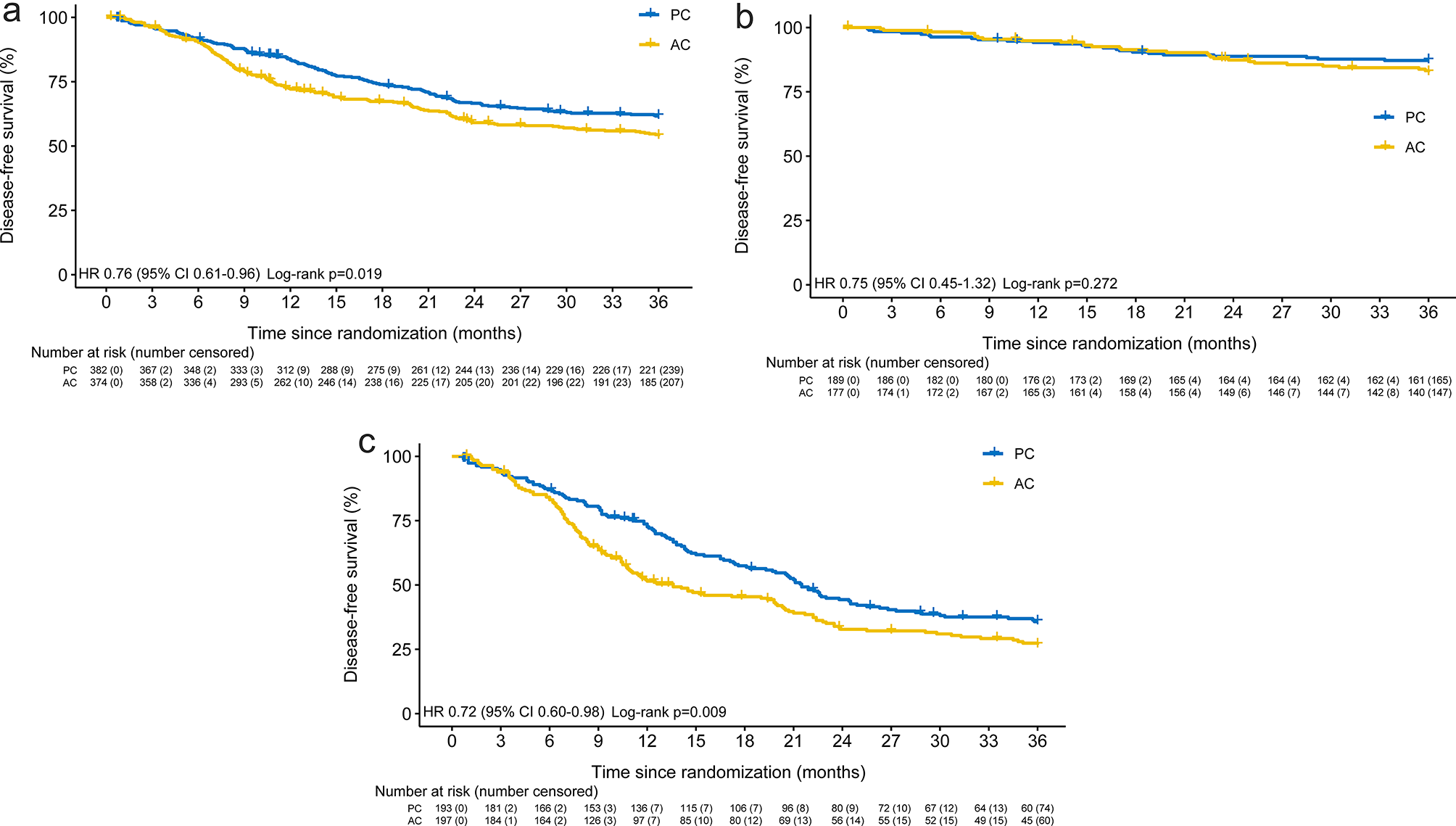
All data in our study were derived from at least three independent experiments, and each experiment was conducted in triplicate. Data were processed with GraphPad Prism 7 and were described as the means ± SD. Unpaired Student’s t test and one-way analysis of variance were applied to analyze the difference between two or multiple groups. The log-rank test was performed to compare survival. Statistical significance was considered at P < 0.05.


留言 (0)