
記住我


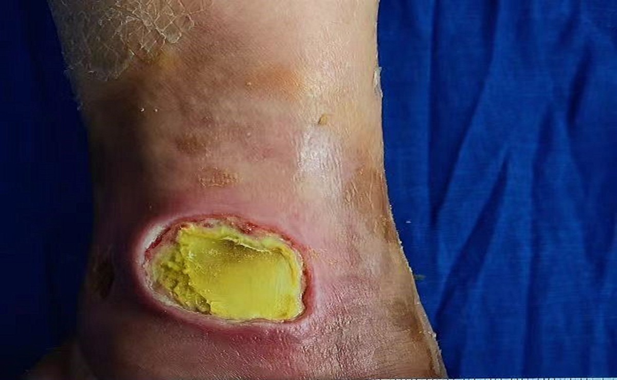


The occurrence of diabetic foot ulcers with underlying osteomyelitis is a frequent complication of diabetic foot syndrome and the most common diabetic foot infection (DFI).1,2 Diabetic foot osteomyelitis (DFO) is found in 50% to 60% of severe DFI cases3 and is defined as any foot infection with associated systemic manifestations of the systemic inflammatory response syndrome.4 Serious sequelae, such as minor and major amputations, are associated with DFO.3 Major amputations result in higher mortality (48% mortality 2 years after amputation and 68% mortality 5 years after amputation).5
Several imaging modalities for characterization and diagnosis of DFO are available, including plain X-rays, MRI, computed tomography, bone scintigraphy, single-photon emission computed tomography, positron emission tomography, and ultrasonography.6,7 The diagnostic accuracy of these imaging tests has been reviewed in the literature.7,8X-ray is an important diagnostic tool for DFO because it has several advantages, including ease of use, wide availability, relatively low cost, and minimal harm to patients.4,7–9 In addition, X-ray and MRI are both commonly used to evaluate complications in patients with DFI, especially when an X-ray shows radiologic changes.7
Although X-rays offer many advantages and utility for DFO diagnosis, there is a lack of research analyzing the role of serial X-rays to diagnose or monitor DFO. Serial X-rays in patients with DFO could provide useful information for diagnosing and detecting long-term DFO-related complications. In this study, researchers analyzed the radiologic changes shown on plain X-rays in patients with DFO and noted the development of complications in a long-term follow-up.
METHODSIn a specialized diabetic foot unit, the researchers conducted a prospective observational study between November 2014 and November 2018 that included 115 patients with DFO. The selection criteria consisted of the following: patients with diabetes mellitus who were 18 years or older, who demonstrated ulcer healing after surgical or antibiotic treatment for DFO management, and who signed written consent agreeing to participate in the study. Patients were excluded if they had critical limb ischemia, were pregnant or lactating, or refused to sign the informed consent. At the baseline visit, researchers collected patients’ medical histories and demographic data, confirmed a DFO diagnosis, and performed vascular and neurologic examinations.
Diabetic foot osteomyelitis was clinically suspected after using a combination of a probe-to-bone (PTB) test and plain X-ray.10 The international guidelines for the management of diabetic foot currently recommend the diagnosis of DFO through a combined initial testing of PTB, the erythrocyte sedimentation rate (or C-reactive protein and/or procalcitonin), and plain X-rays to diagnose osteomyelitis.4 The PTB test was performed with metal Halsted mosquito forceps and was considered positive when the researcher could feel a gritty or hard surface. Each PTB test was performed by the same investigator, who has 10 years of experience in diabetic foot management. Researchers obtained X-rays (two standard views) and considered the images to be positive when the bone showed periosteal elevation, cortical disruption, gross bone destruction, a sequestrum, and/or involucrum. All patients received a DFO diagnosis based on positive bone culture or histology result.4,11 Bone biopsies were taken for histologic analysis when possible; otherwise, a microbiological bone culture was performed.
During vascular examination, researchers noted the presence of peripheral arterial disease when the patient fulfilled the following criteria: absence of distal pulses (posterior tibial and dorsal pedis) and ankle-brachial index (ABI) <0.9. In cases with an ABI >1.4 (arterial calcification), peripheral arterial disease was considered with a toe-brachial index <0.7 and a transcutaneous oxygen pressure (TcPo2) of <30 mm Hg (TCM4 transcutaneous monitor; Radiometer Medical, Brønshøj, Denmark).12,13 Researchers documented the presence of critical limb ischemia when the patient met the following criteria: absence of distal pulses and ankle systolic pressure < 70 mm Hg or ABI <0.5 or toe systolic pressure <50 mm Hg.13,14 The neurologic examination was performed by Semmes-Weinstein 5.07/10-gauge monofilament (Novalab Ibérica, Alcala de Henares, Madrid, Spain) and bio-tensiometer (Me.Te.Da. S.r.l., San Benedetto del Tronto, Italy). Researchers noted the presence of neuropathy when the patient felt no sensation during one of the two tests.
The patients received either surgical or medical treatment, according to the previously published recommendations.15 Patients who received surgical treatment underwent conservative surgery, defined as procedures in which only infected bone and nonviable soft tissue were removed, but no amputation of any part of the foot.16 All surgeries were performed by the same surgeon, and bone samples were sent for microbiology and/or pathology analysis for diagnostic confirmation of DFO. For the first week after surgery, all patients received antibiotic treatment according to the results of the antibiogram;17 in cases of negative culture results, researchers prescribed empirical antibiotics according to Infectious Disease Society of America guidelines.18 Patients undergoing medical treatment received empiric antibiotic treatment, which was modified as needed according to the bone culture results.17,18 Antibiotic treatment lasted 90 days.19,20 Patients were evaluated twice per week until healed. In accordance with the department’s wound care protocol, both groups received the same local treatment with dressings selected based on exudate control, comfort, and cost. A removable cast walker was used as off-loading for every patient.
After healing, patients were evaluated for a 1-year period. Authors performed four follow-up visits: visit 1 (healing), visit 2 (1-month follow-up), visit 3 (6-month follow-up), and visit 4 (12-month follow-up). Authors considered ulcer healing to be complete epithelialization of the ulcer, with the skin remaining intact after 2 weeks.21 During the follow-up visits, researchers obtained X-rays (two standard views) and analyzed the following features for radiographic image changes: affected bone marrow (focal loss of trabecular pattern or marrow radiolucency), active periosteal reaction (periosteal reaction or elevation), sequestrum (devitalized bone with radiodense appearance separated from normal bone), cortical disruption (loss of bone cortex, with bony erosion or demineralization), and other types of signs. The authors analyzed the association between X-ray changes and the development of complications after healing during the 1-year follow-up period.
The researchers considered several events to be complications during follow-up, including DFO recurrence, new cases of DFO, soft tissue infection, ulcer recurrence, reulceration, minor or major amputation, death, and other diabetic foot disease-related events. All patients were followed for 1 year or until any adverse event that caused early termination or death.
The authors obtained ethical approval (14/485-E) and completed the study in accordance with the ethical standards of the responsible committee and the code of ethics of the Declaration of Helsinki.22 Each patient provided informed consent.
The researchers performed statistical data analyses using SPSS version 20.0 for iOS (SPSS, Inc, Chicago, Illinois). Descriptive analyses were performed. The authors calculated the means and SDs for quantitative variables and frequency distributions and percentages for qualitative variables. The χ2 test was used to identify differences in qualitative variables, and odds ratios (ORs) were determined for statistical significance associations. Differences were considered significant at P < .05 for a confidence interval of 95%. A log-rank test was performed to compare survival time without complications between the presence and absence of radiologic changes on the X-ray images.
RESULTSOf the 115 patients with DFO included in the study, during the follow-up visits, 10 patients (8.7%) showed radiologic changes at visit 1, 6 at visit 2 (5.2%), 9 at visit 3 (8.3%), and 8 at visit 4 (7.9%). The radiologic changes are summarized in Table 1. During the 1-year follow-up, 85 patients (73.9%) developed complications distributed as follows: 34 patients (29.8%) showed complications at visit 2, 62 at visit 3 (56.4%), and 54 at visit 4 (51.4%; Figure 1). The main baseline characteristics of the patients enrolled in the study are summarized in Table 2.
Table 1 - OBSERVED RADIOLOGIC CHANGES Observed Change Visit 1 Figure 1:
 FLOWCHART OF DISTRIBUTION OF COMPLICATIONSAbbreviation: DFO, diabetic foot osteomyelitis.
Table 2 - BASELINE PATIENT CHARACTERISTICS Variable n (%) Sex Male 95 (82.6) Female 20 (17.4) Type of DM Type 1 12 (10.4) Type 2 103 (89.6) PAD 48 (41.7) Neuropathy 115 (100.0) Ulcer location Forefoot 106 (92.2) Midfoot 5 (4.3) Hindfoot 4 (3.5) Treatment for DFO Surgical 96 (83.5) Medical 19 (16.5) Variable Mean ± SD Age, y 63.0 ± 10.1 DM duration, y 17.6 ± 12.3 HbA1c, % 8.1 ± 6.0 BMI, kg/cm2 28.4 ± 5.6 Duration from ulcer, wk 15.8 ± 32.3 Time until healing, wk 15.9 ± 9.8Abbreviations: BMI, body mass index; DFO, diabetic foot osteomyelitis; DM, diabetes mellitus; HbA1c, glycosylated hemoglobin; PAD, peripheral arterial disease.
The researchers observed that changes in X-ray images after ulcer healing (visit 1) had a significant association with development of complications during the 1-year follow-up (P = .049; OR, 1.4 [95% confidence interval, 1.2–1.6]). Moreover, all patients who showed radiologic changes at visit 1 developed complications during follow-up. When the authors analyzed the association between the presence of radiologic changes and development of complications at the different follow-up visits, they observed additional significant associations (P = .043 [OR, 5.2; 0.9–29.9] at visit 2; P = .046 [OR, 6.7; 0.8–55.3] at visit 3; and P = .025 [OR, 8.1 [1.0–68.8] at visit 4). In patients with radiologic changes on X-ray images, the percentage of complications increased during the 1-year follow-up, with 11.8% of these patients experiencing complications at visit 2, 12.9% at visit 3, and 14.0% at visit 4. In contrast, during the same follow-up period in patients without radiologic changes, the percentage of complications decreased, with 2.5% experiencing complications at visit 2, 2.2% at visit 3, and 2.0% at visit 4. Moreover, as observed in Figure 2, patients with radiologic changes on X-ray images had lower survival probabilities in comparison with patients who did not show radiologic changes.
 Figure 2:
 SURVIVAL FUNCTION FOR THE DEVELOPMENT OF COMPLICATION DURING THE 1-YEAR FOLLOW-UP ACCORDING TO THE ABSENCE OR PRESENCE OF RADIOLOGIC CHANGES
DISCUSSIONComplications frequently develop in patients with DFO. In the present study, almost 74% of participants developed complications during the first year after healing. The results revealed that X-ray is a useful tool for detecting and confirming complications and that patients exhibiting radiologic changes on X-ray images have a high probability of developing a complication. All patients who had radiologic changes after the ulcer healed developed a complication during follow-up.
X-ray is a commonly used diagnostic imaging technique for the diagnosis of DFO and is widely recommended by international guidelines.4,18X-ray is recommended as a first test to confirm advanced DFO and to rule out other disorders.4,9 As stated in the Infectious Disease Society of America guidelines,18 if an X-ray shows radiologic changes compatible with DFO, the recommendation is to initiate treatment for suspected osteomyelitis. Moreover, the recent update of the International Working Group of Diabetic Foot guidelines4 suggests that X-rays showing signs of bone healing and no further bone destruction should be considered an improvement. However, if no radiologic changes are seen on the X-ray and no suspected infection of the bone exists, the recommendation is to assess the patient every 3 weeks using control X-rays.1
Currently, DFO is considered in remission when diagnostic tests suggest infection improvement; DFO should probably not be considered cured until no recurrence occurs at 1 year posttreatment.23,24 Although X-ray is easy to use, widely available, relatively cheap, and associated with minimal harm,4,9 its utility has not been studied for monitoring or following up patients during the DFO remission period. The present results demonstrate the utility and necessity of using plain X-rays to monitor the DFO remission period because radiologic changes are associated with the development of long-term complications.
In a recent prospective study with a follow-up period of 1 year, Tardáguila-García et al25 examined the relationship between blood parameters and the development of long-term complications. They found that serial blood testing (lymphocytes, erythrocyte sedimentation rate, C-reactive protein, albumin, and glycemia) is a useful diagnostic tool for detecting patients who could develop complications. The authors recommended assessing patients during the DFO remission period by performing serial blood testing. Moreover, a recent article published by experts in the management of DFO also recommended following the patients for at least 1 year after DFO.26,27 Based on the present findings, the authors add that monitoring patients for at least 1 year after ulcer healing by performing serial X-rays to detect radiologic changes could indicate complication development.
The main limitation of this study is that it did not include a control group, which would be an interesting comparison for future studies. In addition, the authors did not compare data regarding X-ray changes and complications during the follow-up period between surgical versus nonsurgical management of DFO. However, this is the first study to analyze the utility of X-ray in monitoring patients with DFO and its long-term follow-up (1 year).
CONCLUSIONSThe present results support inclusion of preventive models during the DFO remission period and monitoring patients using plain X-rays. The use of X-rays after healing in patients who had DFO could be useful in early detection and diagnosis of complications that could appear 1 year later. The authors recommend serially following healed DFUs at 1, 6, and 12 months to monitor for complications.
The presence of radiologic changes during DFO remission period is related to the development of complications. Plain X-rays could be useful in selecting patients with DFO who are likely to develop a complication after healing.
References 1. Lázaro-Martínez JL, Tardáguila-García A, García-Klepzig JL. Diagnostic and therapeutic update on diabetic foot osteomyelitis. Endocrinol Diabetes Nutr 2017;64:100–8. 2. Lázaro Martínez JL, García-Álvarez Y, Tardáguila-García A, García-Morales E. Optimal management of diabetic foot osteomyelitis: challenges and solutions. Diabetes Metab Syndr Obes 2019;12:947–59. 3. Lipsky BA. Bone of contention: diagnosing diabetic foot osteomyelitis. Clin Infect Dis 2008;47:528–30. 4. Lipsky BA, Senneville E, Abbas ZG, et al. Guidelines on the diagnosis and treatment of foot infection in persons with diabetes (IWGDF 2019 update). Diabetes Metab Res Rev 2020;36Suppl 1:e3280. 5. Schofield CJ, Libby G, Brennan GM, et al. Mortality and hospitalization in patients after amputation: a comparison between patients with and without diabetes. Diabetes Care 2006;29:2252–6. 6. Bires AM, Kerr B, George L. Osteomyelitis: an overview of imaging modalities. Crit Care Nurs Q 2015;38:154–64. 7. Lauri C, Leone A, Cavallini M, Signore A, Giurato L, Uccioli L. Diabetic foot infections: the diagnostic challenges. J Clin Med 2020;9(6):1779. 8. Senneville E, Lipsky BA, Abbas ZG, et al. Diagnosis of infection in the foot in diabetes: a systematic review. Diabetes Metab Res Rev 2020;36Suppl 1:e3281. 9. Llewellyn A, Kraft J, Holton C, Harden M, Simmonds M. Imaging for detection of osteomyelitis in people with diabetic foot ulcers: a systematic review and meta-analysis. Eur J Radiol 2020;131:109215. 10. Aragon-Sanchez J, Lipsky BA, Lázaro-Martínez JL. Diagnosing diabetic foot osteomyelitis: is the combination of probe-to-bone test and plain radiography sufficient for high-risk inpatients?Diabet Med 2011;28:191–4. 11. Tardáguila-García A, Sanz-Corbalán I, García-Morales E, García-Álvarez Y, Molines-Barroso RJ, Lazaro-Martinez JL. Diagnostic accuracy of bone culture versus biopsy in diabetic foot osteomyelitis. Adv Skin Wound Care 2021;34:204–8. 12. Kalani M, Brismar K, Fagrell B, Ostergren J, Jorneskog G. Transcutaneous oxygen tension and toe blood pressure as predictors for outcome of diabetic foot ulcers. Diabetes Care 1999;22:147–51. 13. Norgren L, Hiatt WR, Dormandy JA, et al. Inter-society consensus for the management of peripheral arterial disease (TASC II). J Vasc Surg 2007;45Suppl S:S5–67. 14. Hinchliffe RJ, Forsythe RO, Apelqvist J, et al. Guidelines on diagnosis, prognosis, and management of peripheral artery disease in patients with foot ulcers and diabetes (IWGDF 2019 update). Diabetes Metab Res Rev 2020;36Suppl 1:e3276. 15. Lipsky BA. Treating diabetic foot osteomyelitis primarily with surgery or antibiotics: have we answered the question?Diabetes Care 2014;37:593–5. 16. Aragón-Sánchez J. Treatment of diabetic foot osteomyelitis: a surgical critique. Int J Low Extrem Wounds 2010;9(1):37–59. 17. Tardáguila-García A, Lázaro-Martínez JL, Sanz-Corbalán I, García-Álvarez Y, Álvaro-Afonso FJ, García-Morales E. Correlation between empirical antibiotic therapy and bone culture results in patients with osteomyelitis. Adv Skin Wound Care 2019;32:41–4. 18. Lipsky BA, Berendt AR, Cornia PB, et al. 2012 Infectious Diseases Society of America clinical practice guideline for the diagnosis and treatment of diabetic foot infections. Clin Infect Dis 2012;54(12):e132–73. 19. Lázaro-Martínez JL, Aragón-Sánchez J, García-Morales E. Antibiotics versus conservative surgery for treating diabetic foot osteomyelitis: a randomized comparative trial. Diabetes Care 2014;37:789–95. 20. Tone A, Nguyen S, Devemy F, et al. Six-week versus twelve-week antibiotic therapy for nonsurgically treated diabetic foot osteomyelitis: a multicenter open-label controlled randomized study. Diabetes Care 2015;38:302–7. Diabetes Care 2015;38:735. 21. Van Netten JJ, Bus SA, Apelqvist J, et al. Definitions and criteria for diabetic foot disease. Diabetes Metab Res Rev 2020;36Suppl 1:e3268. 22. World Medical A. World Medical Association Declaration of Helsinki: ethical principles for medical research involving human subjects. JAMA 2013;310:2191–4. 23. Vouillarmet J, Morelec I, Thivolet C. Assessing diabetic foot osteomyelitis remission with white blood cell SPECT/CT imaging. Diabet Med 2014;31:1093–9. 24. Gariani K, Lebowitz D, von Dach E, Kressmann B, Lipsky BA, Uckay I. Remission in diabetic foot infections: duration of antibiotic therapy and other possible associated factors. Diabetes Obes Metab 2019;21:244–51. 25. Tardáguila-García A, García-Álvarez Y, García-Morales E, Álvaro-Afonso FJ, Sanz-Corbalán I, Lázaro-Martínez JL. Utility of blood parameters to detect complications during long-term follow-up in patients with diabetic foot osteomyelitis. J Clin Med 2020;9(11):3768. 26. Tardáguila-García A, Sanz-Corbalán I, García-Alamino JM, Ahluwalia R, Uccioli L, Lázaro-Martinez JL. Medical versus surgical treatment for the management of diabetic foot osteomyelitis: a systematic review. J Clin Med 2021;10(6):1237. 27. Tardáguila-García A, García-Álvarez Y, García-Morales E, López-Moral M, Sanz-Corbalán I, Lázaro-Martínez JL. Long-term complications after surgical or medical treatment of predominantly forefoot diabetic foot osteomyelitis: 1 year follow up. J Clin Med 2021;10(9):1943.
留言 (0)