Disrupted white matter integrity in the brain of type 1 diabetes is associated with peripheral neuropathy and abnormal brain metabolites
Aims
We aimed to quantify microstructural white matter abnormalities using magnetic resonance imaging and examine their associations with 1) brain metabolite and volumes and 2) clinical diabetes-specific characteristics and complications in adults with type 1 diabetes mellitus (T1DM) and distal symmetric peripheral neuropathy (DSPN).
Methods
Diffusion tensor images (DTI) obtained from 46 adults with T1DM and DSPN and 28 healthy controls were analyzed using tract-based spatial statistics and were then associated with 1) brain metabolites and volumes and 2) diabetes-specific clinical characteristics (incl. HbA1c, diabetes duration, level of retinopathy, nerve conduction assessment).
Results
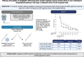
Adults with T1DM and DSPN had reduced whole-brain FA skeleton (P = 0.018), most prominently in the inferior longitudinal fasciculus and retrolenticular internal capsule (P < 0.001). Reduced fractional anisotropy (FA) was associated with lower parietal N-acetylaspartate/creatine metabolite ratio (r = 0.399, P = 0.006), brain volumes (P ≤ 0.002), diabetes duration (r = −0.495, P < 0.001) and sural nerve amplitude (r = 0.296, P = 0.046). Additionally, FA was reduced in the subgroup with concomitant proliferative retinopathy compared to non-proliferative retinopathy (P = 0.03). No association was observed between FA and HbA1c.
Conclusions
This hypothesis-generating study provided that altered white matter microstructural abnormalities in T1DM with DSPN were associated with reduced metabolites central for neuronal communications and diabetes complications, indicating that peripheral neuropathic complications are often accompanied by central neuropathy.


留言 (0)