The study was approved by the local ethical committee and registered on ClinicalTrials.gov (NCT04904640).
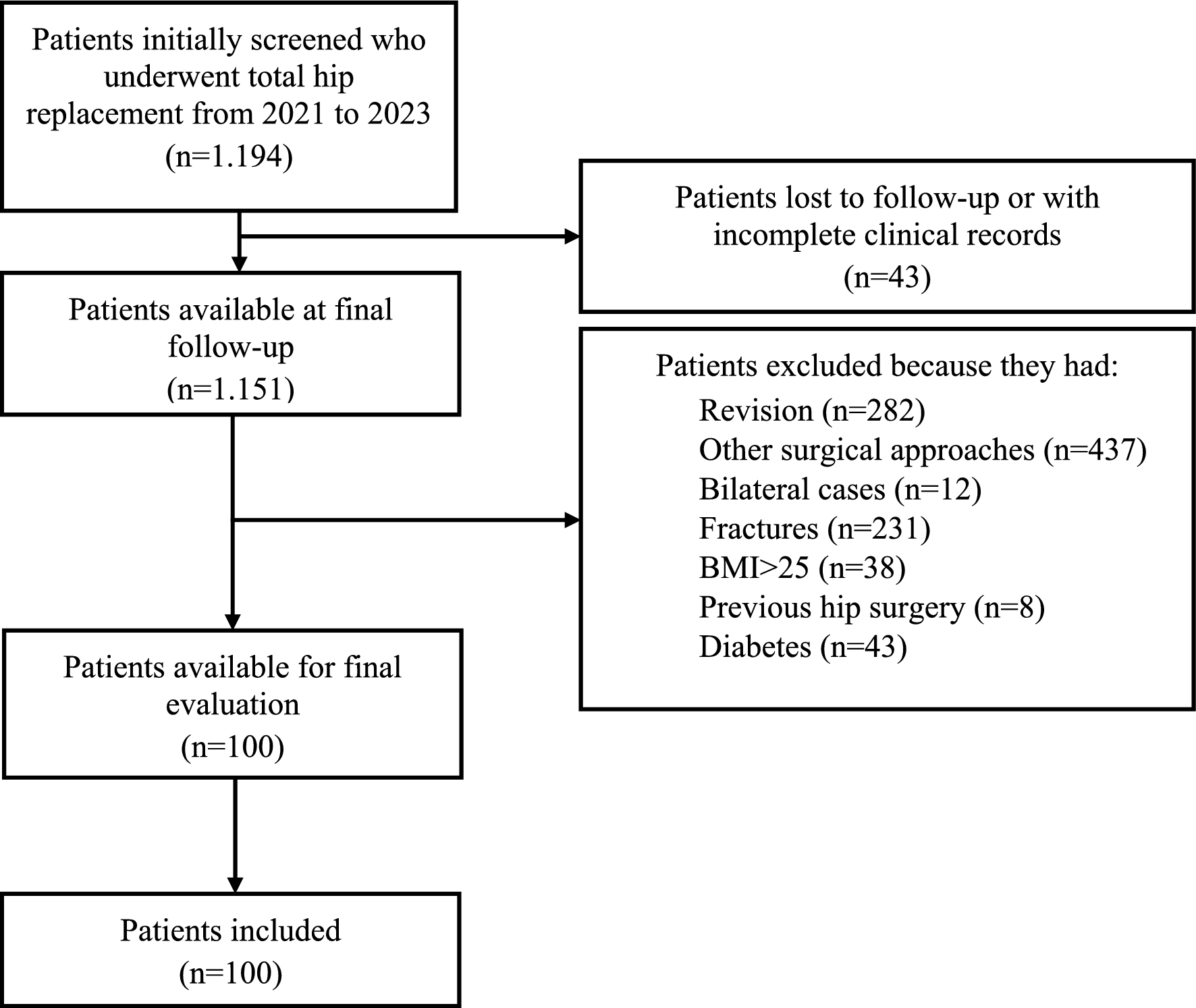
The hospital database was screened for pelvis CT scans that included a native dysplastic hip and were performed during 2000–2018 for pre-operative THA planning or painful THA diagnosis, with the aim being to achieve 200 eligible hips.
The inclusion criteria were:
Definition of DDH according to Wiberg (center edge angle < 20°) [7]
Pre-operative CT scan extended from the fourth lumbar vertebra to the tibial plateau.
Hips with other congenital or acquired pathologies, arthroplasties or inadequate CT scans were excluded.
200 hips in 150 CTs (150 Caucasian patients) were randomly selected. The first author simulated the implantation of three different stem designs per hip using a 3D CT-based pre-operative planning software (Hip-Op) after an appropriate assessment of the native femoral morphology.
3D CT-based pre-operative software for surgical planning: Hip-Op
The software reproduced a 3D CT-based planning environment [8, 9]. The user-friendly graphical user interface is based on a multimodal display visualization. The user can choose the components from a library of selected implants: the implants and the patient anatomy are rendered in each view. The planner may evaluate the implant type, size and position by interactively moving and rotating the components in the view area. The validity of Hip-Op software as a pre-operative planning aid has previously been assessed [9].
Implant features and planning technique
The planning was performed by a single arthroplasty surgeon (the first author) in every case. Three non-modular stems were selected based on their design and classification according to Khanuja et al. [10]: CLS (Zimmer Biomet, Warsaw, USA) single wedge tapered stem, type 1; Aptafix (Adler Ortho, Milan, Italy) anatomical stem, type 6; Wagner Cone (Zimmer Biomet) conical tapered stem, type 3B [11,12,13]. The CLS had 13 stem sizes with three CCD angles (125°, 135° and 145°) [11]. The Aptafix had a standard version and a offset solution (7.5 mm lateralization), both with a CCD angle of 135°: eight possible stem sizes are available [12]. The Wagner Cone had 12 stem sizes with two CCD angles, 125° and 135° [13]. The acetabular cup was the Continuum (Zimmer), which was adopted for every single stem simulation.
In all the dysplastic hips, the surgical planning aimed to reproduce the native center of rotation every possible time (a high hip center was only accepted in cases of very severe superolateral bone deficit). All the cups were implanted using a conservative circumferential reaming technique, admitting slight medialization in shallow acetabula. Cup inclination was set at 40–45°, with no more than one-third superior undercoverage. Cup anteversion was determined avoiding an anterior overhang (or reducing it to a minimum), aiming for 10–20° of anteversion. The stems were positioned with the aim of achieving an acceptable combined anteversion according to Dorr and Widmer, no offset reduction, adequate canal filling, the most neutral positioning in the coronal and sagittal planes, and leg length equalization, avoiding over-lengthening superior to 3 cm. For all the stems, the surgical planning was performed according to the instructions provided by the manufacturers and the experience of the planner.
Reliability of the simulations and comparison with post-operative measurements
Fifteen hips with a post-operative CT scan performed for contralateral hip planning and achieving a 2-year excellent clinical outcome with good signs of radiographic osseointegration were selected. In all cases, an anatomical stem was implanted. The first author, blinded to the post-operative component positioning, performed the simulation on the 15 hips. Another simulation was performed by the same author on the same hips after 4 weeks. All the tests were compared to the post-operative component positioning as provided by the CT scan. The outcomes and the reference values were the same as explained below.
Demographics of the cohort and assessment of the native hip morphology
The demographics of the patients were collected. On the CT scan, the DDH pathology was classified according to Crowe et al. and Hartofilakidis et al. [14]. The native femoral morphology was assessed in the coronal and axial planes [1,2,3, 15, 16] (Table 1).
Table 1 The native hip morphology was described using several CT-based measurements (mean ± standard deviation; median in parentheses): the features that are correlated with the Crowe classification are shown in bold (LT lesser trochanter)Outcome measurements
In all the simulations, the following measurements were taken (cup measurements were unique to every case; stem measurements were performed for every stem in every case): cup anteversion, stem anteversion, acetabular offset, femoral offset, sagittal and coronal tilt, and canal filling at the mid-third of the stem [1, 11, 17, 18].
When planning, the following five outcomes had to be matched for the stem to be considered acceptable:
Combined anteversion (according to Dorr et al.) between 25° and 50° and combined anteversion (according to Widmer et al.) < 37° (both the targets had to be matched) [11];
Global offset (acetabular + femoral offset) loss not inferior to 12% of the native global offset [19];
Coronal and sagittal stem tilt < 5° [17];
Canal filling > 80% [18];
Leg lengthening < 3 cm [20].
Statistical analysis
The analyses were performed using SPSS 14.0 (SPSS Inc, Chicago, IL, USA). Quantitative data are reported as average values, standard deviations and minimum to maximum ranges. Qualitative data are expressed as frequencies and percentages and were tested using the chi-squared test. The reliability of the simulations was assessed using Fisher’s test (categorical variables). Correlations between parameters were assessed using Kendall, Spearman or Pearson coefficients, depending on the data type. The correlation strength was evaluated according to the current medical literature [21]. Threshold for significance: p = 0.05.


留言 (0)