
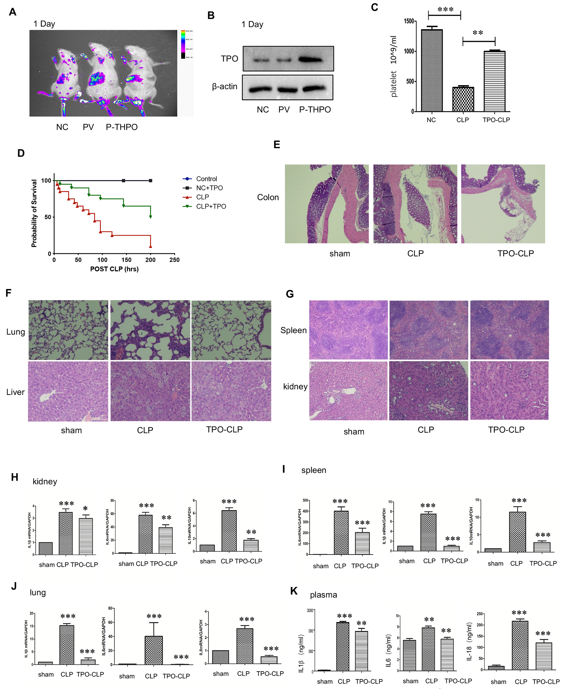
記住我


Chronic exposure to inhaled irritants including cigarette or biomass smoke and/or environmental pollutants are the principle causes of chronic obstructive pulmonary disease (COPD). The Global Burden of Disease analysis has recently reported that COPD caused 3.2 m deaths annually and also has a considerable health-care and societal burden globally [1]. These numbers are likely to rise as people are now living longer. Currently, 2 billion people are smokers or are exposed to second-hand smoke, over 2 billion people are affected by biomass fuel use and ∼1 billion of the population are exposed to the detrimental consequences of outdoor air pollution [1]. As a consequence, in 2019 there were almost 550 m people globally with a chronic respiratory disease, mostly due to COPD, with only cardiovascular disease (CVD) and cancer causing more deaths annually [1]. Across the EU, the annual health-care costs, including primary and inpatient health care costs, for chronic respiratory diseases was €380 billion in 2019 [1].
COPD is considered a chronic immunoinflammatory disease of the airway, which drives severe airflow limitation due to subsequent remodelling of the small airways together with mucus hypersecretion and/or alveolar wall destruction or emphysema [2]. The inflammatory response in COPD is linked to the recruitment and activation of neutrophils and alveolar macrophages and dysregulation of structural cells such as epithelial cells with enhanced mucus production (bronchitis) and attenuation of ciliated epithelial cell function [2]. The release and function of the numerous inflammatory cytokines, chemokines, growth factors and other mediators that are elevated in COPD is exacerbated by the presence of endogenous and exogenous oxidative stress [2] (Fig. 1). There is increasing evidence for a role of oxidant stress-induced autoimmunity against modified self-antigens in COPD as well as the recognition of premature senescence occurring in the lungs and airways of COPD patients [3] (Fig. 2). Bacterial and viral infections result in acute exacerbations of COPD due to an altered innate immune system and this is a major cause of death in COPD [4]. The global initiative for chronic obstructive lung disease (GOLD) guidelines now assesses severity of COPD according to symptoms and the frequency of exacerbations [5]. Current therapies for COPD are directed towards palliative effects on lung function and none of the current therapeutic agents reverse inflammation and prevent disease progression [2].
Fig. 1
Sources of reactive oxidant species (ROS) within the lung. The high levels of ROS reported in the lung and airways of COPD subjects is derived from both exogenous and exogenous sources. Exogenous sources include cigarette smoking, environmental pollution, pathogens and inflammation. ROS is generated endogenously through mitochondria including mitochondrial ROS (mtROS), peroxisome activation, hypoxia and inflammation. ROS affects the function of many intracellular organelles such as proteasome, inflammasome, lysosome and the endoplasmic reticulum (ER) via the unfolded protein response (UPR) to elicit detrimental effects on cellular functions
Fig. 2
Molecular and cellular targets of reactive oxygen species (ROS) in the airways and lungs of COPD patients. ROS directly affects the activity and/or expression of redox-sensitive kinases, transcription factors, mitochondria, anti-oxidant pathways, iron (Fe) biology and innate immune systems such as complement and autoantibody production. Modulation of these processes promotes cell proliferation/survival and cellular senescence which is associated with enhanced inflammation. Enhanced oxidant pathways and reduced anti-oxidant activity affects mucosal defence against bacteria and viruses including reduced phagocytosis, whilst oxidative stress actions on catalase and Fe allow excess bacterial growth. ROS also causes post-translational modifications of DNA, RNA, lipids and proteins to affect cellular function and reveal neo-epitopes for auto-antibody induction. The generation of oxidised phospholipids (OxPLs) can further drive mitochondria dysregulation and activate the inflammasome. Abbreviations: AP-1: activator protein-1; ARE, anti-oxidant response element; ERK, extracellular signal-regulated kinase; ETC, electron transport chain; GSH, glutathione; GPX, glutathione peroxidase; GRX, glutaredoxins; HIF1α, hypoxia-Inducible Factor 1α; JAK-STAT, Janus kinase-signal transducer and activator of transcription; KEAP, Kelch-like ECH-associated protein; MEK, mitogen-activated extracellular signal-regulated kinase; mtDNA, mitochondrial DNA; NADPH, nicotinamide adenine dinucleotide phosphate; NF-κB, nuclear factor κB; NOS, nitric oxide synthase; Nrf2, Nuclear factor-erythroid factor 2-related factor 2; p38 MAPK, p38 mitogen activated protein kinase; PI3K, phosphoinositide 3-kinase; RNS, reactive nitrogen species; TRX, thioredoxins
The mechanisms that drive COPD pathogenesis are not well understood beyond the recognition of the key importance of oxidative stress, but the increasing understanding and identification of pathways involved in disease phenotypes may provide new therapeutic opportunities.
Reactive oxygen species (ROS) and oxidative stressOxygen is essential for the supply of energy to eukaryotes but it also forms detrimental ROS and the related nitrogen species (RNS) following both enzymatic and non-enzymatic processes [6]. This leads to protein, lipid and DNA damage and must therefore be kept under tight regulatory control by the presence and activity of antioxidants located within cells and in the lung epithelial lining fluid [6]. External factors such as infection, air pollution or cigarette smoke exposure can overcome the local antioxidant capacity whilst internal sources of ROS include inflammatory cell activation and disease. Persistent ROS is associated with several airway diseases including asthma, COPD and lung cancer [6]. Indeed, it is recognised that COPD pathogenesis involves a disturbed balance between antioxidant defences and enhanced oxidative stress [7].
Glutathione is a major redox buffer that acts as an antioxidant defence mechanism to protect the lung from oxidative stress [8]. More recently, glutathione is recognized for its ability to induce S-glutathionylation which can change the structure and function of the target protein [8]. S-glutathionylation also allows the protein to be regenerated enzymatically as it protects them from irreversible oxidation. Glutathione S-transferases and glutaredoxins catalyze this process [8].
There are increased levels of ROS in COPD due to enhanced numbers of superoxide anions (O− 2), hydroxyl radicals (•OH), hydrogen peroxide (H2O2) and suppression of antioxidant and antiinflammatory gene expression. Key factors include heme oxygenase (HO)-1, glutathione peroxidase (GPx) and thiol metabolism-associated detoxifying enzymes (glutathione S-transferases, GSTs) together with antioxidant transcription factors including nuclear factor erythroid 2-related factor (Nrf)2 [7]. In addition, oxidative stress in the presence of nitric oxide (NO) results in the formation of various RNS including peroxynitrite (ONOO −) which cause cell damage and disruption of biological processes by inducing protein and DNA nitration that impacts upon DNA damage/repair, mitochondrial respiration and inflammation. Myeloperoxidase (MPO) activation as well as H2O2 can also promote nitration of proteins following nitrite (NO2 -) oxidation [7] (Fig. 2).
COPD is often considered as a disease of premature lung ageing which itself is associated with abnormal responses to oxidative stress [9]. Age-related changes in cell quality control systems are linked particularly with a reduced ability to undertake redox and protein homeostasis. This age-related redox imbalance may act as an initial cellular ‘hit’ that induces cell adaptive stress-response pathways, increases oxidative stress with the resulting enhancement of lung injury leading to COPD [9]. Thus, a number of ‘secondary hits’ including smoking and environment-related pollution together with infections could the primed or dysregulated adaptive defence and repair pathways with further enhanced redox stress that results in the onset and progression of COPD [9].
Mitochondrial ROS and COPDMitochondria are critical components of redox signaling and their aberrant function is linked to abnormal oxidative stress and metabolic dysfunction [10]. In addition to signaling by ROS, mitochondria regulate many cellular processes including cellular survival, control of anabolic and metabolic pathways and of innate immune signaling factors that are all altered in COPD [10]. Furthermore, in some patients with COPD, there is a hypoxic drive to their disease due to ventilation:perfusion mismatch which will also impact on mitochondrial function [11]. Hypoxia, which is sensed by the transcription factor HIF1α, results in further ROS production and oxidative damage, heightened inflammation and a metabolic switch towards a more glycolytic state [11]. The overall change in mitochondrial function and metabolic adaptation to an altered local microenvironment results in the production of distinct metabolic intermediates that can modulate the inflammatory response by acting as signaling molecules [11] (Fig. 2).
Enhanced levels of mitochondrial ROS (mtROS) as well as decreased mitochondrial membrane potential (ΔΨm) and mitochondrial superoxide dismutase (SOD2) levels were observed in bronchial biopsies from COPD patients [12]. ΔΨm in lung samples significantly correlated with forced expiratory volume in 1 s (FEV1, % predicted), 6-min walk test, lung carbon monoxide transfer factor (TLCOC % predicted) and maximum oxygen consumption. Increased total ROS and mtROS were found in the quadriceps muscle of the same patients with no effect on ΔΨm, SOD2 or levels of electron transport chain (ETC) complex proteins [12]. The data suggest that the mitochondrial changes observed in the quadriceps muscle are likely to result from spill over from the lung.
Monocyte-derived macrophages (MDMs) and alveolar macrophages from COPD patients are less effective at bacterial phagocytosis than cells from healthy control subjects [13]. This leads to impaired responses against exacerbation triggers in COPD and heightened airway inflammation in these subjects [14]. For example, COPD alveolar macrophages secrete excess proinflammatory mediators and proteases and express an altered pattern of surface and intracellular markers [15]. Phagocytosis of Haemophilus influenzae and Streptococcus pneumoniae by COPD cells, but not cells from healthy smokers or non-smokers, increased early mtROS levels and decreased ΔΨm [13] (Fig. 2). Furthermore, exogenous ROS decreased the phagocytic activity of control alveolar macrophages [13] suggesting that drugs that restore mitochondrial dysfunction may improve the defective phagocytic response seen in COPD macrophages. Indeed, MDMs exposed to cigarette smoke displayed inhibited bacterial phagocytosis via a reduction in alveolar macrophage cystic fibrosis transmembrane conductance regulator (CFTR) expression [16]. The effects of cigarette smoke on phagocytosis were attenuated by the free radical scavenger N-acetylcysteine [16].
Type II alveolar epithelial (ATII) cells from emphysematous subjects generate high levels of mitochondrial superoxide, exhibit mtDNA damage with associated mitochondrial dysfunction [17]. The degree of impaired mitochondrial fission/fusion as suggested by reduced expression of mitofusin 1 (MFN1), optic atrophy 1 (OPA1), Fission, Mitochondrial 1 (FIS1) and phosphorylated dynamin-related protein 1 (p-DRP1) correlated with level of emphysema [17]. An interesting study that compared the ATII like cell line A549 with mitochondria to those without mitochondria (A549-Rho-0), showed the loss of mitochondria resulted in enhanced pro-inflammatory mediator release, reduced epithelial repair functions and a loss in corticosteroid sensitivity [18] (Fig. 2). This latter effect appeared to be dependent upon glycolytic reprogramming and altered phosphoinositide-3-kinase (PI3K) activity [18]. This study highlights the importance of mitochondrial homeostasis on lung epithelial cell responses and how these may contribute to COPD pathogenesis.
Cigarette smoke-induced mitochondrial dysfunction (mtROS, oxidative phosphorylation or OXPHOS protein expression, structural changes and ΔΨm) in airway epithelial cells isolated from human lung can result in cellular senescence [19]. Cigarette smoke exposure also altered mitochondrial respiration as indicated by markers of oxygen consumption rate including maximum respiration, production of ATP and oxygen spare capacity) in BEAS-2B cells and NHBE cells [19]. Mechanistically, these changes were associated with reduced mitochondrial Rho GTPase 1 (MIRO1) and PTEN Induced Kinase 1 (PINK1) expression highlighting their potential role in the pathogenesis of COPD [19]. In primary human airway fibroblasts from COPD patients, the mitochondrial-associated senescence phenotype was reversed by pharmacologic induction of HO-1 [20]. HO-1 up-regulation in COPD cells enhanced the replicative capacity and attenuated the senescence and inflammatory capacity following restoration of ‘normal’ mitochondrial respiration, glycolysis and ATP levels and a reduction in the enhanced mtROS production and restored mitophagy [20].
The development of non-small cell lung carcinoma is increased in COPD patients and their disease has a worse prognosis [21]. Carcinogenesis is driven, at least in part, by abnormal mitochondrial function enhanced oxidative stress and the expression of phosphoglycerate mutase family member 5 (PGAM5), a mitophagy regulator, was highly expressed by alveolar macrophages from COPD patients and in malignant and pre-neoplastic epithelial cells [21]. Macrophage PGAM5 levels trended towards being greater at the periphery of the cancer in patients with COPD. Together these data suggest that PGAM5 expression is associated with patient mortality and that this may be linked to abnormal mitochondrial function in specific subsets of macrophages [21].
Cellular biosynthetic and redox pathways are influenced by changes in fatty acid oxidation (FAO) and glycolysis and these extensive metabolic changes play an important function in innate immunity in COPD [4]. COPD airway smooth muscle (ASM) cells possess an aberrant mitochondrial function and a specific metabolic phenotype that is associated with enhanced growth [22]. For example, their energy production is abnormal with enhanced generation of lactate, glutamine, fatty acids and amino acids compared to cells from healthy subjects under both stimulated and unstimulated conditions. In addition, FAO capacity was attenuated at baseline in COPD ASM cells which was restored by stimulation with transforming growth factor-β (TGFβ)/foetal calf serum [22]. This was accompanied by elevated flux through the pentose phosphate shunt and of nucleotide biosynthesis. Together, this suggests that differences in glycolysis, glutamine and fatty acid metabolism occur in COPD ASM cells resulting in increased biosynthesis and redox balance which switch the cellular phenotype towards supporting cell growth in COPD [22].
As an alternative to reversal of COPD-associated mitochondrial defects in airway smooth muscle cells by small molecule drugs, Li and colleagues used mesenchymal stem cells (MSCs) to deliver ‘healthy’ mitochondria to COPD cells and in vivo to ozone-exposed animals [23]. Culture of MSCs with airway smooth muscle cells attenuated cigarette smoke-induced increased mtROS and reduction in ΔΨm loss which was associated with apoptotic cell death [23]. In these experiments, transfer of healthy mitochondria from MSCs to smoke-exposed airway smooth muscle cells occurred via tunnelling nanotubes. These results were recapitulated in vivo in murine lungs where MSCs reduced ozone-induced mitochondrial dysfunction, inflammation and airway hyperresponsiveness. The authors indicated that an MSC-based therapeutic approach may be useful in COPD [23].
Mitochondrial dysfunction in COPD may be greater in females than males [24] particularly in COPD patients with an emphysema-predominant disease [25]. Using an integrated network interference approach to analyse transcriptomic datasets from COPD and healthy controls, it was possible to identify gene sets involved in mitochondrial function and energy metabolism as being sexually dimorphic [24]. This data is supported by a recent analysis of the Emphysema versus Airways Disease project (EvA) whereby in a large study of COPD patients (312 subjects) with transcriptomic data from bronchial brushings there was a difference in the number of differentially expressed genes in males (n = 40) and in females (n = 73) between healthy and COPD [25]. Male COPD patients particularly those with an emphysema phenotype expressed a signature of mitochondrial-encoded functional genes.
Iron (Fe), oxidative stress and COPDDisrupted iron homeostasis is linked to severity of stable COPD and during acute exacerbations of COPD (AECOPD) possibly as a result of iron regulatory protein (IRP)-2 polymorphisms and independent of anaemia [26]. In a small observational study of exhaled breath condensate there was an attenuated capacity to respond correctly to cigarette smoke-induced iron handling and excretion (production of redox active iron) in patients with COPD [27] (Fig. 2) with iron responsive element binding protein 2 (IREBP2) protein being raised in COPD lung [28]. The usual cigarette smoke-induced COPD-phenotype is not observed in mice deficient in IREBP2 due to prevention of mitochondrial dysfunction. Interestingly, mice fed a mitochondrial iron chelator also did not demonstrate the cigarette smoke-induced COPD-phenotype [28]. In addition, the expression of the iron regulatory peptide hepcidin is reduced in COPD and this is recapitulated in mice after cigarette smoke exposure [29]. Murine cigarette smoke-exposed alveolar macrophages and human alveolar macrophages from smokers also have elevated levels of ferroportin. This dysregulated hepcidin/ferroportin axis contributes to reduced phagocytosis of bacteria and an enhanced response to Streptococcus pneumoniae infection [29] (Fig. 2).
Bronchoalveolar lavage (BAL) fluid (BALF) levels of iron and ferritin were elevated in subjects from the SPIROMICS (Subpopulations and Intermediate Outcomes in COPD Study) study and were raised to a greater extent in COPD patients with more exacerbation [30]. However, BAL levels did not correlate with systemic iron markers [30]. Overall, enhanced iron retention in the airways and lungs of patients with COPD can contribute to the oxidative stress-induced cellular damage and microbial virulence [27]. Overall, there is a link between iron levels, oxidative stress and the host-airway microbiome [31, 32].
Both oxygen and iron (Fe) are important in the formation of ROS [33]. There were significantly lower serum levels of antioxidant carotenoids which regulate Fe levels in COPD subjects (n
留言 (0)