
記住我


Infectious Agents and Cancer 2022, 17(Suppl 1): PL4
Background: Human telomerase reverse transcriptase (hTERT or TERT) is the catalytic subunit of the telomerase enzyme, which elongates telomeres at the ends of chromosomes during embryonic development and in stem cells. TERT gene is repressed in somatic cells and telomeres undergo progressive shortening at each division (Hayflick limit) until senescence. Nevertheless, telomerase is re-expressed in up to 90% of human cancers through several mechanisms, including somatic mutations, genetic and epigenetic alterations and virus integration. In HPV-related cancers, TERT gene is always expressed in cells infected by high risk HPVs at diverse body sites, such as cervix, vulva, vagina, anus, penis and head-and-neck cancers. Several studies have shown that high risk HPV E6 oncoprotein is a main regulator of TERT expression for its ability to transactivate the TERT promoter. Nevertheless, in cervical neoplasia, telomerase expression was found to be significantly correlated with the histological severity of cervical lesions regardless of HPV E6 RNA levels.
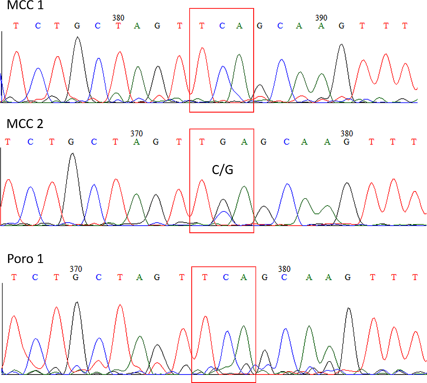
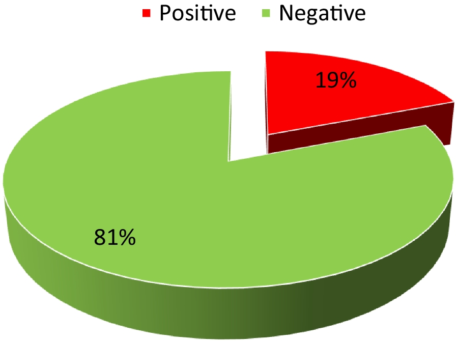
Results: An HPV-independent activation of telomerase is caused by hot spot changes at position -124 (G > A) and -146 (G > A) in the core promoter region of the TERT gene. We have identifed, by Sanger sequencing and droplet digital PCR, the TERT promoter mutations -124A and -146A in 17% of cervical, 53% of vulvar and 54% of penile squamous cell carcinoma (SCC) as well as in 60% of oral carcinoma. Overall TERT promoter mutations were more frequent in tumours negative for HPV infection. TERT promoter mutations were not detected in cervical intraepithelial neoplasia grade 3 (CIN3). In HPV-positive cervical cancer cases TERT gene expression was significantly higher in TERT promoter mutated than in non-mutated tumours regardless of HPV16 E6 levels. The expression profile of p53-related genes showed that telomerase overexpression driven by promoter mutations is associated with the activation of extratelomeric functions involving among others the IGF1R/AKT signalling axis in cervical SCC as well as in TERT promoter mutated SiHa cell line.
Discussion and conclusions: Our results suggest that in cervical neoplasia the promoter mutations have a much stronger effect in TERT activation than HPV E6 oncoprotein. We can hypothesize that the role of telomerase in HPV-related cancers is dual: (1) telomerase expression is reversibly regulated by viral E6 protein in the early stages of tumorigenesis, and (2) telomerase is irreversibly and highly activated by genetic alterations and promoter mutations in TERT gene in progressing cervical neoplasia. The knowledge of non-canonical functions activated by highly expressed telomerase would be valuable to identify new actionable targets in SCC of the lower genital tract as well as in many TERT mutated tumours.
Acknowledgements: This work was supported by research grants funded by Ricerca Corrente (M1-3) and Ministero della Salute (RF-2018-12366163). We thank Vincenzo Gigantino for assistance in sample selection, retrieval and experimental procedures.
References
1. Annunziata C, Pezzuto F, Greggi S, Ionna F, Losito S, Botti G, Buonaguro L, Buonaguro FM, Tornesello ML. Distinct profiles of TERT promoter mutations and telomerase expression in head and neck cancer and cervical carcinoma. Int J Cancer. 2018 Sep 1;143(5):1153–1161. https://doi.org/10.1002/ijc.31412.
2. Tornesello ML, Buonaguro FM. Human Papillomavirus and Cancers. Cancers (Basel). 2020 Dec 15;12(12):3772. https://doi.org/10.3390/cancers12123772.
3. Cerasuolo A, Annunziata C, Tortora M, Starita N, Stellato G, Greggi S, Maglione MG, Ionna F, Losito S, Botti G, Buonaguro L, Buonaguro FM, Tornesello ML. Comparative analysis of HPV16 gene expression profiles in cervical and in oropharyngeal squamous cell carcinoma. Oncotarget. 2017 May 23;8(21):34070–34081. https://doi.org/10.18632/oncotarget.15977.
PL5. Chronic inflammation in HIV-1 infection, are elite controllers different?Nicolas Noel*Service de Médecine Interne et Immunologie Clinique, GHU Paris Saclay, AP-HP BICÊTRE, Faculté de Médecine Paris Saclay; INSERM /CEA 1184 (Immunologie des maladies virales, autoimmunes, bactériennes et hématologiques);Presenting author: Nicolas Noel (nicolas.noel@aphp.fr)Infectious Agents and Cancer 2022, 17(Suppl 1): PL5
The Human Immunodeficiency Virus is responsible for a chronic viral infection during which antiretroviral therapy is usually mandatory to maintain an undetectable viremia and to preserve immune functions.
Immune activation is a hallmark of this pathology, occurring in the early stages of infection. Multiple sources of immune activation have been described: viral replication and production, antigen presentation, microbial translocation of mucosal origin, activation of the interferon pathway, viral co-infections, etc. Moreover, immune activation is linked with the control of the HIV reservoir and viral latency, as well as with the risk of clinical events such as cardiovascular or neurocognitive diseases.
Antiretroviral treatments reduce chronic immune activation but not always normalize inflammatory parameters. In this context, HIV-1 controllers allow an interesting study of the causes and consequences of inflammation. These rare patients are defined on a virological basis, controlling viral replication spontaneously without any antiretroviral treatment. This lecture will present the evidence of persistent immune activation in HIV-controller patients in comparison with controlled patients under treatment, and discuss the risk of evolution and the research perspectives in this field.
PL6. Mechanisms of nucleic acid vaccines for therapy of chronic viral infections and cancerMargaret A. Liu 1,2,3 1ProTherImmune, Lafayette, CA, USA; 2Karolinska Institutet, Stockholm, Sweden; 3University of California, San Francisco, USAPresenting author: Margaret A. Liu (Liu@ProTherImmune.com)Infectious Agents and Cancer 2022, 17(Suppl 1): PL6
Immunotherapies for chronic viral infections and cancer have utilized approaches including monoclonal and bispecific antibodies, immunomodulatory agents (cytokines and checkpoint inhibitors), vaccines (utilizing various delivery systems), and adoptive cell therapies. The efficacy of certain antibodies, immunomodulators, and cell therapies for certain cancers have demonstrated their potential. This has led to increased efforts to develop immunotherapeutic vaccines targeting tumor and viral antigens. But challenges remain even after selecting an appropriate antigen to target, whether a viral protein or a tumor antigen. One reason for this is that tumors and chronic infections can affect the immunological milieu resulting in tolerance or immune suppression. This talk will describe the use of nucleic acid vaccines as potential vectors for therapeutic vaccines for both chronic infections and cancer. The focus will be on the immune mechanisms stimulated by these vaccines, including innate immunity and how these innate immune responses may contribute to both the specific immune responses against an encoded antigen as well as for non-specific immune benefits including immune fitness.
PL7. The path towards the cure of chronic HBV infectionFabien Zoulim1,2 1Hepatology Department, Hospices Civils de Lyon; 2Cancer Research Center of Lyon, Lyon, FrancePresenting author: Fabien Zoulim (fabien.zoulim@inserm.fr)Infectious Agents and Cancer 2022, 17(Suppl 1): PL7
Hepatitis B virus (HBV) affects more than 250 million people worldwide, and is one of the major aetiologies for the development of cirrhosis and hepatocellular carcinoma (HCC). In spite of universal vaccination programs, HBV infection is still a public health problem, and the limited number of available therapeutic approaches complicates the clinical management of these patients. Thus, HBV infection remains an unmet medical need that requires a continuous effort to develop new individual molecules, treatment combinations and even completely novel therapeutic strategies to achieve the goal of HBV elimination. Progress in our understanding of HBV persistence and pathobiology have allowed to characterize novel antiviral and immune targets for therapy, as well as novel biomarkers to assess the intrahepatic viral reservoir and host antiviral immune responses. These discoveries are now translated to chronic hepatitis B patients with clinical trials of combination therapy with direct acting antivirals and immune modulators. These clinical and translational studies show promising results that may pave the way for a functional cure of HBV and the prevention of HCC development. It will be exciting to see how the drug discovery and biomarker pipelines will feed the clinical development of novel treatments and guide clinical investigations to treat all facets of the disease.
O6. Activity of long control region of human papilloma virus as determinant of oncogenicityRiaz Y. Seedat1, Catharina E. Combrinck2, Yuri Munsamy2, Felicity J. Burt2,3* 1Department of Otorhinolaryngology, University of the Free State, Bloemfontein, South Africa; 2Division of Virology, University of the Free State, Bloemfontein, South Africa; 3National Health Laboratory Service Universitas, Bloemfontein, South AfricaPresenting author: Felicity J. Burt (burtfj@ufs.ac.za)Infectious Agents and Cancer 2022, 17(Suppl 1): O6
Background: Human papilloma viruses (HPV) have been associated with benign and malignant diseases. Approximately 150 HPV genotypes have been described, including high risk or carcinogenic viruses such as HPV 16 and 18 associated with cervical cancers, high risk HPV 31 associated with head and neck squamous cell carcinoma, and low risk types such as 6 and 11 more frequently associated with benign lesions and are the aetiological agents of recurrent respiratory papillomatosis (RRP). The HPV genome is organized into early (E) and late (L) open reading frames (ORFs), based on the position of these genes on the genome, and includes a noncoding region, or long control region (LCR) comprising crucial elements necessary for control of viral replication and transcription as well as the origin of DNA replication. The influence of mutations within the LCR is not clear and warrants investigation. The aim of this study was to use a reporter gene system to determine if mutations identified in the non-coding LCR of two HPV clinical isolates (type HPV 31 and HPV 6) had an influence on transcriptional activity.
Methods: Mutations in the LCR were identified for two HPV isolates during phylogenetic studies. The complete genome of an HPV isolate from a laryngeal squamous cell carcinoma was determined with sequence variations relative to the HPV 31 prototype. The complete genome of an HPV 6 isolate from a patient with aggressive RRP was determined and a 170 bp duplication was detected in the LCR. In each instance the non-coding region was amplified and cloned upstream of a heterologous reporter gene and the activity of the reporter gene product determined using transfected cells.
Results: Reporter gene activity from an HPV 6 isolate derived LCR region with a 170 bp duplication was significantly higher than a control with no duplication. Similarly enhanced transcriptional activity was observed for a reporter gene system constructed using HPV 31 derived LCR with nucleotide variations in the p97 promotor region. Enhanced transcriptional activity was observed with the mutant that possessed a single nucleotide change within the YY1 transcription binding site.
Conclusion: Sequence variation within the LCR may have a functional effect on viral promotor activity. The results suggest that HPV isolates with mutations in the non-coding region warrant further investigation for potential biomarkers of aggressive disease.
Acknowledgements: Research was funded by the National Research Foundation. South Africa Society of Otorhinolaryngology Head and Neck Surgery Research Fund.
O7. Comparative characteristics of TC-1 and novel 4T1-based cell lines expressing HPV 16 oncoproteins E6 and E7 in ability to reproduce HPV-associated carcinogenesis in a mouse modelDarya Avdoshina1*, Alesja Dudorova2,3*, Svetlana Gebrila2, Alla Kondrashova1, Ekaterina Bayurova1,4, Tatiana Gorodnicheva5, Olga A. Kuznetsova6, Ekaterina M. Olyushina6, Alexander Artyukhov7, Erdem Dashinimaev7, Dmitry Kostyshev8,9, Anastasia Kostysheva8, Amir I. Tukhvatulin4, Dace Skrastina10, Sergejs Isajevs11,12, Juris Jansons2,10, Ilya Gordeychuk1,4,13, Maria G. Isaguliants2,4,14 1Chumakov Federal Scientific Center for Research and Development of Immune-and-Biological Products of Russian Academy of Sciences, Moscow, Russia; 2Riga Stradins University, Riga, Latvia; 3Paul Stradins University Hospital, Riga, Latvia; 4NF Gamaleya Research Center of Epidemiology and Microbiology, Moscow, Russia; 5Evrogen, Moscow, Russia; 6Medical Academy for Continuous Professional Education, Moscow, Russia; 7Center for Precision Genome Editing and Genetic Technologies, Pirogov Russian National Research Medical University, Moscow 127994, Russia; 8National Medical Research Center for Tuberculosis and Infectious Diseases, Moscow, Russia; 9Scientific Center for Genetics and Life Sciences, Division of Biotechnology, Sirius University of Science and Technology, 354340 Sochi, Russia; 10Latvian Biomedical Research and Study Center, Riga, Latvia; 11Department of Pathology, Faculty of Medicine, University of Latvia, Riga, Latvia; 12Centre of Pathology, Riga East University Hospital, Riga, Latvia; 13Sechenov First Moscow State Medical University, Moscow, Russia; 14Peoples Friendship University of Russia (RUDN University), Moscow, RussiaPresenting authors: shared first co-authorship Darya Avdoshina (avdoshina_dv@chumakovs.su), Alesja Dudorova (alesja.dudorova@rsu.lv)Infectious Agents and Cancer 2022, 17(Suppl 1): O7
Background: Prophylactic HPV vaccination prevents nearly 90% of infections and HPV-associated neoplasia, however, vaccine coverage is insufficient. Besides, prophylactic vaccination does not clear HPV post-infection, requesting development of therapeutic HPV vaccines to cure chronic infections and associated cancerous lesions. Such development relies on small animal models. A widely used murine model today is based on primary lung epithelial cells co-transformed with the HPV16 E6/E7 and activated c-H-Ras TC-1 [1] and its subclones carrying Luciferase (Luc), syngenic to C57Bl/6 mice. Models in other mouse strains are few, and are costly to establish and use [2, 3]. The aim of the study was to create model of HPV-associated cancer syngenic to BALB/c mice based on murine mammary gland adenocarcinoma 4T1luc2 cells, compare their in vitro and in vivo properties with established TC-1 model.
Material and methods: DNA encoding E6 and E7 from HPV16 carrying plasmid (ATCC) was recloned into lentiviral vector pLJM1 (Addgene). Resulting lentivirus was used to transduce 4T1luc2 cells (“Bioware Ultra Cell Line 4T1luc2,” Caliper) generating subclones overexpressing E6/E7 of HPV 16 (n = 9). Cell line TC-1/luc2 was kindly donated by Prof TC Wu (John Hopkins University, USA). Expression of E6/E7 mRNA was assessed by real-time RT-PCR. Genetic stability of cells was assessed by counting γ-H2AX foci after staining with anti-γ-H2AX MAb26350 (Abcam) Alexa Fluor594 anti-mouse Ab150116 (Abcam) and nuclear staining with Hoechst. Cell cycle analysis after propidium iodide staining was performed on BD FACSCantoII cytometer (BD Biosciences). 4T1 subclones were ectopically implanted into BALB/c and TC-1, into C57bl/6 mice; tumor growth was monitored by in vivo, and metastatic activity, by ex vivo (organ) bioluminescence imaging (BLI) (Lumina, PerkinElmer) as described [4, 5]. Formalin-fixed paraffin-embedded (FFPE) tumors and organs were prepared [4, 5] to grade tumors, and assess metastatic activity. For this, FFPE blocks were sectioned, stained with H&E, scanned on digital scanner (Leica). Number and size of metastases were quantified using ImageScope software (Leica, Germany). Data were analysed using nonparametrical statistics (STATISTICA 11, Tibco; Graphpad); p values < 0.05 were considered significant.
Results: TC-1 cells carry > 500 E6/E7 gene copies [1], while 4T1luc2-based subclones had < 1. All 4T1 subclones expressed E6/E7 mRNA, levels of E6 and E7 mRNA were correlated (r = 0.9, p < 0.005). Expression of E6/E7 mRNA in 4T1luc2 induced expression of TERT (for E6 r = 0.8, for E7 r = 0.7, p < 0.05). E6/E7-expressing 4T1luc2 demonstrated signs of G0/G1 arrest, with increase in G0/G1 population inversely correlated to population in S-phase (r = -0.8, p = 0.005), which was in turn correlated to E7 mRNA levels. For TC-1, on the contrary, cells accumulated in S-phase, accumulation positively correlated with expression of E7 mRNA. Both TC-1 and E6/E7-expressing 4T1luc2 subclones exhibited γ-H2AX foci indicating dsDNA damage attributed to production of ROS. Implanted into mice, both TC-1 and E6/E7-expressing 4T1luc2 cells formed solid tumors of similar size and growth rate (p > 0.1). Ex vivo BLI assessment of infiltration of tumor cells into organs demonstrated preference of lungs in 4T1- and of spleen in TC-1 models, with similar levels of infiltration into the liver. TC-1 and E6/E7-expressing 4T1luc2 cells did not differ in the number and size of liver metastases, both were inferior to the parental 4T1luc2 cells (p < 0.05). Size and number of metastases caused by implanted E6/E7 expressing 4T1luc subclones did not correlated with the level of expression of E6/E7 mRNA.
Conclusions: Parameters of tumor growth and metastatic activity of E6/E7-expressing 4Tluc2 cells were similar to TC-1. E6/E7 expression in 4T1 cells did not cause enhanced tumor growth, or increased metastatic activity, although the main affected organ were the lungs, not the spleen as in the TC-1 model. Interestingly, in 4T1luc2 cells, as in squamous cell carcinomas in humans, expression of E6/E7 caused overexpression of TERT. New subclones could be useful for testing HPV vaccines in BALB/c mice.
Acknowledgements: LZP-2018/2-0308 to M.I., LZP2020/2-0376 to A.D., J.J. and M.I., NAWA to M.I., RFBR 20-015-00442 to D.K., A.K, and RFBR 19-04-01034.
References
1. Lin K.Y. et al., Cancer Res 1996, 56, 21–26.pmid:8548765.
2. Doorbar J. J Pathol. 2016 Jan;238(2):166–79. https://doi.org/10.1002/path.4656.
3. Henkle T.R. et al. Cancer Res. 2021 Sep 1;81(17):4560–4569. https://doi.org/10.1158/0008-5472.CAN-21-0399.
4. Bayurova E. et al., Oxid Med Cell Longev. 2019 Dec 2;2019:6016278. https://doi.org/10.1155/2019/6016278.
5. Jansons J. et al., Vaccines (Basel). 2020 Jun 18;8(2):318. https://doi.org/10.3390/vaccines8020318.
O8. Ongoing hepatitis D virus epidemics in Tuva despite 20 years of vaccination against hepatitis BKaren K. Kyuregyan1,2,3*, Anastasiya A. Karlsen1,2,3, Olga V. Isaeva1,2, Oleg V. Kuzmin2, Ilya A. Potemkin1,2, Fedor A. Asadi Mobarhan2, Eugeniy V. Mullin2, Victor A. Manuylov4, Andrei A. Pochtovyy4, Vladimir A. Gushchin4, Anna A. Saryglar5, Lyudmila Yu. Ilchenko2, Mikhail I. Mikhailov1,2 1Russian Medical Academy of Continuous Professional Education, Moscow, Russia; 2Mechnikov Research Institute of Vaccines and Sera, Moscow, Russia; 3Peoples' Friendship University of Russia (RUDN University), Moscow, Russia; 4N.F. Gamaleya National Research Center for Epidemiology and Microbiology, Moscow, Russia; 5Kyzyl Hospital of Infectious Diseases, Kyzyl, RussiaPresenting author: Karen K. Kyuregyan (karen-kyuregyan@yandex.ru)Infectious Agents and Cancer 2022, 17(Suppl 1): O8
Background: Hepatitis delta virus (HDV) causes severe rapidly progressive chronic hepatitis in patients co-infected with hepatitis B virus (HBV). HDV infection can be indirectly prevented by vaccination against HBV. Due to this, HDV is believed to be a vanishing infection in countries with successful HBV vaccination programs. We aimed to estimate evolutionary and epidemiological dynamics of HBV and HDV in Tuva, the region of the Russian Federation that has been highly endemic for HBV in pre-vaccination era and had one of the highest rates of liver cirrhosis and liver cancer among Russian regions in recent years.
Materials and methods: Time-scaled phylogeographic Bayesian analysis was performed separately for 73 HDV complete genome sequences obtained in Tuva in 2017–2019 and for 64 HBV sequences presenting the 676 nt long fragment of S-gene collected in Tuva in 2008–2009 supplemented with 25 HBV sequences obtained in 2021. The Skyline methods were used to extract data on past population dynamics of HBV and HDV in Tuva from phylogenetic trees.
Sera of 1170 healthy volunteers from Tuva collected in 2019, i.e., 22 years after the start of the universal HBV vaccination of newborns in the region, were tested for HBsAg and anti-HDV using commercially available ELISA kits (Vector-Best, Novosibirsk, Russia). Ten age groups were enrolled: less than 1 year, 1–4 years, 5–9 years, 10–14 years, 15–19 years, 20–29 years, 30–39 years, 40–49 years, 50–59 years, and over 60 years; each age group included roughly 123 people.
Results: Bayesian analysis has shown that HBV had a long history of circulation in Tuva with the time to most common ancestor (MRCA) for predominant genotype HBV-D dated back to 972 (95% HPD: 535-1253) for subtype D1, 1274 (95% HPD: 936-1384) for D2 and 1173 (95% HPD: 1005-1618) for D3. However, HDV circulation in Tuva started much later and resulted from two waves of introduction of HDV genotype 1 (HDV-1). First wave was associated with HDV-1 sequences from Central Asia and its estimated TMCA is dated 1811 (95% HPD: 1741–1834). The second wave of HDV-1 introduction was associated with strains from Russia, its TMCA is dated in 1960s (95% HPD: 1953–1979). SkyGrid reconstruction of population dynamics showed an increase in the intensity of the spread of HDV since the 1990s with its peak in the 2010s. The reproduction number (Re) for HDV in Tuva calculated based on the population dynamics predicted using Birth–Death Skyline analysis increased rapidly after 2010s, reaching the plateau of about 3 cases of infection from one source in recent years. We also observed the rise in predicted Re values for HBV in Tuva, from less than 1 before 2000s to 5 after year 2000. Serosurvey of healthy volunteers have shown the average detection rate of HBsAg with anti-HDV to be 1.0% (95% CI: 0.57%–1.81%), which was significantly lower compared to data from 2008 serosurvey conducted in similar age cohorts of healthy volunteers (2.3% (26/1154, 95% CI: 1.53%–3.30%), p = 0.0218, Fisher exact test). No anti-HDV positive samples were detected in 2019 among participants under 30 years. HBsAg/anti-HDV positivity rate peaked at 7.4% in age group 50–59 years, which was significantly higher compared to similar age cohort surveyed in 2008 (1.6%, p < 0.0001).
Conclusions: Results of Bayesian analysis together with data from serosurvey have shown an increase in the intensity of the spread of HDV in Tuva in recent decades, which is apparently associated with HBV circulation in non-vaccinated adults and a large number of people living with HBV who are susceptible to HDV superinfection. Such silent HDV epidemics explains the high rates of liver cirrhosis and hepatocellular carcinoma observed in Tuva. This situation urges for implementation of HDV screening and prevention measures in the region, together with development of chemo- and immunotherapy aimed at HBV and HDV eradication.
Acknowledgements: This research was funded by a grant of the Russian Science Foundation (ID-20-15-00148).
O9. MicroRNAs profiling in Senegalese HBV-infected patientsRayana Maryse Toyé1,2*, Marie-Laure Plissonnier1, Gora Lô2, Maud Lemoine3, Yasmina Chouik 1,4,5, Souleymane Mboup2, Fabien Zoulim1, Massimo Levrero 1,4,5, Coumba Touré-Kane2 ** and Isabelle Chemin1** 1Institut national de la santé et de la recherche médicale (Inserm) U1052, CRCL, Lyon, France; 2Institut de recherche en santé, de surveillance épidémiologique et de formation (Iressef), Diamniadio, Sénégal; 3Imperial College London, St Mary’s Hospital Campus, London, UK; 4Université Claude Bernard Lyon 1 (UCLB1), Lyon, France; 5Hospices Civils de Lyon (HCL), Lyon, FrancePresenting author: Rayana Maryse Toye; ** Principal Investigators (isabelle.chemin@inserm.fr)Infectious Agents and Cancer 2022, 17(Suppl 1): O9
Background: Among the 257 million hepatitis B virus (HBV) carriers worldwide, approximately 70 million live in Africa. The feared complications of the infection are cirrhosis and hepatocellular carcinoma (HCC). In Senegal, HBV is endemic and HCCs are often diagnosed at an advanced stage. Various factors appear to influence liver carcinogenesis, including viral genotype, exposure to aflatoxin B1 (AFB1) and regulation of microRNAs (miRNAs). MiRNAs are small noncoding RNAs that regulate gene expression and impact carcinogenesis. The aim of this work was to profile circulating miRNAs in chronic HBV carriers (CHB) and HCCs in Senegal.
Materials and methods: Serum samples were collected between 2013 and 2016 as part of the Prolifica project. Approximately 800 circulating miRNAs were profiled using NanoString's nCounter® technology in a retrospective cohort of 34 Senegalese patients (17 CHB and 17 HBV-related HCCs).
Results: A total of 404 miRNAs were detected. Principal component analysis (PCA) of the circulating miRNAs revealed that 58 miRNAs were quantified in at least 75% of patients. No differences were observed according to age, viral load, HBe status or clinical parameters such as ALT, AST. Nine miRNAs (let-7 g-5p, miR-122-5p, miR-181a-3p, miR-210-3p, miR-2682-5p, miR-300, miR-451a, miR-514a-3p and miR-519c-3p) were significantly differentially detected (p < 0.05) in the HBV-related HCCs as compared to CHB. Six of these miRNAs (let-7 g-5p, miR-122-5p, miR-210-3p, miR-300, miR-451a and miR-519c-3p) shared a network of regulatory pathways and target genes (e.g. MYC, BCL2L1, HIF1A, and BMI1) involved in the regulation of cell proliferation and immune system.
Discussion and conclusions: This study highlights the modification of the expression of several miRNAs in HBV-related HCC in Senegalese patients. In particular, miR-122-5p is strongly downregulated in HCCs and might represent a good candidate biomarker to be combined in a composite clinical diagnosis/prognostic score. The potential of these differentially detected circulating miRNAs for HCC prediction in CHB needs to be evaluated in a larger, longitudinal cohort.
O10. Dynamics of LILRB2 immune-checkpoint in HIV and SIV infectionsBenoit FavierCenter for Immunology of Viral, Auto-Immune, Hematological and Bacterial Diseases (IMVA-HB/IDMIT), Université Paris-Saclay, Inserm, CEAPresenting author: Benoit Favier (benoit.favier@cea.fr)Infectious Agents and Cancer 2022, 17(Suppl 1): O10
Dendritic cells (DCs) play an important role in initiating and regulating adaptive immune responses leading to the control of viral infections. However, previous reports have shown that HIV infection dysregulates DC functions that may account for viral persistence. Further studies indicate that this dysregulation is induced by the interaction of the Leukocyte immunoglobulin-like receptor subfamily B member 2 (LILRB2) inhibitory receptor with its MHC-I ligands and is associated with the rate of progression of the disease. In this context, we investigated the dynamics of the LILRB2/MHC-I inhibitory axis in DCs during the different phases of the infection. Our results show that patients in the primary phase of HIV infection show a strong increase of LILRB2 and MHC-I expression on the surface of DCs. We then used a macaque model of SIV infection to better characterize the dynamics of LILRB2 and MHC-I in the early phase of infection in blood and tissues. Our data show an up-regulation of LILRB2 and MHC-I on DCs but also macrophages, from one week after the onset of SIV infection. Overall, our results identify LILRB2 as a potential target to improve DC functions and thus anti-viral adaptive immune responses. We also discuss our latest tools to assess the role of LILRB2 in vivo and the potential of our strategy for the development of new immunotherapies against HIV.
O11. The CD4 + CCR6 + T cell compartment is unique in EC compared to long-term ART treated individualsSara Svensson Akusjärvi1*, Shuba Krishnan1, Anoop T. Ambikan1, Soham Gupta1, Jimmy Esneider Rodrigues2, Ákos Végvári2, Maike Sperk1, Anders Sönnerborg1,3, Ujjwal Neogi1,4,5 1Division of Clinical Microbiology, Department of Laboratory Medicine, Karolinska Institutet, ANA Futura, Campus Flemingsberg, 141 52 Stockholm, Sweden; 2Division of Chemistry I, Department of Medical Biochemistry and Biophysics, Karolinska Institutet, Campus Solna, 171 65 Stockholm, Sweden; 3Division of Infectious Disease, Department of Medicine Huddinge, Karolinska Institutet, I73, Karolinska University Hospital, 141 86 Stockholm, Sweden; 4Christopher S. Bond Life Sciences Centre, University of Missouri, Columbia, MO 65211, USA; 5Manipal Institute of Virology (MIV), Manipal Academy of Higher Education, Manipal, Karnataka, IndiaPresenting author: Sara Svensson Akusjärvi (sara.svensson.akusjarvi@ki.se)Infectious Agents and Cancer 2022, 17(Suppl 1): O11
Background: Infection of HIV-1 induces a chronic inflammatory environment in the body that is not restored by antiretroviral therapy (ART). Although suppressive ART reduces infection to a minimal residual disease, the virus persists in different cell subsets in people living with HIV (PLWH) [1]. Persisting HIV-1 in combination with modulated immune activity and function of cell subsets may contribute to HIV-1 pathogenesis and immune dysfunction [2]. Some individuals who naturally control the infection in absence of ART (herein, PLWHEC) have lower levels of inflammatory markers. Even as studies have shown the immune reconstitution during a short-term ART, the effect of long-term therapy is still not known. In this project, we aimed to investigate the role of some key chemokine receptors in PLWHEC compared to PLWH on long-term suppressive therapy (PLWHART).
Materials and methods: The cohort consisted of PLWHEC (n = 14), PLWHART, (n = 54), and HIV-1 negative controls (n = 18). Initial flow cytometry was performed looking at CCR2, CCR3, CCR5, and CCR6 on lymphocytic (CD4 and CD8) cell subsets. Statistical analysis was performed using Mann–Whitney U-test (p < 0.05). Furthermore, CD4+CCR6+ and CD4+CCR6− cell subsets were sorted from PLWHEC (n = 4), PLWHART (n = 6), and HC (n = 3), and TMT proteomics performed. Proteins were searched against the Swiss Prot Human database in Proteome Discoverer v2.5 (false discovery rate 1%). Proteomic data were normalized using Normalyzer DE, missing data imputed by employing nearest neighbour averaging and variations removed using Combat in R. Differential expression analysis was performed using R/Biodonductor package Limma and gene set enrichment using piano. Benjamini–Hochberg adjusted p-value less than 0.2 was considered as significant.
Results: Flow cytometry analysis identified a significant decrease of CCR6 and CCR2 on CD4+ and CD8+ T cells in PLWHEC compared to PLWHART (CD4; p < 0.0001 and p < 0.0001, respectively. CD8; p < 0.0001 and p < 0.0001, respectively) and HC (CD4; p < 0.0001 and p < 0.0001, respectively. CD8; p < 0.0001 and p < 0.0001, respectively). PLWHART also exhibited reduced CCR2 expression on CD8+ T cells compared to HC (p = 0.0292). From the sorted cell populations, CD4+ T cells expressed an enrichment of OXPHOS (adj p < 0.05) and interferon-α response (adj p < 0.05) and decreased glycolysis (adj p < 0.05) in PLWHEC compared to PLWHART. Furthermore, CD4+CCR6+ cells specifically expressed an enrichment of apoptosis (adj p < 0.05) and p53 signalling (adj p < 0.05) in PLWHEC compared to PLWHART not seen in CD4+CCR6− cells.
Conclusions: Flow cytometry analysis revealed a unique expression profile of CCR2 and CCR6 in PLWHEC while PLWHART were more similar to HC on lymphocytic cell populations. The proteomic profile of CD4+ T cells in PLWHEC exhibited reduced proteins involved in glycolysis and increased OXPHOS indicative of altered metabolic profile compared to PLWHART. Furthermore, the enrichment of apoptosis and p53 signalling in CD4+CCR6+ cells from PLWHEC indicates a higher susceptibility towards cell death mechanisms. CD4+CCR6+ T cells have been proposed as highly permissive to HIV-1 and major contributors to the viral reservoir [3]. Therefore, the reduced frequency and metabolic modulation in CD4+CCR6+ cells, together with enrichment of proteins involved in cell death, could potentially aid in facilitating natural control of HIV-1.
Acknowledgements: This study was funded by the Swedish Research Council and Karolinska Institutet Stiftelser och Fonder.
References
1. Cohn, L.B., N. Chomont, and S.G. Deeks, The Biology of the HIV-1 Latent Reservoir and Implications for Cure Strategies. Vol. 27. 2020: Cell Host & Microbe.
2. Zicari, S., et al., Immune Activation, Inflammation, and Non-AIDS Co-Morbidities in HIV-Infected Patients under Long-Term ART. Viruses, 2019. 11(3).
3. Lee, A.Y.S. and H. Korner, CCR6/CCL20 chemokine axis in human immunodeficiency virus immunity and pathogenesis. J Gen Virol, 2017. 98(3): p. 338–344.
O12. Programmed cell death protein 1 (PD-1) and programmed cell death ligand 1 (PD-L1) expression profile in primary and metastatic cervical cancer tissueSona Chowdhury1*, Maxim Vonsky2,3, Maria Isaguliants4,5 6, Teresa M. Darragh7, Joel M. Palefsky1. 1Division of Infectious Diseases, Department of Medicine, University of California, San Francisco, CA, USA; 2D.I. Mendeleev All-Russian Institute for Metrology, St. Petersburg, Russia; 3Almazov National Medical Research Center, St. Petersburg, Russia; 4Gamaleya National Research Center for Epidemiology and Microbiology, Moscow, Russia; 5Peoples Friendship University of Russia (RUDN University), Moscow, Russia; 6Riga Stradins University, Department of Pathology, Riga, Latvia; 7Department of Pathology, University of California, San Francisco, CA, USAPresenting author: Sona Chowdhury (Sona.Chowdhury@ucsf.edu)Infectious Agents and Cancer 2022, 17(Suppl 1): O12
Background: The programmed cell death protein 1 (PD-1) and programmed cell death ligand 1 (PD-L1) axis plays key roles in balancing protective immunity and immunopathology. Cancers exploit PD-L1/PD-1 inhibitory pathway to prevent host immune surveillance. There is limited knowledge regarding the expression profile and function of PD-L1/PD-1 axis in HPV-associated cervical cancer (CC). The aim of this study was to assess the expression of PD-L1 and PD-1 in epithelial cells and immune cell (IC) infiltrates of primary and metastatic squamous cervical carcinomas (SCC).
Methods: We examined 27 cervical tissues including 16 SCC, 1 SCC and high-grade squamous intraepithelial lesion (HSIL), 1 HSIL alone, 1 benign and 8 metastatic lymph nodes (LN), mainly from women living with HIV co-infected with tuberculosis. PD-L1 and PD-1 expression was quantified by immunohistochemistry (IHC) with anti-PD-L1 (Ventana SP263) and anti-PD-1 (Roche NAT105) antibodies.
Results: Of 17 primary SCC, PD-L1 was expressed in epithelial cells in 71% (n = 12), 29% (n = 5) were negative. PD-L1(+)-SCC samples revealed heterogeneous PD-L1 expression patterns in-between tumors and within each tumor. Membranous PD-L1 epithelial staining demonstrated distinct patterns: focal (66%), diffuse (8%), at the center (16.7%) or periphery (16.7%) of invading nests, and at the tumor-stroma interface (8%). LNs (n = 8) consisted of both tumor tissue and non-malignant residual lymph node tissue. 62% of metastatic LNs exhibited PD-L1 expression in epithelium with focal (37%), diffuse (12%) patterns, both in the center (12%) and at the tumor-stroma interface (12%). PD-L1 expression in epithelial cells was observed only in 1 HSIL sample. PD-L1 positive immune cells (ICs) were observed in the stroma of PD-L1(+)-SCC and were manually scored as: few (+ +), in 25%; moderate (+ + +), in 33%; and numerous (+ + + +), in 16.7% of samples. Tumors negative for PD-L1 were all positive for PD-L1 expression in ICs in the stroma. In 50% of metastatic LN samples, PD-L1(+)-ICs were observed in the stroma irrespective of PD-L1 expression in tumor cells.
Epithelial cells did not express PD-1 in either SCC, or HSIL, or LN tissues. PD-1 expression was observed in stromal ICs irrespective of epithelial PD-L1 expression status. We semi-quantitatively scored the numbers of PD-1(+) ICs. 59% of primary SCC had moderate (+ + +) and 12% had numerous (+ + + +) PD-1( +) ICs in the stroma. PD-1( +) ICs were observed in both HSIL samples. In the stroma of LNs, PD-1 expression in IC varied from negative, rare (+) in 12.5% samples, and few (+ +) in 50% of the samples irrespective of PD-L1 expression status in tumor cells. In the benign lymph node, PD-L1( +) and PD-1( +) ICs were clustered mostly in germinal centers of lymph nodes. The manual IHC scoring was further validated by a bioimage analysis software.
Conclusion: Varied PD-L1 expression patterns were observed both in primary and metastatic SCC. PD-L1 expression was seen not only in epithelial cells but also in ICs. In contrast, PD-1 expression was restricted to ICs. Level of PD-L1 expression varied in-between SCC samples and within one sample. This heterogenous pattern of PD-L1/PD-1 expression may contribute to immune-mediated pathogenesis of CC and warrants further investigation.
Acknowledgements: NIH5R01CA217715-03; RFBR17-54-30002.
O13. Comparative gene expression and gene correlation analysis reveals different landscape of genes interaction in colon cancerIvan D. Trotsenko1,2*, Vladimir K. Bozhenko3, Uglesha S. Stanoevich4, Irina V. Stanoevich5, Egor N. Grebenkin4 1Peoples Friendship University of Russia (RUDN University), Moscow, Russia; 2Moscow oncological society; 3Russian Scientific Center of Roentgenoradiology, Moscow, Russia; 4Kursk Oncological Research and Clinical Center named after G.E. Ostroverkhov, Kursk, Russia; 5Oncology department of Kursk State Medical University, Kursk, RussiaPresenting author: Ivan D. Trotsenko (trotsenkoivan@mail.ru)Infectious Agents and Cancer 2022, 17(Suppl 1): O13
Background: The concept of a molecular phenotype, in particular for colon cancer (CC), implies the pooling of samples characterized by stable expression level of a set of genes in one group. Also high expression level is considered as evidence of gene functional activity in a phenotype. At the same time, the mode of gene interactions is not investigated taking into account their correlation coefficients.
Materials and methods: Using RT-PCR we compared expression levels and correlation coefficients of 43 genes, controlling proliferation and differentiation (MYC, NDRG1, KI67, erbB2, GRB7, PTEN, CCNB1, ESR1, STK15 (AURKA), MYBL2, P16INK4A (CDKN2A), IGF1, IGF2, TGFb1, GREM1, LIF, LIFR), apoptosis (BIRC5, BCL2, BAG1, BAX, MMP9), immune response and inflammation (CD68, GNLY, COX-2 (PTGS2), IL2RA, IL12A, IL7, IL15, IL1B, IL10, CD45 (PTPRC), CD69, TLR2, TLR4, TPA (PLAT), HLA-G), matrix remodeling and angiogenesis (VEGFA (subunits 165, 189), MMP11, MMP2, MMP7, PAPPA, LGALS1) in a cohort of 376 tissue samples. Cohort included: 77 normal colon (NC) samples taken from patients with benign colon adenomas, 132 adjacent mucosa (AM) and 167 samples of colon cancer (tumor—T), adjacent mucosa and tumor samples were taken from the same patient in pair. All samples were included into the analysis after morphological examination.
Results: We compared the result of tissue samples classification (T, AM and NC) on the basis of gene expression levels and gene correlation coefficients. Principal component analysis (PCA) of expression profile revealed significant activation of proliferative, martix metalloproteinases, antiapoptotic and angiogenic pathways activation in tumor compared to slight difference between NC and AM. On the PCA plot T samples were separated from NC and AM samples, the last two located in common field (Fig. 1A). These results show that the hallmark cancer pathways are activated in colon cancer as compared to adjacent colon mucosa and normal colon epithelium. Then we compared correlation matrices of gene expression for T, AM and NC samples by the same statistical analyses. According to PCA scores correlations of gene expression in NC differ significantly from correlations in T and AM samples (Fig. 1B) without great difference in last two groups. Cluster analysis revealed that correlation scores of genes in NC were significantly higher than correlation scores in T and AM (Fig. 1C).
Fig. 1 (Abstract O13)
Gene expression and gene correlation profiles for NC, AM and CC. Principal component analysis for gene expression (A) and gene correlation (B) profiles; Heatmap of gene correlation profile (C)
Discussion and conclusions: Gene expression profile demonstrated a significantly higher level of proliferative activity, angiogenesis, apoptosis inhibitors, etc. in CC samples. At the same time, the interaction of these pathways in tumor and surrounding morphologically normal tissue is disrupted. This was demonstrated by comparison of gene correlation coefficients in tumor and normal epithelium. Despite the fact that the level of expression of cancer hallmarks in surrounding tumour mucosa and normal colon epithelium did not differ significantly, changes in intracellular pathways were significantly more coordinated in normal epithelium compared to tumor and adjacent mucosa, which was demonstrated by significantly higher correlation coefficients in NC. Thus, gene correlations analysis revealed violations of gene interaction not only in the tumor, but also in the surrounding morphologically unchanged epithelium.
留言 (0)