
記住我

ChAdOx1 nCoV-19 (AstraZeneca) and Ad26COV2.S (Johnson & Johnson/Janssen) adenoviral vector vaccines have been associated with vaccine-induced immune thrombotic thrombocytopenia (VITT) [1,2,3,4,5]. This condition is similar to spontaneous heparin-induced thrombocytopenia (HIT), which is characterized by immune heparin-independent platelets activation involving anti-platelet factor 4 (PF4) antibodies (autoimmune HIT) [5, 6]. VITT is particularly associated with cerebral venous thrombosis and splanchnic vein thrombosis [1, 2, 5, 7]. However, a minority presents with lower limb ischemia caused by arterial thrombosis [1, 4, 5, 7, 8]. These patients usually require revascularization and management can become particularly challenging. Extrapolation from HIT data suggests they might have a higher bleeding risk due to thrombocytopenia, a potentially higher mortality and arterial thrombosis rate associated with platelets transfusion and higher thrombosis recurrence caused by the underlying immunological thrombotic process [9,10,11]. Optimal perioperative care of patients undergoing vascular surgery in acute VITT is unknown. We are describing 2 cases of lower limb ischemia diagnosed with this condition after receiving ChAdOx1 nCoV-19 vaccine. Both were tested negative for Covid-19. The first one has previously been described in a case series focusing on the role of therapeutic plasma exchange (TPE) in refractory VITT [12]. We are focusing here on perioperative management in VITT and outcomes related to vascular surgery.
CASE 1Patient 1 is a 48-year-old woman with past medical history of invasive ductal breast carcinoma in 2012, considered resolved but for which she is still taking tamoxifen. She consulted 16 days after vaccination, with 24 h of left lower limb pain. She had decreased mobility and sensitivity of the left foot. Her first three toes were cyanotic and distal pulses were absent. An intravenous heparin bolus was given before she was transferred to a tertiary vascular hospital.
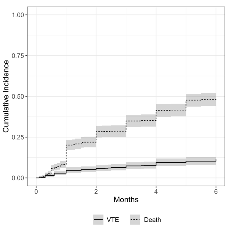
On arrival, platelet count was 37 × 109/L, INR 1.2, aPTT 24.5 s, fibrinogen 1.0 g/L, and D-dimer over 9999 µg/L (Fig. 1).
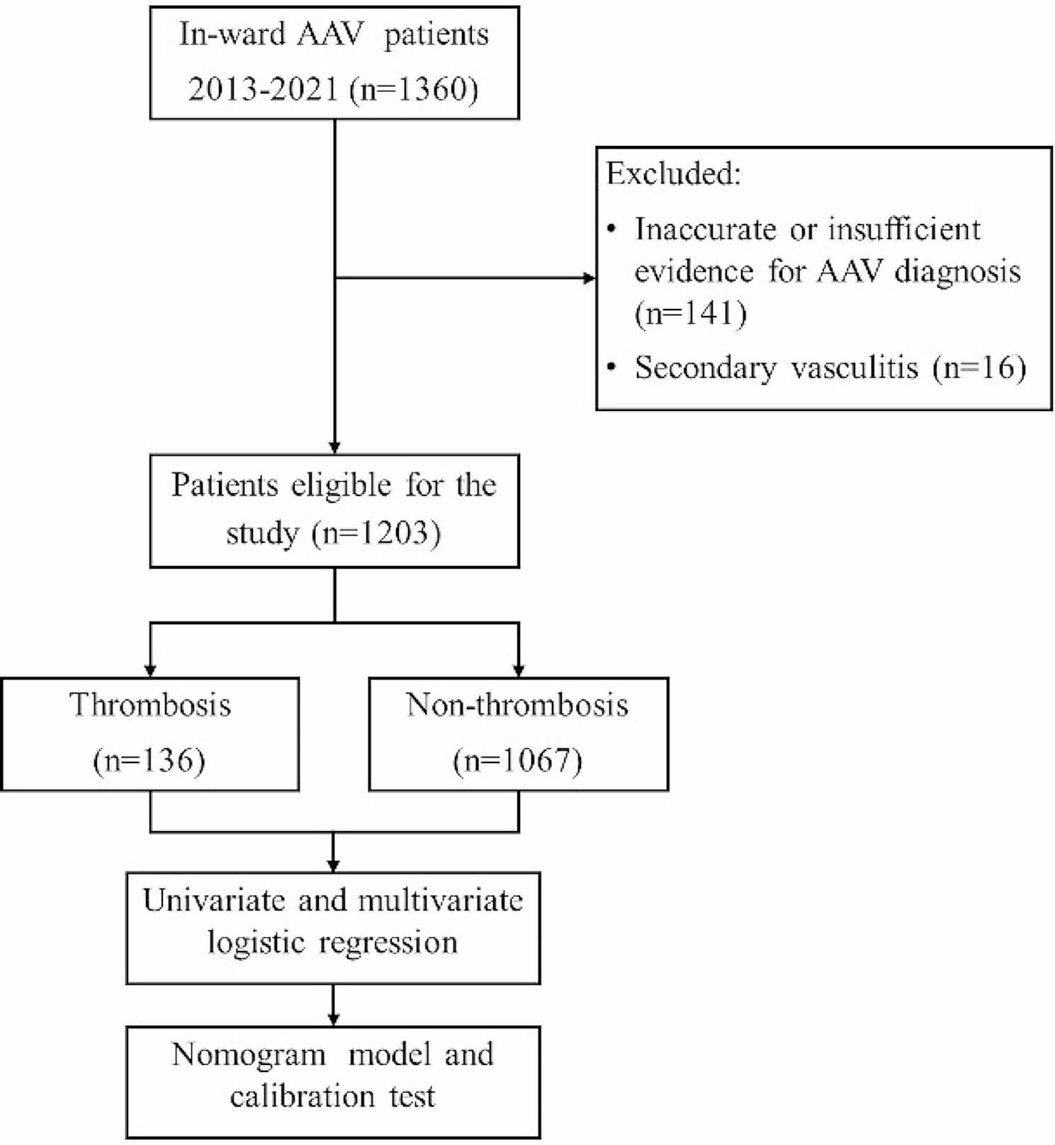
Fig. 1
Platelet count and D-dimer evolution for 2 patients with acute VITT
A CT scan demonstrated left subclavian artery thrombus, thoracic and abdominal aortic thrombus, total occlusion of the right internal iliac artery, and multiple thrombi in the left lower limb arteries without significant atherosclerosis. Troponin-I were 9713 ng/L (normal < 54 ng/L) and echocardiogram showed a 15–20% left ventricular ejection fraction with findings suggesting Takotsubo cardiomyopathy. VITT was suspected and argatroban was started at 0.5 mcg/kg/min, then progressively increased to achieve a target aPTT of 45-55 s (Fig. 1). Ten units of cryoprecipitate were also given as another fibrinogen level came back at 0.7 g/L. Polyspecific anti-PF4 immunoassay (Immucor) result was compatible with VITT (optical density (OD) 2.28). She later had a positive serotonin-release assay with added PF4 (PF4-SRA) performed at the McMaster Platelet Immunology Laboratory, which confirmed the diagnosis. She also received prednisone 1 mg/kg daily and intravenous immune globulin (IVIG) 1 g/kg (Panzyga®, 2 doses of 60 g, weight 68 kg) for 2 days. At this time, a multidisciplinary team of vascular medicine specialists, vascular surgeons, cardiologists and anesthesiologists decided not to proceed with urgent revascularization given cardiac instability and high perioperative risk.
On day 4, her left leg became more ischemic (Fig. 2). The platelet count was 24 × 109/L and the D-dimer were increasing (15 596 µg/L). Reimaging showed new arterial thrombi in both legs and progression of the abdominal aortic thrombosis.
Fig. 2
As this was considered a non-response to IVIG (no significant platelet count increase), TPE was started on day 5 for a total of five treatments. Argatroban perfusion was also empirically increased with aiming a higher aPTT target of 55-65 s. After the first TPE, platelet count increased and D-dimer went down and both continued to improve afterwards. On day 13, 4 days after the last TPE, platelet count was 228 × 109/L and control PF4-ELISA OD was 1.43. At this time, she underwent an above-knee amputation with common femoral artery thrombectomy of her left lower limb without any complication. Argatroban dose during the surgery was 12.83 mcg/kg/min aiming a target aPTT of 65.1–80.0 s, and no bolus was needed. Per surgery, the vascular surgeon found a femoral vein thrombosis that had been previously absent. A few days later, she was switched to rivaroxaban 15 mg twice daily and discharged from the hospital.
CASE 2Patient 2 is a 56-year-old man with a past medical history of hypertension and hypercholesterolemia. He consulted 16 days after the first dose of ChAdOx1 nCoV-19 vaccine with a history of claudication of his left leg for the past 4 days and a constant new right calf pain. He also had a slight headache a few days before. Right leg edema and no distal pulses were noted on the left leg. Left popliteal pulse was preserved.
On arrival, platelet count was 58 × 109/L, INR 1.1, aPTT 24 s, fibrinogen 1.3 g/L and D-Dimer 16 561 µg/L (Fig. 1). A right leg venous doppler ultrasound confirmed a great saphenous vein thrombosis with an extension to the femoral vein. CT angiogram showed an infrarenal aortic thrombus occluding 50% of the lumen and a left popliteal artery thrombosis without significant atherosclerosis. A CT pulmonary angiogram was done and revealed multiple segmental pulmonary embolisms. Brain imaging showed a cerebral vein thrombosis of the left sigmoid sinus with extension to the jugular vein without hemorrhage.
VITT was rapidly suspected and Polyspecific anti-PF4 immunoassay (Immucor) result came back positive (OD 2.13). The PF4-SRA, also performed at the McMaster Platelet Immunology Laboratory, was positive.
Patient was initiated on an argatroban dose of 2.0 mcg/kg/min adjusted to achieve an aPTT of 45-65 s (Fig. 1). He also received prednisone 1 mg/kg daily and IVIG 1 g/kg for 2 days (Panzyga®, 1 dose of 60 g and then 70 g, weight 65 kg). He remained stable after treatment initiation and the platelet count rapidly increased to 162 × 109/L on day 6. On day 10, he had a left popliteal artery thromboembolectomy and patch angioplasty without any complication. The surgery was done aiming an aPTT of 45-65 s and using an argatroban dose of 7.321 mcg/kg/min. One bolus of 100 mcg/kg (6.5 mg) was needed. Since initial IVIG treatment had already been given several days before and because the PF4-ELISA result was still positive (OD 1.736), he received one additional dose of IVIG 1 g/kg (65 g) 12 h preoperatively. This was done even if the platelet count had remained stable (146 × 109/L) and D-Dimer were going down. Four days after surgery, argatroban was switched to rivaroxaban 15 mg twice daily. Six days after the additional IVIG treatment, anti-PF4 was still positive (OD 1.855) but PF4-SRA was not tested. The patient was then discharged home.
留言 (0)