In order to further investigate the biological effects of aberrantlyexpressed lncRNAs and miRNAs in breast cancer, GO enrichment of the target gene were carried out using the GOrilla tool (cblgorilla.cs.technion. ac.il). For each GO term, a list of associated genes is returned with the most optimal at the top of the list. Each gene name is specified by the gene symbol and followed by a short description of the gene.
B. In-vitro analyzesCell culture
The cells were obtained from Iran's National Cell Collection (Pasteur Institute, Iran) and grown according to ATCC guidelines. Two different breast adenocarcinoma cells were employed. At 37 °C in 5% CO2, MCF-HGH and MDA-MB-468 cells were cultivated in Dulbecco’s modified Eagle’s medium (DMEM; Gibco) enriched with 10% FBS (Gibco, USA), 50 U/ml penicillin, and 50 μg/ml streptomycin (Sigma-Aldrich, USA). Normal human breast epithelial cell (MCF-10A) was employed as the Control group.
DNA constructs and gene targeting
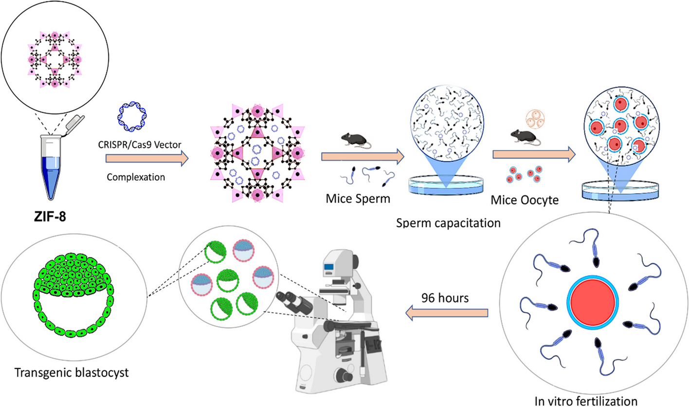
CRISPR/Cas9 technology was utilized to knockout the LINC00511 gene in the human breast cancer cell lines. The LINC00511 gene sequence was found in the GenBank sequence collection of the National Center for Biotechnology Information (National Biosciences, Inc., Plymouth, MN). The CRISPR specially developed CHOPCHOP website (https://chopchop.cbu.uib.no) and (http://crispr.mit.edu/) were used to generate single guide RNA (sgRNA) patterns targeting distinct portions of the LINC00511 gene.
Three vectors were generated: pX459 (involving U6 promoter-sgRNA insertion site-sgRNA scaffold and CAG promoter-Cas9-T2Apuromycin N-acetyltransferase gene-bovine growth hormone polyadenylation signal), pX460–1 (involving U6 promoter-sgRNA insertion site-sgRNA scaffold and CAG promoter-enhanced GFP (EGFP)-bovine growth hormone polyadenylation signal), and pX461–1 (involving U6 promoter-sgRNA insertion site-sgRNA scaffold, and CAG promoter-puromycin N-acetyltransferase (PuroR)-bovine growth hormone polyadenylation signal) were used for sub-cloning of sgRNAs. To achieve this, nucleotide sequences with the sgRNA generating region and steaky ends (Table 1) were produced. To achieve this goal, nucleotide sequences (Table 1) with the sgRNA encoding pattern and steaky ends were produced (Macrogen Inc., South Korea), annealed, phosphorylated, and cloned into BbsI-digested and gel isolated vehicles (using Gel Extraction Kit; DENAzist Asia Co., Iran). The PAM region was also included after the sgRNA coding segment in "CRISPR du-HITI" approaching plasmids. The left homologous arm (546 bp), DsRed2, herpes simplex virus thymidine kinase polyadenylation signal, CMV promoter, PuroR, IRES2, EGFP, SV40 polyadenylation signal, and right homologous arm (546 bp) were all included in the plasmid used for "CRISPR HDR" addressing (832 bp). Table S1 shows that constructs (verified by Sanger sequencing; Macrogen Inc., South Korea) were being used for transfection of breast tumor cell lines using Lipofectamine 2000 chromophore (Thermo Fisher Scientific, USA). Each culture (50 colonies) was observed to proliferate before being chosen by PCR assay two weeks after transfected for "CRISPR excision" and elimination of the LINC00511 exon. The GFP expressing and puromycin dihydrochloride tolerance of cultures was used to qualify them for "CRISPR du-HITI" and "CRISPR HDR" (Sigma-Aldrich, USA). Each cultures colony of "CRISPR excision," "CRISPR du-HITI," and "CRISPR HDR" were evaluated using genetic Material, PCR detection, and Genome Sanger sequence assay (Macrogen Inc., South Korea).
Table 1 List of specific primers used in this researchIsolation and analysis of genomic DNA
The DNA extraction Kit (Cinnacolon, Tehran, Iran) was used to extract DNA Molecules from both wild-type and knockout cell populations, which was then exposed to PCR analysis. After smooth and reaction recuperation, PCR-amplified products were submitted to Sanger Sequencing technology (Macrogen Inc., South Korea).
Detection of mismatched duplexes by T7 endonuclease assay
A mismatch-sensitive T7 endonuclease 1 test (New England Biolabs) was used to confirm that DNA cleavage and targeted sequence disruption occurred at the specified spot. DNA was extracted from the cells using the FavorPrep™ GEL Purification and DNA extraction kit (FAVORGEN Biotech Corp-Taiwan, according to the manufacturer's instructions). In different microtubes, 10 μl (200 ng) of each DNA sample was combined with 2 μl of 10X NE-Buffer 2 buffer and 19 μl of nuclease-free water. At 95 °C for 10 min, the samples were heated. Then, it was allowed to cool at room temperature gradually. 19 μl of each sample were combined with 1 μl of T7 endonuclease I (5 units/μl) and incubated at 37 °C for 15 min before being examined on an agarose gel. Band intensities were measured using Tanon-electrophoretic software (Tanon Science & Technology Co., Ltd., Shanghai, China), and the targeted disruption was seen.
Reverse transcription quantitative PCR
The Y-Tizol RNA extraction Kit was used to extract total RNA from both wild-type and knockout cell populations (Yekta-Tajhiz, Iran). Utilizing capillary electrophoresis and a 2000 Nanodrop spectrograph, the quality, and variety of extracted RNA were determined (Thermo Scientific, USA). With the assistance of random hexamer primers and MMLV reverse transcriptase, 1 μg of total RNA was transcribed reversely and cDNA was synthesized (Thermo Fisher Scientific, USA). Quantitative RT-PCR reactions incorporating Premix Ex-Taq (Probe qPCR) master mix (Takara, Japan), 2 μl cDNA, 500 nM primers, and 100 nM probe (dual-labeled hybridization probes, 5'FAM-3'BHQ1-labeled for LINC00511 and 5'CY5–3'BHQ2 for GAPDH) in a 20 μl reaction mixture were conducted (Qiagen, USA). The following amplifying stages were used: 95 °C for 5 min, then 40 cycles of 94 °C for 30 s, 62 °C for 30 s, and 72 °C for 30 s. Sanger sequencing was used to establish the identification of the PCR products (Macrogen Inc., South Korea). Standard curves were created by subcloning amplified segments and serial diluting. Three PCR assays were run on each concentration, and real-time measurements were taken in duplicate. The log of copy numbers was then compared to cycle threshold (Ct) quantities. Efficiency (E) was estimated for each qPCR reaction based on the computed gradient of standard curves constructed utilizing fivefold serial dilutions vector mixtures, using the following formulae: E = (10–1/slope-1) 100%. In the investigated range, all calibration curves were normal and had a good coefficient of correlation (R2). Based on the corresponding calibration graph, the relative copy number of LINC00511 and GAPDH transcripts was calculated. The amount of the goal transcript (LINC00511) was split by the amount of the reference gene (GAPDH) for 2 different sets of cDNAs, and the results were shown.
Assay for sphere formation
MCF-HGH and MDA-MB-468 breast cancer cells treated with vectors were plated in six-well plates and incubated (Corning, NY, USA). As previously explained [18], cells (2 × 10⁵) were cultivated in serum-free DMEM media with EGF, hFGF (Peprotech, USA), insulin, and penicillin/streptomycin (Gibco) supplemented. Under a light stereomicroscope, clones of spheroids were fixated and dyed with dye solution (crystal violet), then identified (Olympus, Tokyo, Japan).
Assay for CCK-8 proliferation and colony formation
The CCK-8 measurement was carried out with the use of a CCK-8 detection kit (Dojindo Japan). The transfected cells were plated into culture plates, and the cell lines were cultured for 10 h before being treated with the CCK-8 chemical. Absorbance was measured at 450 nm.
Invasion screenings in transwells
Using a 24-well transwell chamber (Corning), breast tumor cells were planted on a member that had been pre-coated with adherent cells (BD Biosciences, San Jose, CA, USA). The individuals on the top surfaces were brushed after 24 h of treatment, and the invaded individuals were fixated with 4 percent paraformaldehyde and labeled with Giemsa. After that, the cells were observed by using optical microscopy.
Assay for luciferase gene reporter
The wild-type and mutant alleles for the E2F1 interaction of the Nanog promoter region were used to generate luciferase reporter constructs. The Lipofectamine 2000 solution was used to co-transfect the plasmids with E2F1 into MCF-HGH and MDA-MB-468 cells (Thermo Fisher, USA). Dual-Luciferase Reporter Assay Kit (Promega) was used to assess the functionality of the Renilla vector (Promega).
MTT assay
The cell viability was validated using the MTT cell viability Kit I (Roche, Switzerland) colorimetric test. In a 96-well flat-bottomed plate, 5 × 10³ cells/well were plated and cultured at 37 °C in a 5% CO2 incubator. Cell viability was measured over three days (24 h, 48 h, and 72 h), with the cells in each well was being washed twice with PBS. Each well was filled with 100 µl of serum-free media and 5 µg/ml Sigma MTT, which were incubated for 4 h at 37 °C in a CO2 incubator. The medium was gradually eliminated, and DMSO was introduced. The ratio of optical density at 570 nm to the background at 690 nm was measured with a State Fax-2100 ELISA plate reader to detect MTT metabolism to generate blue formazan (Awareness Technology, Palm City, FL).
Cell cycle analysis
Absolute ethanol was used to fix the cells for 24 h. The cells were washed twice in PBS before being stained for 15 min with BD Bioscience Pharmingen's PI/RNase staining buffer. FACS flow cytometry was used to determine the DNA content of the cell population. FlowJo V10 software was used to evaluate the cell cycle data (Tree Star, Ashland, OR).
Expression of apoptosis-related genes by quantitative real-time PCR
The expression of the proapoptotic genes P57, P21, Prkca, MDM4, Map2k6 and FADD, as well as the antiapoptotic genes BCL2 and SURVIVIN, was assessed by using a quantitative real-time PCR technique with SYBR green detection. Quantitative real-time PCR was carried out utilizing specific primers (Table 1) and a SYBR® Premix Ex Taq™ II kit (TaKaRa, Japan) based on the manufacturer’s instructions. Relative gene expression levels were quantified by normalizing the respective GAPDH level. Experiments were conducted in duplicates.
Statistical analysis
All studies were done in duplicate and the results were reported as mean SD. One-way ANOVA and independent samples t-tests were used to analyze the differences between groups. SPSS software was used to conduct the analysis, and Graph-Pad Prism was used to create the graphs. Significance was defined as a P value less than 0.05.


留言 (0)