
記住我


Diagnosing food allergies is a difficult and tedious process due to the great variety of symptom-triggering factors and a variety of the symptoms themselves. The standard management in patients with a suspected food allergy is based on a detailed history, physical examination, skin tests (skin prick tests, prick by prick tests, and atopy patch tests), laboratory tests [sIgE, component-resolved diagnostics (CRD), basophil activation test (BAT)], and elimination diets. However, a double-blind placebo-controlled food challenge (DBPCFC) still remains the most important investigation and the gold standard in food allergy diagnostics [1, 2]. The main purpose behind DBPCFC is the need to confirm the causal relationship between the consumption of a certain food and the subsequent hypersensitivity reaction. This test to some extent recreates and mimics the body’s natural response to the given food. A positive result is typically the determining factor for introducing an elimination diet. One important aspect is the fact that every DBPCFC conducted for diagnostic purposes carries the risk of inducing bothersome or dangerous symptoms. Considering the risk of anaphylaxis, any DBPCFC testing should be conducted in a hospital setting [3, 4]. The clinical presentation of food allergies is exceptionally diverse and depends on the type of food, patient age, and individual predisposition. Undoubtedly the most common manifestations are gastrointestinal symptoms, which may come from any segment of the gastrointestinal tract—spanning from the oral cavity (aphthous stomatitis) through the esophagus (eosinophilic esophagitis) and stomach (epigastric pain, nausea, vomiting) to the small and large intestines (enteropathies, eosinophilic enterocolitis) [1, 4]. Other food allergy manifestations include oral allergy syndrome, anaphylactic shock, urticaria, atopic dermatitis, and contact dermatitis. Interestingly, the symptoms of allergic rhinitis and asthma may be also caused by a food allergy [3, 4]. Moreover, oral food challenges have been associated with nasal symptoms, such as itching, sneezing, watery nasal discharge, and nasal congestion [1,2,3]. These observations have been the cornerstone for studies on the use of nasal allergen provocation testing in food allergy diagnostics. Nasal allergen provocation tests are widely used in diagnosing rhinitis, since they show causality, help identify the triggering factors of IgE-mediated nasal hypersensitivity reactions, and confirm the efficacy of medication and allergen-specific immunotherapy in the treatment of allergic rhinitis. Moreover, nasal allergen provocation testing plays an important role in diagnosing local allergic rhinitis and in the differential diagnosis of various types of rhinitis. Nasal allergen provocation testing is a relatively safe procedure, so it may be conducted in an outpatient setting. Any immediate hypersensitivity reactions usually resolve spontaneously within twenty minutes. The results of a nasal allergy provocation test are interpreted in conjunction with the reported symptoms and objective assessments, such as rhinomanometry, acoustic rhinometry, peak nasal inspiratory flow (PNIF), and optical rhinometry [5, 6].
We present the case report of a female patient who underwent a placebo-controlled food challenge and whose local nasal mucosa response was measured with subjective (Total Nasal Score) and objective techniques for assessing nasal obstruction (optical rhinometry and immunoenzymatic assays for measuring tryptase levels in nasal lavage fluid). A clear positive reaction of our patient’s nasal mucosa observed in the course of oral milk allergen challenge demonstrates the potential of intranasal allergen challenges to be used as valuable markers in the process of diagnosing food allergies.
Case presentationIn the year 2021, an 18-year-old female presented at the Allergy Consultation Clinic due to an approximately 10-year history of episodes of abdominal pain and nausea following the consumption of milk-containing products. The patient was a high school student, who was born and lives in a large city. She denied any allergic symptoms in her infancy and early childhood. She was breastfed until the age of 12 months, and reported that neither cow’s milk nor dairy products produced any abdominal discomfort until the age of 8 years. The patient’s mother was diagnosed with cow’s milk allergy a number of years previously and has been on a milk-free diet ever since. No other members of the patient’s family have allergies. Three years earlier, the patient developed abdominal pain, nausea, vomiting, vertigo, weakness, and generalized itchiness approximately 15 min after drinking a glass of warm cow’s milk. At that time, the patient took an antihistamine agent, whose name she cannot remember, and her symptoms resolved 40 min later. The patient did not seek medical attention or visited the Allergy Consultation Clinic. After that episode, the patient limited only the consumption of milk in her diet, but experienced abdominal discomfort following the consumption of cheese, cottage cheese, and yoghurts. Moreover, for the last three years at the end of March and in April, the patient had been experiencing nasal congestion, itching, and watery discharge. She denied cough and wheezing. The patient reported being generally healthy and denied any chronic diseases and long-term medication.
Physical examination findings and differential diagnosticsThe patient underwent a complete physical examination at the Clinic, including a thorough otorhinolaryngological examination. The findings included no relevant abnormalities apart from dry skin and mild inferior turbinate hypertrophy. She also underwent skin prick tests (Alleropharma) with inhaled and food allergens. Positive results were observed for the allergens of cow’s milk 8/16, goat’s milk 4/7, cod 10/20, and birch 10/20, with positive control (histamine) 5/10 and negative control 0/0. Serum allergen-specific IgE levels were also measured (food and inhaled allergen panel) and yielded grade 4 levels of cow’s milk-specific IgE and grade 3 levels of goat’s milk-specific IgE, with negative results for other allergens. CRD revealed alpha-lactalbumin (Bos d 4) levels of 0.63 FIU/mL, beta-lactoglobulin (Bos d 5) levels of 6.12 FIU/mL, and casein (Bos d 8) levels of 22.23 FIU/mL. The material for exfoliative cytology examination of nasal mucosa was collected from the right inferior nasal concha, 1 cm from its anterior edge, using a disposable inoculation loop (nasal curette). The collection technique involved repeated rubbing of the nasal mucosa from its posterior segment towards the anterior segment (scraping method), followed by quick spreading of the material on a microscope slide. After that, the material was fixed with Cytofix aerosol (manufactured by Sanko). Hematoxylin and eosin staining was conducted. The specimen was then assessed using a Delta Microscope Optical Evolution 300 microscope at 400 × magnification. Exfoliative cytology of the nasal mucosa revealed columnar epithelial (61.0%), goblet (5.0%), basal (5.0%), and squamous (62.1%) cells and neutrophils (16.9%). No eosinophils were detected. Additionally, a hydrogen breath test was conducted to exclude lactose absorption problems; the test was negative.
The course of the oral food challenge with cow’s milk allergens accompanied by nasal mucosal response monitoringThe patient, who was in good general condition, underwent the test after the recommended fasting period and having provided her written informed consent (KB 65/2021). This work has been financed by the Medical University of Warsaw grant no. PW/Z/2/2/20(1). The assessment was planned in accordance with the Polish Society for Pediatric Gastroenterology, Hepatology, and Nutrition food allergy division recommendations [7] as well as the European Standard and PRACTALL guidelines [8, 9]. The double-blind food challenge was conducted as follows: ¼ of a muffin, ¼ of a muffin, ¼ of a muffin, ¼ of a muffin, and a whole muffin in 15-min intervals. The active allergen and placebo, which were identical in appearance and taste, had been prepared by a dietician. The muffins had been prepared according the following recipe: (Active product) 250 g of wheat flour, 10 g of baking powder, 25 g of sugar, 50 mL of rape oil, 250 mL of cow’s milk, 1 teaspoonful of vanilla extract, and a pinch of salt. The ingredients were mixed and the mixture was transferred into a baking form and baked for 15 min at 180 degrees Celsius. The quantity of milk per portion was 12.5 mL in a ½ of a muffin and 25 mL in a whole muffin. (Placebo product): 250 g of wheat flour, 10 g of baking powder, 25 g of sugar, 50 mL of rape oil, 250 mL of soy milk, 1 teaspoonful of vanilla extract, and a pinch of salt. The ingredients were mixed and the mixture was transferred into a baking form and baked for 15 min at 180 degrees Celsius. The ingredients in these recipes yielded 10 muffins each.
Four weeks prior to the challenge, the patient was put on an elimination diet, with no cow’s milk products. During that period, the patient used no antihistamines or corticosteroids in any form. The patient’s body weight was 61 kg, height 164 cm. The challenge was conducted by qualified personnel with access to an anaphylaxis kit, in a hospital setting, and outside the birch pollen season. On the day of the challenge, the patient was healthy with no evidence of infection. The physical examination revealed no relevant abnormalities. After each muffin portion was consumed, the patient’s general condition, pulse, blood pressure, and skin were assessed and her chest was auscultated. Blood pressure was 120/70, pulse 72/min, oxygen saturation 99%, the skin was clear, and auscultation revealed normal breath sounds. For organizational reasons the challenge was conducted in two stages 3 h apart. During the first stage, there were no gastrointestinal, dermatological, or respiratory symptoms or any changes in pulse, blood pressure, or oxygen saturation. The patient’s general condition was excellent. Fluctuations in nasal patency, including the physiological nasal cycle, were present throughout the duration of the challenge, with the characteristic variability in nasal cavity diameters. The second stage of the challenge was conducted three hours after the first one. The measurements were conducted in real time during the oral food challenge, and the results were recorded in the assessment report. The nasal obstruction status was assessed every 15 min, in accordance with both oral food challenge and nasal provocation test protocols. The assessment was resumed according to the adopted protocol: after administering ¼ of a muffin, ¼ of a muffin, and ¼ of a muffin (a total of ¾ of a muffin) there were no clinical manifestations; however, seven minutes after the next portion of ¼ of a muffin (one whole muffin in total), the patient reported nausea and abdominal pain. A slight increase in the pulse rate up to 85/min was noted, blood pressure was 110/75, oxygen saturation 99%. She developed mild erythema on the skin of her cheeks without urticaria, dyspnea, or weakness. The abdomen was soft and nontender on palpation; bowel sounds were hyperactive. After another 10 min, the abdominal pain and nausea became exacerbated. The challenge was discontinued, and the result was considered to be positive. Unblinding revealed that the symptoms developed following the administration of one whole muffin containing 25 mL of cow’s milk (active sample). After another 15 min of follow-up, the symptoms gradually subsided and eventually resolved completely.
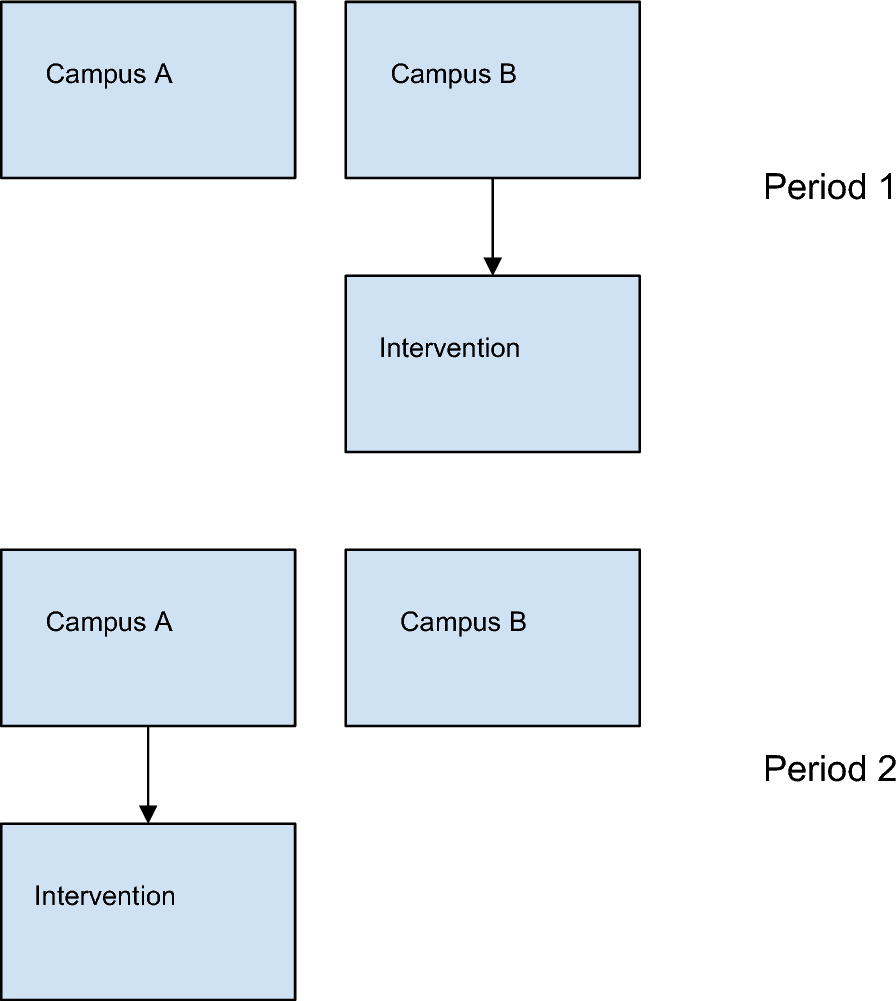
Simultaneously with the food challenge, nasal obstruction was assessed subjectively, with a Total Nasal Score, and objectively, with optical rhinometry (emission spectroscopy, GmbH Rhios, Groerkmannsdorf, Germany) and nasal lavage fluid tryptase (UniCAP, Sweden), which is considered a specific marker of mast cell activation (Fig. 1 Study design). The optical rhinometer is equipped with an optical sensor and a light emitter placed across the bridge of the nose. The light emitter generates 0.2 s pulses of infrared light with the mean wavelength of 600–800 µm, and the sensor continuously and directly measures changes in nasal airway patency (changes in the extent to which the assessed medium slows down and scatters the light beam; in other words optical density (OD) expressed as ∆E [10, 11]). Nasal lavage fluid, collected with Greiff’s technique [12], was centrifuged at 1,000 rpm for 15 min in preparation for an immunoenzymatic assay to detect tryptase, with a sensitivity threshold of 1.0 µg/L. Nasal fluid was collected twice: eight hours prior to the scheduled challenge, in order to minimize the risk of nasal mucosal over reactivity, and after the local nasal mucosal response in the cow’s milk food challenge, which was at hour 2 of the assessment. Nasal lavage fluid was collected with the use of a specially designed tool equipped with two tubes: one for saline administration (administered at room temperature, 8 mL to each nasal opening) and the other for the draining of nasal lavage fluid. Nasal irrigation was performed twice and the obtained biological material was then subjected to further laboratory tests. The subjective and objective assessments were conducted separately, four times, during the placebo phase and after cow’s milk allergen administration, in accordance with the protocol. Optical rhinometry was used to assess the onset of nasal mucosal response (T1), time of maximum response (T2), and optical density. Apart from these objective assessment techniques, a subjective assessment tool was also used (Total Nasal Score). Our optical rhinometry assessments revealed considerable fluctuations in the recorded blood flow during the oral food challenge. Interestingly, there was a significant increase in optical density (up to 0.47 OD) following a cumulative administration of 25 g of cow’s milk, which corresponds to a positive nasal allergen provocation test result (Fig. 2 Nasal mucosal response in the oral food challenge (placebo), Fig. 3 Nasal mucosa response to an oral food challenge with cow’s milk allergens). Tryptase levels in nasal lavage fluid were 278 µL at baseline and 386 µL after the challenge. Changes in the nasal patency recorded during the oral food challenge with cow’s milk allergens were additionally accompanied by upper respiratory symptoms in the form of nasal itching (2 points in a 0–3 point scale) and sensation of nasal obstruction (2 points in a 0–3 point scale). These symptoms were absent during the placebo stage of the oral food challenge. No other relevant symptoms were noted during the assessment.
Fig. 1 Fig. 2
Nasal mucosal response in the oral food challenge (placebo). axis X-length of time in general, axis Y-optical density measured in OD, ΔE-optical density, T1-the beginning of the reaction (time), T2-time to achieve the highest response in the nasal cavity membrane. a The first assessment–placebo; ΔE = − 0.08 OD, T1 = 321 s (5:21), T2 = 658 s (10:58). b The second assessment–placebo; ΔE = − 0.06 OD, T1 = 3 s (0:03), T2 = 32 s (0:32). c The third assessment–placebo; ΔE = − 0.10 OD, T1 = 1752 s (29:12), T2 = 1787 s (29:47). d The fourth assessment–placebo; ΔE = − 0.08 OD, T1 = 2377 s (39:37), T2 = 2422 s (40:22).
Fig. 3
Nasal mucosal response to an oral food challenge with cow’s milk allergens. axis X-length of time in general, axis Y-optical density measured in OD, ΔE-optical density, T1-the beginning of the reaction (time), T2-time to achieve the highest response in the nasal cavity membrane. a The first assessment in the oral food challenge with cow’s milk allergens–(¼ of a muffin); ΔE = − 0.10OD, T1 = 15 s (0:15), T2 = 30 s (0:30). b The second assessment in the oral food challenge with cow’s milk allergens – (½ of a muffin); ΔE = − 0.08 OD, T1 = 709 s (11:49), T2 = 1287 s (21:27). c The third assessment in the oral food challenge with cow’s milk allergens–(¾ of a muffin); ΔE = − 0.07 OD, T1 = 12 s (0:12), T2 = 45 s (0:45). d The fourth assessment in the oral food challenge with cow’s milk allergens–(one whole muffin); ΔE = 0.47 OD, T1 = 1098 s (18:18), T2 = 1402 s (23:22)
The patient was followed up for 2 more hours and discharged home in good general condition. Three days after the assessments, another exfoliative cytology of the nasal mucosa was performed and revealed the presence of eosinophils (53%), columnar epithelial cells (15%), squamous epithelial cells (30%), and neutrophils (12.3%). Due to the positive result of the oral food challenge with cow’s milk allergens, the patient was recommended a milk-free diet and was referred to a dietician, with a view to designing a balanced diet.
留言 (0)