
記住我


The customized anti-PD-L1 Adnectin ADX_5322_A02 was prepared by ChinaPeptides Co. Ltd (Shanghai, China). The sequencing report, protein expression, and sodium dodecyl sulfate–polyacrylamide gel electrophoresis (SDS–PAGE) analysis are shown in Additional file 1: Figs. S1, S2. Maleimide-NODA-GA was purchased from CheMatech (France), and high-purity hydrochloric acid from Merck (Darmstadt, Germany). Other chemicals were purchased from Sigma-Aldrich. The 68Ga/68Ge generator was purchased from ITG Isotope Technologies Garching Gmbh (Germany). PD-10 columns were purchased from GE Healthcare (Chicago, USA). Matrix-assisted laser desorption/ionization time-of-flight (MALDI-TOF) mass spectrometry was performed on a Microflex LT/LRF system (Bruker Daltonics, Billerica, MA, USA). High-performance liquid chromatography (HPLC) /size exclusive chromatography (SEC) was performed on a Waters e2695 system equipped with a Superdex 200 Increase 10/300 GL column (GE Healthcare). Instant thin-layer chromatography (iTLC) was performed on Eckert & Ziegler Mini-Scan/FC (Hopkinton, MA, USA). The samples for binding affinity and biodistribution were measured with a gamma counter (Wizard 2480, Perkin Elmer Instruments Inc, Connection, USA).
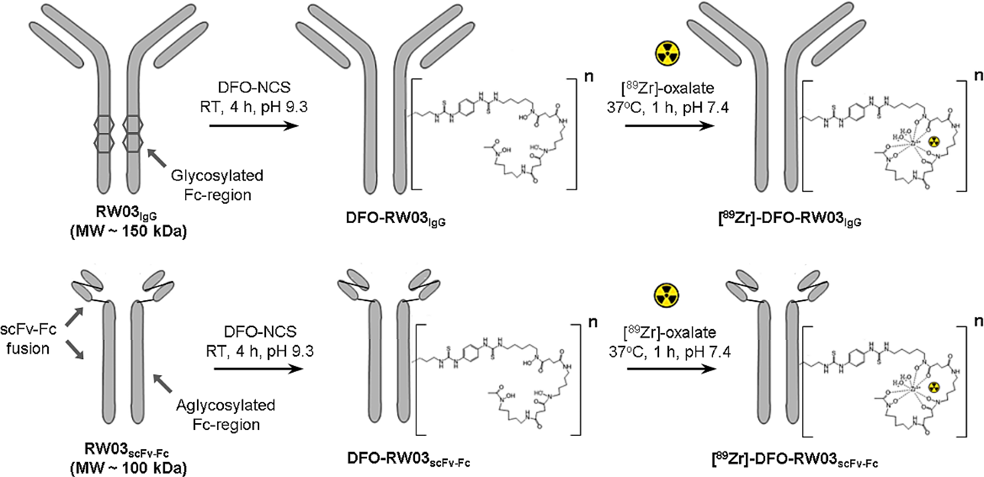
Synthesis and quality evaluation of.68Ga-NODAGA-BMS986192The synthesis scheme of 68Ga-NODAGA-BMS986192 is shown in Fig. 1. Briefly, 50-fold molar excess of maleimide-NODA-GA was first dissolved in PBS (pH 7.4) and added to ADX_5322_A02 (0.2 mg) dissolved in 1 mM tris(2-chloroethyl) phosphate (TCEP) supplemented with 5% Dimethyl sulfoxide (DMSO) [23], followed by incubation at 25 °C for 1 h. The mixture was then purified and concentrated by centrifugal filtration to give NODAGA-conjugated ADX_5322_A02 (NODAGA-BMS986192). Radionuclide 68Ga (370 to 450 MBq) was eluted from a 68Ga/68Ge generator using 0.05 M HCl, mixed with the NODAGA-BMS986192 in sodium acetate, and then incubated at 37 °C for 10 min. The final product, 68Ga-NODAGA-BMS986192, was purified using a PD-10 column using saline as the eluent.
Fig. 1
Synthesis schematic of 68Ga-NODAGA-BMS986192
The radiochemistry yield (RCY) of 68Ga-NODAGA-BMS986192 was measured by the radio-iTLC. The radiochemistry purity (RCP) was measured by the radio-HPLC/SEC chromatography using 0.01 M phosphate buffer (pH 7.4) as the mobile phase at a flow rate of 1 mL/min. In vitro stability was tested by incubation in fresh human serum at 37 °C for 2 h. Cold 69GaCl3 solution was used for the synthesis of 69 Ga-NODAGA-BMS986192 as the standard reference.
Cell lines and culture conditionsHuman glioblastoma cell line LN-229 was purchased from BcNa Culture Collection Co. Ltd (Beijing, China). Mice melanoma cell lines B16-F10 and hPD-L1-gene transfected B16-F10 (hPD-L1-B16F10) were obtained from Ubigene Biosciences Co. Ltd (Guangzhou, China). The real-time qPCR results confirmed that the hCD274 (hPD-L1) RNA expression of hPD-L1-B16F10 is over 5000 times higher than that of the B16-F10 wild type (Additional file 1: Fig. S3). All cell lines were cultured in Dulbecco's Modified Eagle Medium (DMEM) with 10% fetal bovine serum (Gibco) and penicillin/streptomycin (100 U/mL), and incubated at 37℃ in a humidified incubator with 5% CO2.
Binding affinity assayBinding affinity was tested using a PD-L1 positive human glioblastoma cell line LN-229 [21]. Briefly, equal amount of 68Ga-labeled and unlabeled ADX_5322_A02 were mixed, and added to the cells at a total concentration ranging from 10–11 to 10–6 M. After 1 h incubation at 37 °C, cells were washed and centrifuged for 3 times with saline, all of the supernatants were collected as free radioligand. The amount of cell-bound activity (cell pellet) as well as the free radioligand was measured in a gamma counter (Wizard 2480, Perkin Elmer Instruments Inc, USA).
Animal modelsBilateral mouse models were established by subcutaneous inoculation of hPD-L1-B16F10 cells (1 × 10⁶) and B16-F10 cells (1 × 10⁶) [24] at contralateral shoulders of 18–20 g female C57BL/6 mice at 5–7 weeks of age. PET images were acquired once the tumors grew to 500 to 750 mm3 in size [25]. Healthy male cynomolgus weighing 5–7 kg were obtained from TOPGENE BIOTECHNOLOGY Co. Ltd (Wuhan, China).
PET imaging and ex vivo biodistribution in mice modelsAll scans were performed on an uBioEXPLORER PET/CT scanner (United Imaging Life Science Instruments, China). Mice (n = 4/group) were intravenously injected with 3.7–5.6 MBq (100–150 μci) 68Ga-NODAGA-BMS986192. Dynamic PET scans were recorded from 2 to 120 minutes post-injection. For blocking groups, cold ADX_5322_A02 (10 mg/kg) was simultaneously injected with 68Ga-NODAGA-BMS986192. Data were reconstructed in 16 time frames (10 × 2 min, 10 × 10 min) using an ordered subsets expectation maximization (OSEM) algorithm with scattering, attenuation, and decay corrections applied. Three-dimensional regions of interest (ROIs) were drawn over tumors and major organs, and the radioactivities in the regions were measured and quantified as the percentage injected dose per gram (% ID/g).
For ex vivo biodistribution, mice bearing bilateral tumors (n = 5/group) were intravenously injected with 3.7 MBq 68Ga-NODAGA-BMS986192. At 1 h and 2 h post-injection, tumor, blood, and selected organs were collected, washed, and weighed, their radioactivities measured on a gamma counter. The counts per minute (CPM) values for each sample were converted to percent of injected dose per gram of tissue (% ID/g). Data were decay-corrected to injection time and shown as means ± standard deviation (SD). To further improve the rigor of our study, a supplementary biodistribution study of blocking (n = 3/group) was performed by co-administration of unlabeled ADX_5322_A02 (10 mg/kg) with 3.7 MBq 68Ga-NODAGA-BMS986192, tissues were collected at 1 h post-injection.
PET imaging and radiation dosimetry in cynomolgusCynomolgus were first sedated by intramuscular injection of atropine and Shumianling II, and then anesthetized with a mixture of isoflurane and oxygen through endotracheal intubation. Vital parameters were monitored using a portable electrocardiograph (COMEN STAR8000, China). 68Ga-NODAGA-BMS986192 at a dose of 5.6 MBq (~ 150 μci)/kg was injected via the cephalic vein. Dynamic images were collected from the calvarium to the hypogastrium (A Field of View = 484 mm) for two hours, and reconstructed in 58 time frames (30 × 2 s, 10 × 1 min, 10 × 5 min, 6 × 10 min). Three-dimensional ROIs were drawn on major organs and the mean standardized uptake values (SUVmean) were computed to form time-activity curves (TACs). Serial whole-body images were acquired by eight consecutive PET scans (15 min per scan) using a continuous bed motion method (3 individual bed positions, 5 min per position), for up to 120 min post-probe injection.
Data for radiation dosimetry was acquired from whole-body 68Ga-NODAGA-BMS986192 scans at 10, 25, 40, 70, and 115 min after probe injection, respectively. Source organs were chosen based on the highest probe uptake and previously published reports, which consisted of kidneys, bladder, heart, liver, spleen, adrenals, and the remainder of the body [26]. ROIs over the source organs were contoured manually at the 10-min time point and propagated to later scans based on automatic deformable registration between consecutive scans. OLINDA/EXM software, version 2.1 was used to plot and integrate the kinetic organ activity data for the calculation of total-body and organ time-integrated activity coefficients, residence times, and organ-absorbed doses. Effective doses were calculated using radiation weighting factors from the International Commission on Radiological Protection publication 60 [27].
Immunohistochemical stainingHarvested tumors (n = 3) were fixed, embedded in paraffin, and cut into a 4 μm thick sections. The sections were then deparaffinized to retrieve antigen using 10 mM citrate buffer (pH 6.0). Slides were treated with 3% H2O2 for 10 min, blocked with 5% goat serum for 1 h, and then incubated with anti-hPD-L1 mouse monoclonal antibody MIH1 (Catalog Number: 14-5983-80) or anti-mPD-L1 rat monoclonal antibody MIH5 (Catalog Number: 14-5982-81) (ThermoFisher, Shanghai, China) at 4 °C overnight. Subsequently, slides were washed, incubated with biotinylated anti-mouse IgG, and visualized by adding DAB chromogen. Sections were counterstained with hematoxylin.
Biosafety evaluationToxicity tests were performed in accordance with the Organization of Economic Co-operation and Development (OECD) guidelines for testing chemicals with minor modifications. A total of 12 healthy C57BL/6 mice (16–18 g, 5–6 weeks old females) were randomly assigned into saline control, acute toxicity (24 h post-injection), and subchronic toxicity (7 d post-injection), in which the treated groups were injected with a single dose (37 MBq) of 68Ga-NODAGA-BMS986192. Mice were observed daily for physical appearance (skin, hair, eye, weight), behavior patterns (moving, eating, and sleeping), and signs of injury, pain, and illness. Blood samples were collected for routine blood test as well as cardiac, hepatic, and renal functions. Heart, liver, spleen, lung, and kidney organs were also collected for histopathological examination.
Statistical analysisAll data were presented as mean ± standard deviation from three to five independent replicates. Statistical analyses were performed using GraphPad Prism 5.0 (GraphPad Software, San Diego, CA, USA), and a p value < 0.05 was considered to be statistically significant. Differences between groups were evaluated using Student's t test or one-way analysis of variance followed by post hoc Tukey multiple comparisons.
留言 (0)