
記住我
A 71-year-old man with a history of recurrent deep vein thromboses in multiple sites dating from 1993 was diagnosed with triple-positive APS in 2006. Since then, the patient started antithrombotic prophylaxis with warfarin and acetylsalicylic acid (ASA). In 2013, he developed spontaneous ecchymoses and epistaxis with a platelet count of 6000/mm3. Bone marrow biopsy excluded other hematologic disorders and a diagnosis of secondary ITP was made. The first episode of thrombocytopenia was managed with corticosteroid therapy. However, from that moment the patient had recurrent episodes of clinically relevant thrombocytopenia. Therefore, chronic administration of azathioprine was started to maintain normal platelet count. In 2019, the patient developed acute myocardial infarction complicated by cardiac arrest, which was treated with multiple stenting and dual antiplatelet (ASA and clopidogrel) therapy and warfarin until November 2019, when ASA was discontinued.
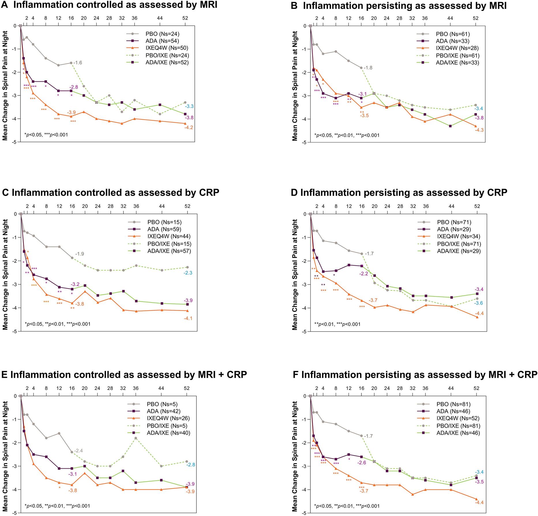
On April 9, 2021, the patient was admitted to the emergency department of our hospital for cough, fever, malaise, nausea, and dizziness. He tested positive for SARS-CoV-2 with a molecular nasopharyngeal swab and was hospitalized in our COVID-19 medical ward. At admission, the patient’s physical examination was unremarkable except for the presence of crackles in the right pulmonary base. A chest X-ray was performed, revealing thickening of the peribronchovascular interstitium, while arterial blood gas test and ECG were normal. The patient did not present visible hemorrhages, petechiae, or hematomas, and he did not report melena. Blood tests showed severe thrombocytopenia (1000/mm3), while the remaining blood tests were as follows: Hb 11.6 g/dl, WBC 5380/mm3, INR 3.5, aPTT ratio 3.48, creatinine 1.26 mg/dl. The autoimmune screening showed a positive lupus anticoagulant test (although in the course of warfarin treatment) associated with the presence of anti-cardiolipin IgG and IgM, and anti-β2 glycoprotein 1 IgG and IgM. In addition, ANA with at the titer of 1/160 with fine speckled pattern, with positive Ab anti-Ro60 (89.4 U/ml) and anti-Ro52 (21.7 U/ml) were detected, whereas other anti-extractable nuclear antigens (ENA), anti-PF4 and anti-dsDNA were negative. C3 levels were low and C4 were within the lower normal range. A comparison of aPL antibodies and complement between baseline and the moment of admission is presented in Table 1. Systemic corticosteroids (prednisone 1 mg/kg) and sublingual vitamin K were started and clopidogrel was discontinued. In addition, therapy with intravenous immunoglobulins (IVIg) 500 mg/kg o.d. for the following 4 days was administered with a slight improvement of the platelet count to 7000 cells/mm3. During the following days, the patient developed gastrointestinal bleeding with blood in feces along with a gradual reduction in hemoglobin levels (8 g/dl on April 14). Hence, warfarin was discontinued, and four units of red cell transfusions were administered. A negative total body CT scan was carried out, while colonoscopy and esophagogastroduodenoscopy were initially not performed due to hemoglobin stabilization and respiratory worsening. Given the concurrent SARS-CoV-2 infection, immunosuppressive therapy with azathioprine was discontinued. On April 15, schistocytes in peripheral blood smear were detected, along with normal bilirubin levels (0.62 mg/dl), high LDH levels (305 U/l), normal renal function (creatinine 0.97 mg/dl), INR 2.31, aPTT ratio 2.18, fibrinogen 626 mg/dl and D-dimer 512 mcg/l. Furthermore, ADAMTS13 functional activity was within the normal range. A therapeutic trial with PEX (three sessions) was started, with the subsequent improvement of platelet count and hemoglobin levels, which reached, respectively, 153.000/mm3 and Hb 10 g/dl on April 23. During hospitalization, the patient developed moderate respiratory failure (PaO2/FiO2 ratio nadir 193) secondary to COVID-19 pneumonia, requiring support with high flow nasal cannula. Except for oxygen supplementation and corticosteroids, the patient did not receive any specific treatment for COVID-19. Then, from April 22, patient’s respiratory function improved and we progressively reduced oxygen supplementation, which was discontinued shortly after. On April 23, an esophagogastroduodenoscopy was performed, excluding upper gastrointestinal sources of bleeding, while in the colonoscopy a diverticulosis was found. The patient was discharged on May 3 and maintained a platelet count within the normal range during the following weeks (230,000/mm3 in July). The blood count trend during hospitalization is shown in Fig. 1.
Table 1 Baseline and admission comparisonFig. 1
留言 (0)