
記住我

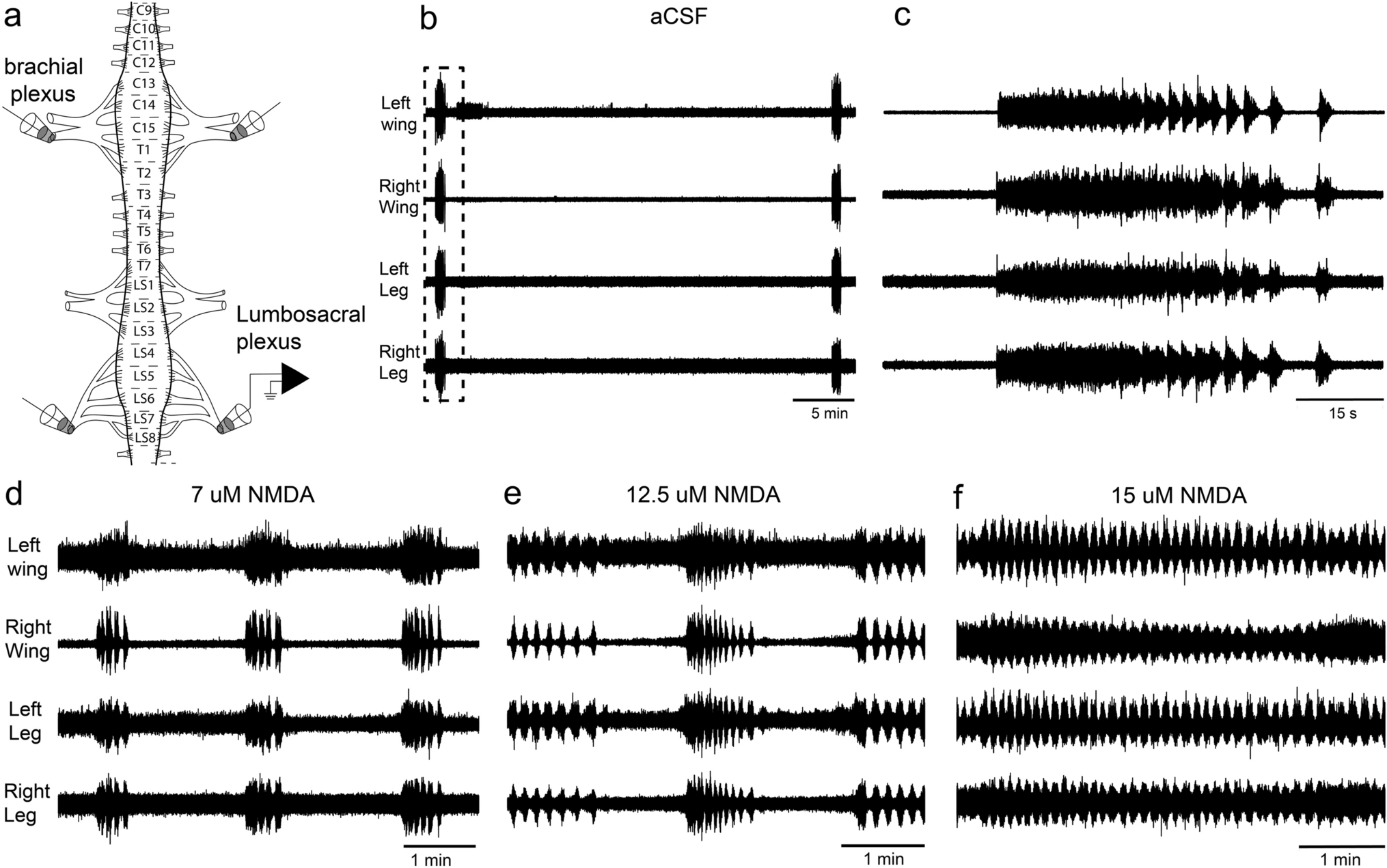


In total, we analyzed 5357 slow-phase segments from the responses of six birds. The data resulted from an array of conditions: the individual birds (Table 1), clockwise and counter-clockwise stimulation, binocular and monocular stimulation, stimulus velocity and age of the birds, given in PHDs.
Table 1 Distribution of number of cases in respect to individual birdsResponses to clockwise and counter-clockwise stimulation were equivalent in binocular adults (Wagner et al. 2021). Therefore, we pooled the responses in these two conditions for the further analyses. Binocular stimulation contributed 1380 data points, monocular stimulation in the T–N direction 2335 data points, and monocular stimulation in the N–T 1642 data points. With respect to age, we attempted to record data at certain PHDs for most velocities. At the remaining PHDs we only recorded data for stimulus velocities of 10, 15 and 30 deg/s. Thus, the number of cases at the different PHDs (Table 2) and the different velocities (Table 3) differ. As consequence, we have much more data for the stimulus velocities 10, 15 and 30 deg/s than for the other stimulus velocities. The earlier data appear to provide the most reliable results and may serve as critical benchmarks for interpretation. We also present the data from the other stimulus velocities below, because no other data from juvenile owls are available. Moreover, they illustrate the development more broadly. In this sense, we regard them as supplementary data that complete the picture (for more discussion see below). With respect to individual birds, we concentrated on certain velocities for certain birds (owl I: 10 deg/s; owls G + H: 15 deg/s, owls J + K: 30 deg/s). Owl F was tested with all velocities.
Table 2 Distribution of number of cases in respect to ageTable 3 Distribution of number of cases in respect to velocityIn the following, we first describe general observations of the juveniles in the stimulus set-up during the recordings. We then present the temporal development of binocular responses, and finally report responses to monocular stimulation.
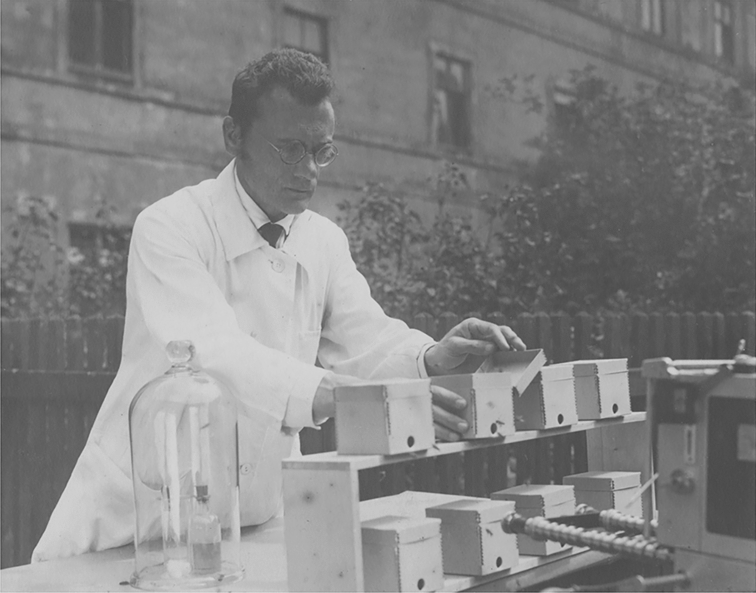
General observations of juvenile barn owls during recordingTests with three owls started before the birds showed a reaction to the stimulus, and before they presumably opened their eyes. The eye lids are closed at birth. Then, a small slit can be seen, but it is not clear whether the birds really see something. The latter can only be inferred from behavioral reactions or through invasive methods, which we did not use. Initially, we used behavioral testing with several stimuli apart from the wide-field stimulus later used for recording optocollic data. Amongst these were stimulation with a moving stick or a moving hand. During these attempts, the owls were typically sitting in the drum on different platforms. Stimulation always lasted several minutes. When the birds were not being tested, they were maintained in a comfortable environment close to the experimenters. Therefore, tests with the very young birds could be repeated several times a day.
Owl K did not react to the optomotor stimulus on P9 (see video 1 in supplements) and P10. It showed the first following behaviors on P11. During the recording on P11, the bird was sitting in a beaker in the drum and was stimulated by wide-field motion (see video 2 in supplements; Fig. 3a–c). Likewise, owl F did not follow stimulus motion on P11, but did so on P12. Thus, in these two birds the very first reactions to the wide-field stimulus could be documented. Owl J was tested every day from P10 on. It first reacted to the stimulus on P13, but the first data available are from P14. In the other three birds, testing started also on P13 or P14. All six owls showed persistent reactions from P14 on (see video 3 in supplements). Interestingly, the periods during which the birds followed the stimulus were typically interrupted by periods during which the birds did not react (see video 3 in supplements). Also, apart from the rotational movements, we sometimes observed translational movements of the head (see video 3 in supplements). The latter were not further analyzed. Across owls, our data set consists of quantitative measurement from P11 to P65.
Fig. 3
Examples of OCRs of juvenile barn owls. a-o The responses to different stimulus types at different ages and different velocities for different birds as notified in the insets or on top of the middle plots are shown. Dashed lines represent a reference position on the wide-field pattern, plotted in the range between ± 100 degrees. Note that the dashed lines from + 100 to -100 degrees and the saw-tooth-like appearance of stimulus position are due to wrapping. Solid lines signify the position of the owl’s head in azimuth. The numbers close to the individual slow-phase segments specify the gain during the respective segment. The arrows in g and m point to rare events as explained in the text
Typically, very young birds were placed in a staining dish or a beaker, and supported by soft paper for comfort, but otherwise free to move during the recording (see video 3 in supplements). It was obvious that very young birds (approximately up to P13) had difficulty in stabilizing their head. Nevertheless, the birds exhibited high-gain responses. The head was above the upper rim of the dish, with the lower jaw often touching the rim. In this situation, the head rotated and followed the rotation of the pattern. The birds could hold up their head from about P14 on (Fig. 2a). Although the birds were not yet standing on their feet, now the head did no longer touch the rim of the staining dish. The birds were calm and typically followed the stimulus. After a few more days (around P20), the birds became able to stand (Fig. 2b). At this time, the birds became more agile (Fig. 2c), and sometimes started to negotiate the staining dish. During testing, we moved the birds to a beaker adapted to the size of the birds. Note that the birds were free to move in the beaker and not restrained in any way. While the birds tolerated being seated in the beaker, their responses became more variable after P30, which typically begins a period of motor development, and exploration of the nest. Untrained birds were more easily distracted and sometimes showed no interest in the stimulus pattern (see video 4 in supplements). Nevertheless, it was possible to record data after P30 and up to P65, the last day of juvenile life covered in this work.
Binocular optocollic responses of juvenile barn owlsThis report includes binocular data from all owls and for all stimulus velocities (Table 4). Binocular stimulation with both wide-field patterns very reliably elicited the OCR in juvenile owls of all ages. The birds showed consistent reactions to all stimulus velocities tested (Fig. 3a, d, g, j, m). Binocular gains were adult-like from the first day of response for all stimulus velocities tested (Fig. 4). In the following, we first discuss five typical examples that provide a picture of the variability of the responses (Fig. 3a, d, g, j, m). Afterwards, we present a quantitative analysis (Figs. 4, 5).
Table 4 Distribution of the number of cases on different conditions (binocular, N–T, T–N)Fig. 4
Dependence of binocular gains on age. Median data (triangles) and 1st to 3rd quartiles (lines) are shown for different days of recording (x-axis) and different stimulus velocities, including all (a, c, e) or only data of an individual bird (b, d, f). The respective fit function is shown by the dotted line. Adult data (Wagner et al. 2021) are documented for comparison in each plot on the right. The numbers specify the number of cases for each condition
Fig. 5
Comparison of juvenile and adult binocular OCRs. The median data together with the 1st and 3rd quartiles are shown. Note that the rotational speeds are not plotted on a linear axis. The numbers between 0 and 20% gain specify the respective numbers of cases. The data of juveniles and adults are not different (ns) for 6 out of 7 velocities and highly significantly different (****) for 10 deg/s. For 80 deg/s, only juvenile data were available
The typical reaction of an owl to visual wide-field stimulation was to follow the stimulus by head rotation. Stimulus movement in the counter-clockwise direction elicited a counter-clockwise head rotation during the slow-following phase (Fig. 3d). Opposite (clockwise) head turning occurred with opposite (clockwise) stimulus movement (Fig. 3a, g, j, m). A slow-phase segment ended with a saccadic turn in the opposite direction to the slow-phase movement. While the owl followed the stimulus, the angular velocity of the head was almost constant. This may be concluded from the almost linear change of head azimuth with time (Fig. 3a, d, g, j, m). Gains were often 80% or higher. Only one out of nineteen slow-phase segments shown for binocular stimulation in Fig. 3 had a gain below 70% (see numbers close to the single slow-phase segments in Fig. 3a, d, g, j, m and Fig. 4).
Before analyzing the typical behavior of the birds presented so far, we point to some rare behavior. For example, a special situation is shown in Fig. 3g. Here, the first following movement had a high gain. A low-amplitude saccade followed. Then, the owl ceased to follow the stimulus for about 3 secs, before it started the next following movement (see arrow in Fig. 3g). The period during which the owl was not following the stimulus was not included in the analysis. This may be seen from the gain values noted in Fig. 3g (83.6 and 72.2). Another peculiarity occurred in the sequence shown in Fig. 3m. Here, a return saccade started at 3.68 s. After this saccade, the head movement was initially much faster than the stimulus movement for more than half a second (3.92–4.64 s, see arrow in Fig. 3m). Then a movement in the opposite direction occurred with a low velocity (4.72 to 4.96). Finally, the bird started to follow the stimulus with a gain of 86% at 5.12 s. Both, the fast head rotation from 3.92 to 4.64 s, and the movement in the opposite direction were not included in the analysis. In the other 3 examples (Fig. 3a, d, j), the owl followed the stimulus during the total time sequence. This was the typical behavior that occurred in the vast majority of cases. Note, however, that the amplitudes of the following movements varied considerably. We did not further analyze amplitudes and durations of the slow-phase segments. Instead, in this study, we concentrated on the development of gain.
The quantitative analysis of the data sets for stimulus velocities of 10, 15, and 30 deg/s (Fig. 4) demonstrated that adult-like gain values were reached very early. The median gains reached an adult-like value from the first day of response. For example, the first day of response for 30 deg/s was on P11 in owl K (Fig. 4f). Already at this age, the gain was not statistically different from the gain at P33 (Mann–Whitney U test, number of cases P11: 6, P33: 11 (U = 32, z score = 0.05025, p = 0.96012). Non-significant differences were also observed for the first and last days for which we have data in the other two owls (Mann–Whitney U test, number of cases owl F: P19: 17, P26: 14 (U = 99, z score: 0.7647, p = 0.4444); owl J: Mann–Whitney U test, number of cases P14: 23, P40: 8 (U = 49.5, z score = − 1.8967, p = 0.05787)). Data from all owls (owls F, K, J) were similar (Fig. 4e). The data recorded during the whole juvenile period were pooled and tested against the data from adult birds as published in Wagner et al. (2021). There was no difference between the two data sets (Mann–Whitney U test, number juveniles: 407, number adults: 73, U = 15,073, z score = 0.1989, p = 0.8424; see also Fig. 5). The time course of development was fitted by a sigmoidal function (which was chosen as it describes also the monocular data (Figs. 6, 7), see Material and methods). The function fitting the 30 deg/s data demonstrated that the 90%-PHD corresponded to the first day of response (Fig. 4e, f).
Fig. 6
Dependence of monocular gains on age. a–n All monocular data shown as recorded for different velocities (row) and either N–T (left column) or T–N (right column) stimulation together with the fit functions (dotted lines). Specifications are as explained in the legend to Fig. 4. Note the lower gains and the delayed development for N–T responses compared with T–N responses
Fig. 7
Quantification of asymmetry. a Fit parameter “asymptotic upper value”, b First day of response and 90%-PHDs. The “first day of response” (for a definition see text) refers to both T–N and N–T conditions and is documented for each stimulus velocity. The 90%-PHDs are separately plotted for T–N and N–T stimulation. d, e Data from three developmental periods (P11–P18, P19–P25, P26–P65). c, f Asymmetry factors T–N/N–T in juveniles (juv) derived from the fits (c) and from the data (f) in comparison to the measured adult factors (ad)
Similar observations were made for a velocity of 15 deg/s for which data from owls G and H were available. The earliest recording in owl H, at P13, already yielded data (median gain value 87.8) that was statistically not different from the data at P32 (median gain value 91.1) (Mann–Whitney U test, number of cases P13: 15, P32: 20, U = 141, z score = 0.28333, p = 0.77948). These observations were supported when the data of owls G and H were pooled (Fig. 4c). Again, juvenile and adult data were not different (Mann–Whitney U test, number juveniles: 211, number adults: 11, U = 1046.5, z score = − 0.5224, p = 0.6015; see also Fig. 5).
The data for a stimulus velocity of 10 deg/s were mainly based on recordings with owl I (Fig. 4b). Some data came also from owls F, J, and K (Fig. 4a). Again, the very first recordings, on P14, showed a median gain (84.3) close to that measured at P29 (85.4) or P 33 (92.2). These median gains were much higher than that determined at P56 (72). For 10 deg/s stimulus velocity, the juvenile data yielded significantly lower gains than measured in adults (Mann–Whitney U Test, number juveniles: 634, number adults: 64, U = 9867, z score = − 6.7781, p = 1.218*10– 11; see also Fig. 5). The reason for this difference is not clear. For all other stimulus velocities tested, the juvenile and the adult responses were not different (Fig. 5).
Median gains with binocular stimulation were close to 100% for velocities up to 20 deg/s (Fig. 5). The median gains decreased to 70% for velocities up to 60 deg/s and to about 40% for a stimulus velocity of 80 deg/s (Fig. 5). Gain values did not change during development. There were some extraordinary recording days, with median values either below (Fig. 4a, b, P17 and P18) or above (Fig. 4e, P36) the rest of the values. The differences between the 1st and the 3rd quartiles were between 14.3 and 24.2 percent of gain in absolute terms. The differences amounted to 15–29%, determined relative to the median gain values. In summary, binocular gains measured in juvenile birds were not statistically different from adult gains for 6 out of 7 stimulus velocities tested that ranged from 5 to 60 deg/s (Fig. 5).
Monocular optocollic responses of juvenile barn owlsThe response pattern for monocular stimulation was more complex than the responses to binocular stimulation. Major differences occurred in the responses to N–T and T–N stimulation. The first PHD at which the birds responded was P11 (in owl K, stimulus velocity: 30 deg/s). The responses to both T–N and N–T stimulation were short and of low gain at this PHD (Fig. 3b, c; see Fig. 6i, j for a quantitative analysis of the reaction with a stimulus velocity of 30 deg/s). This changed fast for the responses to T–N stimulation, both for 10 deg/s (Fig. 3n, Fig. 6d), and for 30 deg/s (Fig. 3e, h, k; Fig. 6j). By contrast, gains to N–T stimulation remained low for several days. These gains gradually increased during development. At P19 responses to N–T stimulation were of high gain for a stimulus velocity of 10 deg/s (Fig. 3o: single gain values 81.5 and 85, quantitative analysis in Fig. 6c: median gain: 76.8). At this PHD, gains were still low for 30 deg/s (Fig. 3i: single gain values: 26 and 31, quantitative analysis in Fig. 6i: median gain: 47). At P27, gains for N–T stimulation had increased also for a stimulus velocity of 30 deg/s (Fig. 3l: single gain values: 71, 56, 71, 70.7, 64.5, quantitative analysis in Fig. 6i: median gain: 66).
The fitting of the responses provided insights into the duration of development. The inflection points of the fit function (parameter b in Eq. 2) were all between P9 and P13, which suggested that the development began at similar times for all velocities and conditions. The duration of development may be derived from the 90–50 differences and the 90%-PHDs. These two parameters are related to the factor c of the fitting function. They varied a lot with stimulus velocity (e.g., range 10–26 days for 90%PHD, Fig. 7b). They yielded highly correlated values over the 7 stimulus velocities used (7 data points, correlation coefficient: 0.988, p < 0.00003). In the following, we arbitrarily use the 90%-PHDs as a measure for the duration of the development (Fig. 7b). The 90%-PHDs for N–T stimulation were 26, 17, and 24 for stimulus velocities of 10, 15, and 30 deg/s, respectively (Fig. 7b). The responses to T–N stimulation were high from very early on. The 90%-PHDs for T–N stimulation were between P11 and P14 for all stimulus velocities tested (Fig. 7b). In other words, the 90%-PHD was reached almost immediately after the first day of response (Fig. 7b). The longest time necessary to reach 90% of the final values with T–N stimulation was three days. This occurred for a stimulus velocity of 30 deg/s (Fig. 7b).
The fitting of the data not only made it possible to quantify the duration of development, but also provided insight into the differences in upper gain values for the different stimulus types (binocular, monocular T–N, monocular N–T). For binocular stimulation, sufficient data for fitting were available for 10, 15 and 30 deg/s. The comparisons showed that the upper values for binocular stimulation were very close to the upper values for T–N stimulation (compare dashed and dotted lines in Fig. 7a). Larger differences were seen between the responses to binocular and T–N stimulation on the one and the responses to N–T stimulation on the other side (Fig. 7a). The upper values for N–T responses were significantly lower than the upper values for T–N responses (7 pairs of upper values, Wilcoxon Matched Pairs signed rank test, z = − 2.418; p = 0.016).
Figure 7a shows an increase of the differences in the upper values for T–N and N–T responses with stimulus velocity. This resulted in an increase of the T–N/N–T factors with stimulus velocity (Fig. 7c). The juvenile T–N/N–T factors derived from the fits are very similar to the measured T–N/N–T factors in adults for velocities up to 20 deg/s. The earlier factors are slightly higher than the latter factors for higher velocities.
The upper values of the fits yielded data that reflected the final result of development. Additionally, it was also interesting to examine the temporal change of the gains and specifically the T–N/N–T factors during development. To this end, we pooled data from three distinct age periods (P11–P18, P19–P25, and P26–P65). We compared the results from the juveniles with those from the adults (Fig. 7d–f). We are aware that the pooling coarsened the time resolution of the data compared to the data shown in Fig. 6. However, the resulting curves are smoother, and allow better insight into the underlying mechanisms than the plots shown in Fig. 6. Figure 7d demonstrates that the T–N gains were high from early on. T–N gains for a stimulus velocity of 60 and 80 deg/s decreased in the last period (Fig. 7d). Note, however, that the latter data points are based on low numbers (Table 3). Gains for N–T stimulus did not change much for stimulus velocities up to 20 deg/s and also not for 40 and 60 deg/s (Fig. 7e) in the course of development. The gain for a stimulus velocity of 30 deg/s increased in the last period ranging from P26-P65 compared to the gains in the earlier two periods and reached an adult-like value (Fig. 7e). Figure 7f summarizes the data shown in Fig. 7d and e. This plot demonstrates that the measured factors T–N/N–T for velocities up to 20 deg/s were close to 1 and adult-like from the first period on. By contrast, there were developmental changes of the factors T–N/N–T for velocities above 20 deg/s. The values were larger than the adult values for the first two time-averaging periods from P11 to P18 and P19 to P25. The T–N/N–T factors derived from the measured gain data reached adult-like values for the last analysis period (P26–P65) (Fig. 7f). This observation is consistent with the T–N/N–T factors derived from the fitted data shown in Fig. 7c.
留言 (0)