
記住我


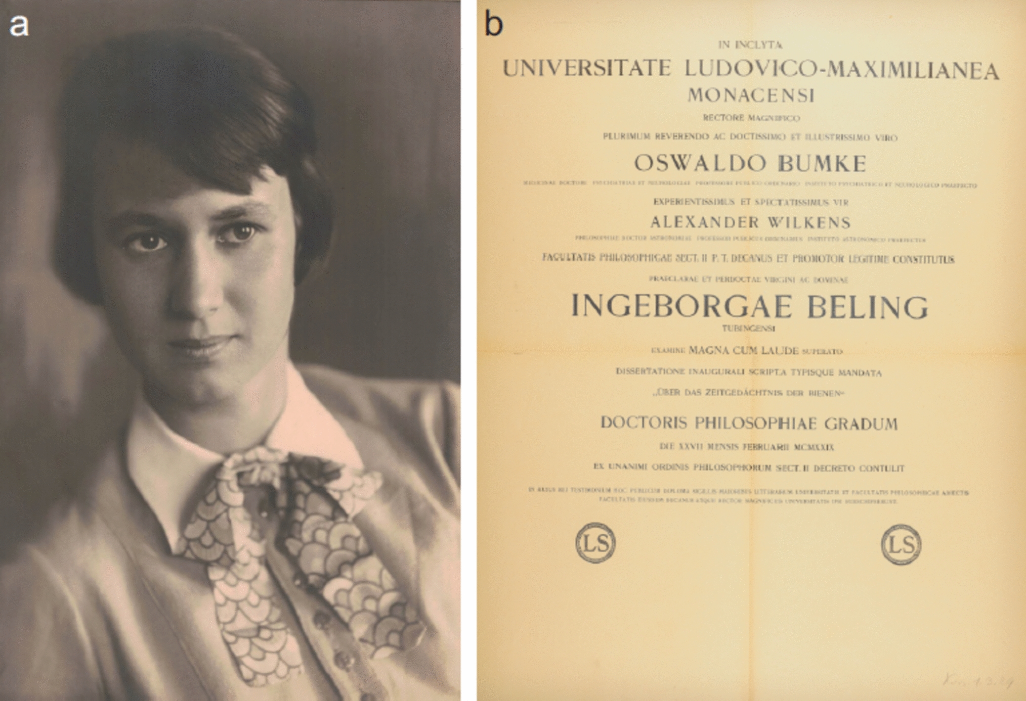


Adults of C. elaterii (Fig. 1a) were collected on wild plants of Ecballium elaterium (L.) (Cucurbitaceae) in the field close to Perugia (Italy) in June 2020 and reared in the laboratory inside net cages (300 mm × 300 mm × 300 mm) (Vermandel, Hulst, The Netherlands) on plants of Cucumis melo L. (Cucurbitaceae) obtained from seeds. Adults of H. axyridis (Fig. 2a) (from laboratory culture, Dipartimento di Scienze Agrarie, Alimentari e Ambientali, University of Perugia, Italy) were reared from larvae kept inside net cages (300 mm × 300 mm × 300 mm) (Vermandel, Hulst, The Netherlands) and fed using aphids Aphis fabae Scopoli (Hemiptera: Aphididae) reared on young Vicia faba L. (Fabaceae) plants. Adults of C. herbacea (Fig. 3a) were collected on wild mint plants in the field close to Perugia in June 2020.
Fig. 1
Female of Chnootriba elaterii under stereomicroscope (a, b) and tarsal attachment devices in the cryo-SEM (c–e). a, Insect attached to the force sensor by means of the polyethylene thread; b, details of the interlocking between the claws and a single trichome of Cucurbita moschata leaf; c, latero-ventral view of the tarsus with the attachment organs represented by a pair of pretarsal bifid dentate claws (C) and two hairy pads (HP) situated on the ventral side of the first and second tarsal segments; d,e, details of the pair of claws in frontal (d) and lateral (d) view. Each claw is bifid (arrows) and has a basal tooth (asterisk) separated from the claw by a deep cleft (arrow head)
Fig. 2
Female of Harmonia axyridis under stereomicroscope (a, b) and tarsal attachment devices in the cryo-SEM (c–e). a, Insect attached to the force sensor by means of the polyethylene thread; b, details of the interlocking between the claws and a single trichome of Cucurbita moschata leaf; c, ventral view of the tarsus with the attachment organs represented by a pair of pretarsal dentate claws (C) and two hairy pads (HP) situated on the ventral side of the first and second tarsal segments; d, e, details of the pair of claws in frontal (d) and lateral (d) view. Each claw has a basal tooth (asterisk) separated from the claw by a deep cleft (arrow head)
Fig. 3
Female (fig. c to be replaced with the female) of Chrysolina herbacea under stereomicroscope (a, b) and tarsal attachment devices in the cryo-SEM (c-e). a, Insect attached to the force sensor by means of the polyethylene thread; b, details of the interlocking between the claws and a single trichome of Cucurbita moschata leaf; c, ventral view of the tarsus with the attachment organs represented by a pair of pretarsal divergent claws (C) and three hairy pads (HP) situated on the ventral side of the first, second, and third tarsal segments; d,e, details of the two curved divergent claws in frontal (d) and lateral (d) view
The three insect species were kept in a controlled condition chamber (14 h photoperiod, temperature of 23 ± 1 °C, and a relative humidity of 60 ± 10%). Only females were used in the experiments.
PlantsTwo plant species having either visually smooth, shiny leaves (cherry laurel P. laurocerasus 'caucasica') or hairy leaves (squash or pumpkin C. moschata cv “zucca lunga gigante di Napoli”) were used in the experiments. C. moschata was obtained from seeds and P. laurocerasus was collected in the field. The adaxial side of the leaf surface was used in the experiments.
Leaf replicasNegative moulds of the adaxial side of the leaf surface of C. moschata were prepared using the silicone elastomer President light body (PLB; Coltène® Whaledent AG, Altstätten, Switzerland; using the automatic mixing device). PLB was stored in a freezer at – 18 °C to extend the handling time. A leaf was removed from the plant and silicon elastomer was immediately applied and spread onto the adaxial side of the leaf blade. A Petri dish was gently pressed down upon the silicon elastomer to remove air bubbles and fill folds and asperities of the original substrate. After polymerization (approximately 5 min), the plant surface was carefully peeled off. The negative mould was subsequently filled out with liquid Epon-Araldite resin mixture (Sigma-Aldrich) and polymerized overnight at 60 °C. To hold the liquid resin during polymerisation, a 3 mm-high edge of silicone was created around the negative mould. The hardened positive resin replica of the leaf was removed from the silicone negative mould.
Cryo-scanning electron microscopySamples of (1) adult tarsi of the three coleopteran species, (2) the adaxial side of the tested plant leaves, and (3) resin leaf replicas were studied in a scanning electron microscope (SEM) Hitachi S-4800 (Hitachi High-Technologies Corp., Tokyo, Japan) equipped with a Gatan ALTO 2500 cryo-preparation system (Gatan Inc., Abingdon, UK). For details of sample preparation and mounting for cryo-SEM, see Gorb and Gorb (2009). Insect tarsi and plant leaves were sputter-coated in frozen conditions and the resin replicas in warm conditions with gold–palladium (thickness 10 nm) and examined at 3 kV acceleration in the SEM.
Force measurementsThe friction force of the females of the three tested insect species on the natural and artificial surfaces and the trichome stiffness of natural and artificial substrates were measured using a Biopac force tester (Biopac Systems Ltd, Goleta, CA, USA). For a better interpretation of the results obtained with insects using the Biopac force tester, a centrifugal force tester was applied to measure the attachment ability of females of H. axyridis and C. elaterii to hydrophilic glass [water contact angle of 32.49 ± 4.17° (mean ± SD)] and the adaxial side of the C. moschata leaf. These two different techniques were used, because the Biopac force tester measures the force generated by the insect during locomotion, whereas the centrifugal experiment measures the force generated by the insect when an external force (in the natural situation—e.g., wind, predator) is applied to an insect.
Prior to the force measurements, insects were weighed on a micro-balance (Mettler Toledo AG 204 Delta Range, Greifensee, Switzerland). Experimental insects were anaesthetized with CO2 for 60 s and made incapable of flying by gluing their forewings together with a small droplet of melted bee wax. Before starting the experiments, the treated insects were left to recover for 30 min. All the experiments were performed during the daytime at 25 ± 2 °C temperature and 50 ± 5% RH.
Biopac force tester experiments. The Biopac force tester consisted of a force sensor FORT-10 (10 g capacity; World Precision Instruments Inc., Sarasota, FL, USA) connected to a data acquisition unit MP 160 (Biopac Systems Ltd, Goleta, CA, USA). Data were recorded using AcqKnowledge 5.0 software (Biopac Systems Ltd, Goleta, CA, USA).
(1) Insect attachment forces. One end of a fishing thread Gel Spun Polyethylene 0.02 mm diameter (Berkley Spirit Lake, Iowa, USA) about 10 cm long was fixed with a droplet of molten wax to the insect thorax. The insect was attached to the force sensor by means of the thread (Figs. 1a, 2a, 3a) and was allowed to move on the test substrate in a direction perpendicular to the force sensor (and parallel to the substrate). The force generated by the insect walking on artificial [hydrophilic glass with water contact angle of 51.71 ± 2.22° (mean ± SD) and resin replicas of leaves with water contact angle of 107.81 ± 8.20° (mean ± SD)] and natural surfaces (the adaxial side of C. moschata and P. laurocerasus leaves) was measured. Insect pulled on the plant leaf walking from its proximal to distal portion. Force–time curves were used to estimate the maximal pulling force produced by tethered running insects (traction, friction).
To test the role of claws in the insect attachment ability to trichomes, ablation experiments were carried out. The females were anaesthetized with CO2 for 120 s and immobilized with Patafix (UHU Bostik, Milano, Italy) under the stereomicroscope. Claws were removed by cutting off the distal portion of the last tarsal segment with microscissors. Insects used as a control (without ablations) were handled in exactly the same way as the ablated individuals. The insects were left to recover for 24 h before conducting the experiments, to avoid any negative effect due to the manipulations and to the possible bleeding observed just after the ablations. In total, 11 females of C. elaterii, 14 females of H. axyridis, and 18 females of C. herbacea were tested in both conditions, with claws (intact) and without claws (ablated).
(2) Trichome stiffness. To measure the trichome stiffness, a special hook (4 mm in its vertical part, 1.4 mm in its horizontal part) made with the tip of a metal pin was connected to a thin copper lamina attached to the force sensor (Fig. 4a). The force sensor was fixed to a motorized micromanipulator (Narishige MMO-203). A leaf of C. moschata was fixed with double-sided adhesive tape to a glass plate with the adaxial side facing up. Under visual control with the help of a stereomicroscope Leica MZ6, the hook attached to the sensor was approached to the upper side of the leaf. The hook could contact a single trichome in the middle of its length (Fig. 4b), without touching the leaf surface. The force sensor was moved (speed, 75 µm/s) over the leaf from its distal to proximal side in such a way that the hook could contact the trichome, to push and bend it until finally loose a contact with it.
Fig. 4
Experimental set-up for testing the trichome stiffness on the adaxial side of the Cucurbita moschata leaf. a, Metal hook (H) connected to a thin copper lamina attached to the force sensor (FS) fixed to the motorized micromanipulator (M). The hook could contact a single trichome (T) in the middle of its length (see detail under stereomicroscope in b and was moved over the leaf from its distal to proximal side (arrow) to push and bend the trichome until loosing contact with it
Centrifugal force tester experiments. The centrifugal force tester (Gorb et al. 2001) consists of a metal drum covered by a substrate disc to be tested. The metal drum is driven by a computer controlled motor. Just above the disc, the fibre-optic sensor monitored by the computer is situated. After the positioning of the insect on the horizontal disc, the centrifuge drum started the rotation at a speed of 50 rev min–1 (0.883 rev s−1). The position of the insect on the drum was monitored using a combination of a focused light beam and a fibre-optical sensor. The drum speed continuously increased until the insect lost its hold on the surface under centrifugal force. The rotational speed at contact loss, the last position of the insect on the drum (radius of rotation), and the insect weight were used to calculate the maximum frictional component of the attachment force. To test the insects on the adaxial side of the C. moschata leaf, a portion of the leaf was fixed with tape to the metal drum of the centrifuge. In total, 27 females of H. axyridis and 26 females of C. elaterii were tested on hydrophilic glass and 22 females of H. axyridis and 22 females of C. elaterii were tested on the leaf of C. moschata.
Statistical analysisThe stiffness (force of the trichome acting against the moving force sensor) of the non-glandular trichomes of different length (long, medium, and short) covering the adaxial side of C. moschata leaf and the resin leaf replicas was analyzed using two-way analysis of variance (ANOVA), considering the surfaces and the trichomes length as factors.
The safety factor (force divided by insect weight) of insect attachment on glass and the normalized friction force (friction force reported as percentage of friction force recorded by each insect on glass) on the different surfaces (the adaxial leaf surface of C. moschata, its resin replica, and the adaxial leaf surface of P. laurocerasus) of the females of the three insect species with claws and without claws were analyzed using two-way repeated-measures analysis of variance (ANOVA), considering the insect condition (with and without claws) and the insect species as main factors. When necessary, after all the ANOVA tests, for significant factors, Tukey unequal N HSD post hoc test for multiple comparisons between means was performed (Statistica 6.0, Statsoft Inc. 2001). In the centrifugal force tester experiments, the Mann–Whitney U test was used for the comparison between H. axyridis and C. elaterii, both for the friction force and safety factor. Before the parametric analysis, all the data were subjected to Box–Cox transformations, to reduce data heteroscedasticity (Sokal and Rohlf 1998).
留言 (0)