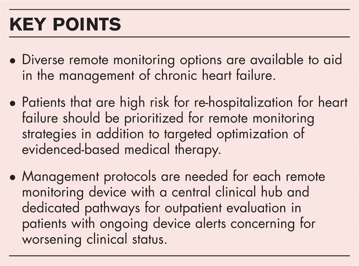
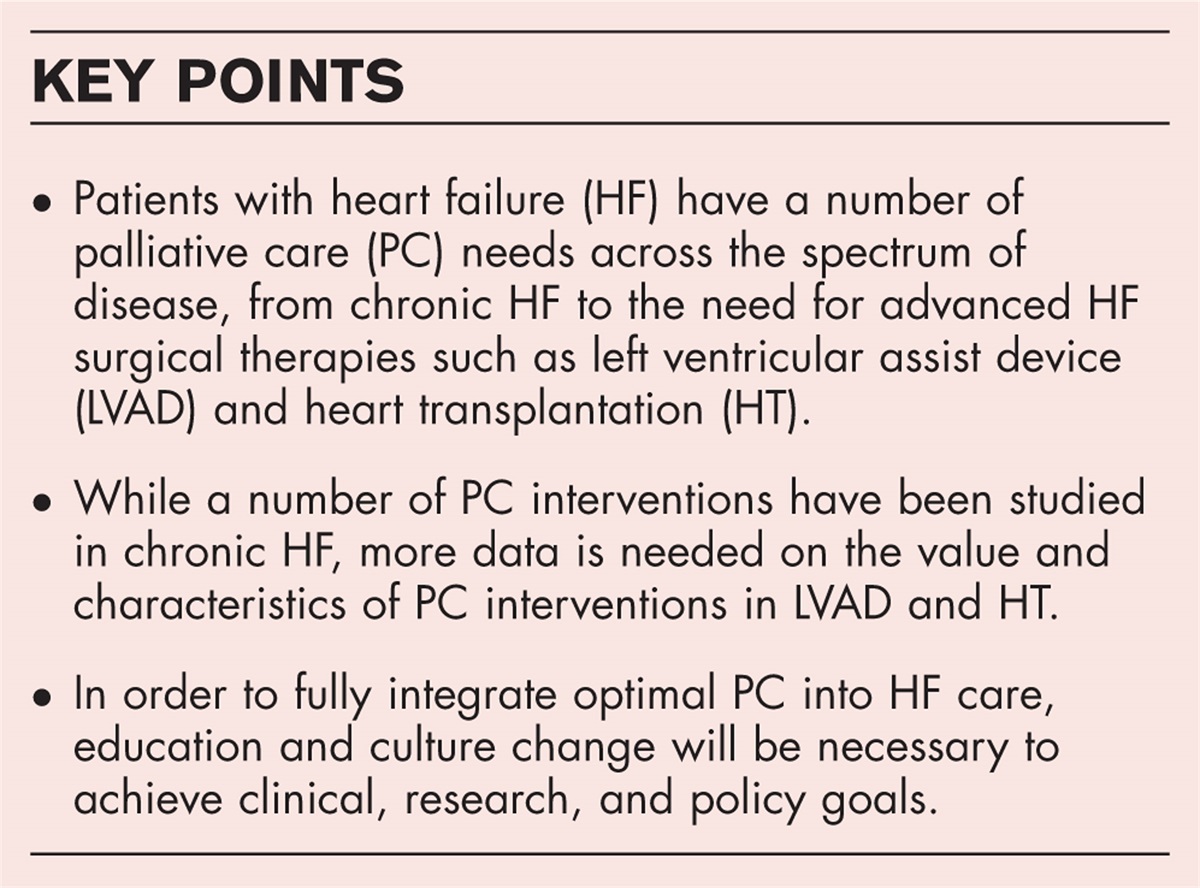
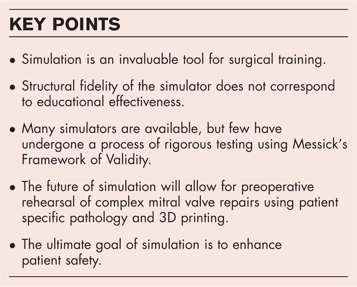
The role of multimodality imaging in patients with heart failure with reduced and preserved ejection fraction
Purpose of review
The burden of clinical heart failure, both heart failure with a reduced ejection fraction (HFrEF) and with a preserved ejection fraction (HFpEF), continues to increase both nationally and globally. This review summarizes the expanding role of multimodality imaging techniques in the evaluation and management these patients.
Recent findings
Echocardiographic assessment for heart failure continues to expand and should include a robust hemodynamic and strain assessment. Nuclear techniques have also continued to evolve and advances including computed tomography attenuation correction for single photon emission-computed tomography positron-emission tomography increase diagnostic accuracy as well as provide information such as myocardial blood flow and viability assessment. Computed tomography imaging, already well established in the assessment of coronary and valvular disease, has increasing utility in the characterization of myopathy, and cardiac magnetic resonance imaging (MRI) continues to expand its role in tissue characterization to a wider breadth of diseases, including right ventricular cardiomyopathy and left ventricle noncompaction.
Summary
Although heart failure remains a clinical diagnosis based on history and examination, early imaging is critical for further assessment. Due to its widespread availability, affordability, and safety, transthoracic echocardiography has long been the mainstay tool for both initial evaluation as well as for periodic surveillance of heart failure patients, but advances in multimodality imaging are occurring at a rapid pace and promise to provide an increasing wealth of data to help manage such patients.


留言 (0)