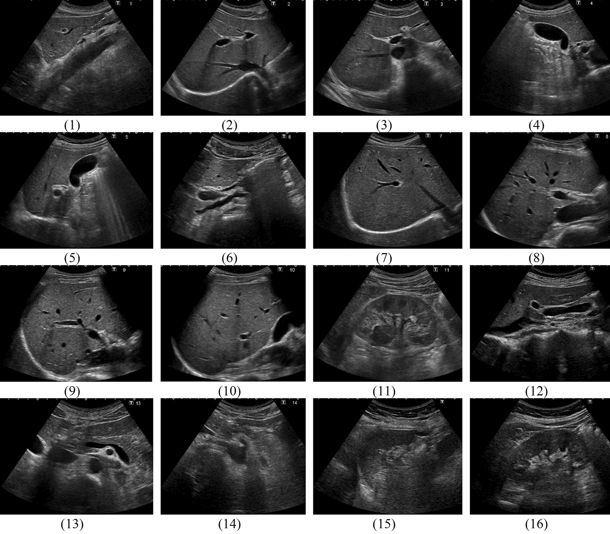
MpMRI (3 T) showed a high ability to visualize PCA at the localization of PSM, which occurred mostly apical and/or posteriorly at the capsule or at the apical urethra. In 6 patients with PSM the tumor was invisible on mpMRI. 35% of patients with PSM had organ-confined PCA. Best clinical parameter to predict PSM was the postoperative T-stage. LCC was the best MRI predictor for PSM at the capsule, which performed better than the clinical parameters. Nevertheless, in tumors with PSM at the apical urethra, UD was the best MRI parameter. Highest accuracy was documented for UD ≤ 3.5 mm, indicating a high risk for PSM at the urethra and for LCC ≥ 22.5 mm, indicating a high risk for PSM at the capsule. Using these MRI parameters PCA localizations at risk for PSM might be pre-operatively determined.
Park et al. developed and validated a scoring system for MRI to predict PSM inducing the PI-RADS score, tumor location on posterolateral side or at the apex, and length of capsular contact, archiving an AUC value of 0.80 [19]. Compared to our results the score was slightly better than LCC alone, but inferior to UD for apical tumors. There are some aspects of this score that needs to be discussed. First, PI-RADS score was weighted more heavily compared to tumor localization. Second, PI-RADS category 3 (clinically significant cancer is equivocal) and PI-RADS category 4 (clinically significant is likely to be present) have the same impact on the score, although in clinically consequence is different. Third, capsular contact ≤ 14 mm was rated with 0. Other studies showed that the risk for EPE was already increased at LCC ≥ 11 mm [4]. Main limitation is that risk factors for PSM depend on the localization of PSM and therefore should be assessed separately for either risk of PSM at the apical urethra or at the capsule.
The diagnostic performance of mpMRI is influenced by the different prevalence of EPE in different risk stratified cohorts [20]. High negative predictive values (88%) are only reached in low-risk cohorts, where patients could benefit if they were selected for nerve sparing surgery by the prior mpMRI. Positive predictive value was highest (89%) in high-risk cohort, which could help to reduce the risk of PSM. In a prospective randomized single-center trial preoperative MRI could only reduce PSM in low-risk PCA [21]. According to the authors a main limitation is lacking communication between radiologists and urologists, which is crucial to adopt surgical approaches.
A recent meta-analysis showed that mpMRI had a considerable impact on the extent of resection during RPE, but modifications of neuro-vascular-bundle preservation did not influence PSM rates [22]. However, apart from relatively small number of studies, mostly retrospective design, different MRI protocols, scanner, and field strength, a further reason for these results might be the lack of standardized MRI reading.
This study is limited by the retrospective design and the single-center evaluation. Although PCA is usually a slow growing tumor, the time interval between MRI and operation might have influenced the results. MRI was acquired at 3 T scanners and reading was performed by subspecialized experts in consensus, but we did not access interreader variability, so less experienced readers may perform differently. Furthermore, PSM at the capsule and at the urethra may have different clinical impact, therapeutic consequence, and risk for biochemical recurrence (BCR). However, this study focuses on the MRI visibility and prediction of PSM.
In conclusion, mpMRI (3 T) was an excellent tool to visualize PCA and could help to identify patients at risk for PSM, which occurs mostly apical and/or posteriorly at the capsule or at the apical urethra. Best predictive parameters were LCC at the capsule and UD for apical located tumors. Since communication with the surgeon is crucial to adopt the surgical strategy, EPE, LCC and UD should be highlighted in the structured report.


留言 (0)