
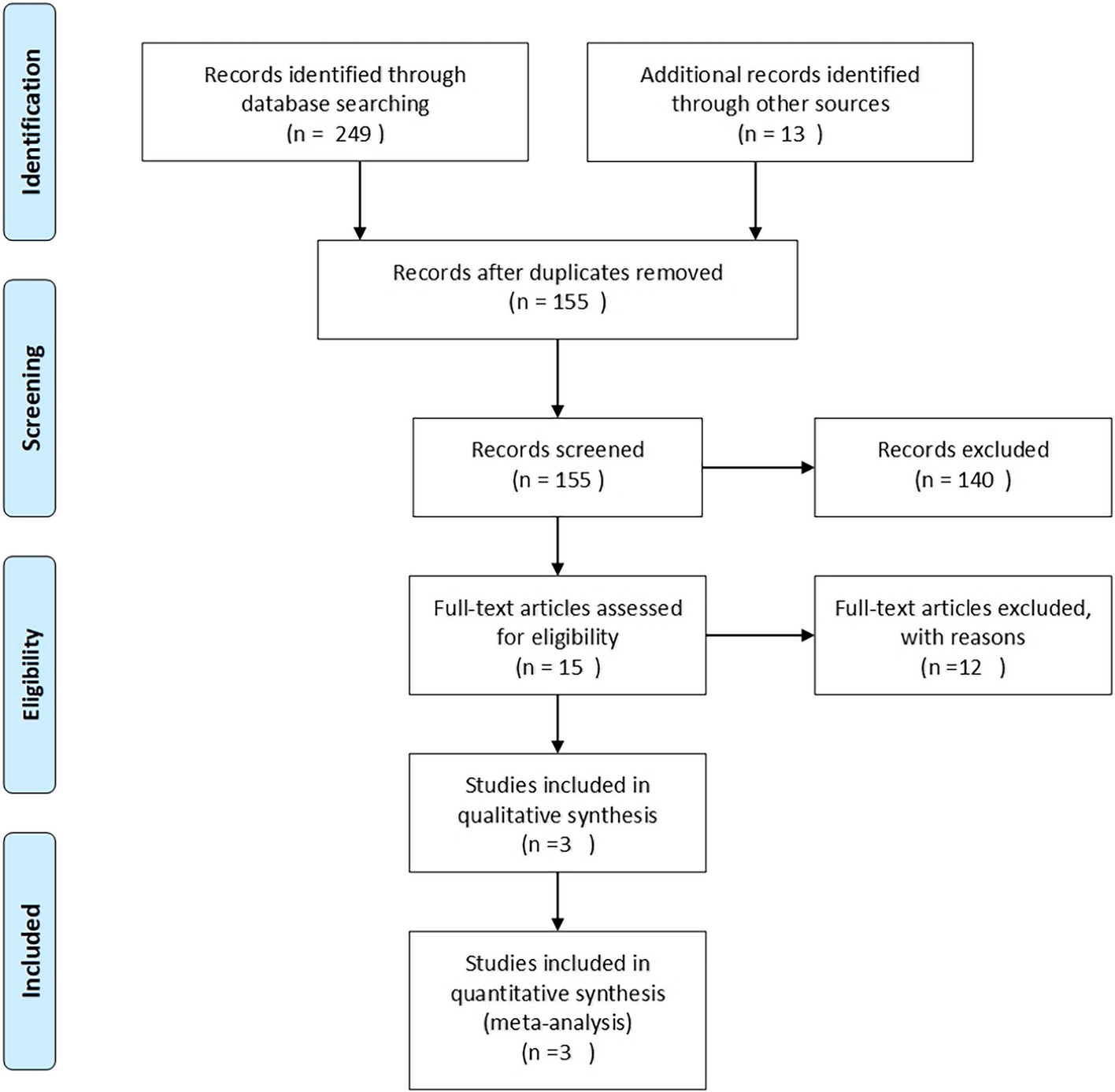
記住我


A 65-year-old Colombian woman with a history of hypertension, presented with 8 days of asthenia, retroorbital pain, frontoparietal headache in location, rated 6/10, joint and muscle pain. 2 days before admission, he added dry cough, sore throat, and sensation of dyspnea without any other associated symptoms. She was observing quarantine and denied contact with cases suspicious or confirmed of COVID-19 infection as well as mosquito bites. The patient had self-medicated with paracetamol, which provided temporary relief; however, her condition was persistent, prompting consultation.
Her vital signs revealed a body temperature of 38.9 °C, a respiratory rate of 26 breaths/minute, a pulse rate of 110/minute, and a blood pressure of 130/80 mmHg. Oxygen saturation at presentation was 90% in room air. Physical examination only showed bibasilar crackles and a petechial rash. Analysis revealed leukopenia with lymphopenia, thrombocytopenia, moderate D-Dimer, transaminases, C-reactive protein (CRP), and elevation of LDH (Table 1).
Table 1 Timeline events of DENV and SARS-CoV-2 coinfection casesA chest CT scan was performed, and scattered ground glass images were shown in both lung fields, compromising 50–60% of the lung parenchyma due to probable viral pneumonia (Fig. 1).
Fig. 1
Axial CT scan view showing scattered ground glass in both lung fields, with 50% lung involvement (red arrows)
She was hospitalized with supportive treatment, dexamethasone (after the recovery trial), IV fluids, paracetamol, additional oxygen with nasal cannula, and close monitoring. The rapid dengue test revealed a positive nonstructural protein 1 (NS1) with positive immunoglobulin (Ig) M and IgG and a nasopharyngeal swab for SARS-CoV-2 real-time reverse transcriptase (RT-PCR) was taken.
This treatment did not improve her symptoms and has gradually worsened. ABG tests were performed which showed severe hypoxemia (partial pressure of oxygen [PaO2]: 36 mmHg, PaO2/fraction of inspired oxygen [FiO2] 68 mmHg. A repeat of the complete blood count showed a sudden drop in the platelet count to 20,000/mm3 without any visible bleeding. Therefore, dengue RT-PCR was requested due to doubtful diagnosis and DENV serotype 2 (DENV2) was detected. RT–PCR for SARS-CoV-2 was positive, confirming the diagnosis of dengue with warning signs associated with severe COVID-19.
She was transferred to the ICU, for ventilatory support due to progression to acute respiratory distress syndrome and refractory hypoxemia that requires invasive mechanical ventilation. Clinical characteristics were attributed to SARS-CoV-2 infection. On subsequent days, increasing trends in the number of platelets and leukocytes were observed, and clinical symptoms improved. However, extubation was not achieved; she required a tracheostomy and was discharged to a chronic care unit for pulmonary rehabilitation.
Case 258-year-old Colombian male, without known medical history, complained of persistent fever of 39 °C, diarrhea, dyspnea, asthenia, myalgias, and dry cough that lasted 3 days; he had tested positive for SARS-COV 19 by RT-PCR. Due to the worsening of cough, dyspnea, and shortness of breath, he consulted an online clinic where he was referred to the hospital for evaluation. On examination, he appeared dehydrated, with peripheral cyanosis, somnolent but arousable with marked respiratory effort and bibasilar crackles. Vital signs with a pulse rate of 108/minute, respiratory rate of 32 breaths/minute, blood pressure of 100/70 mmHg. Oxygen saturation at presentation was 84% in room air, without any other findings on physical examination.
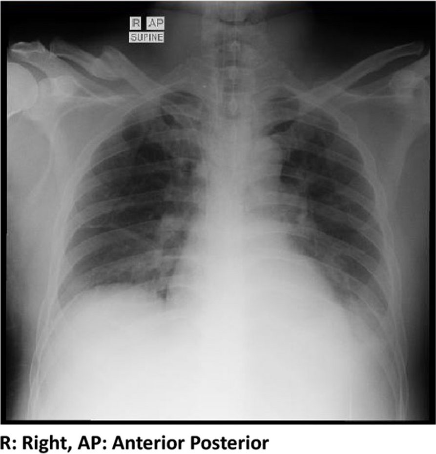
Immediately, a portable chest radiograph was performed showing multiple radiopacities of interstitial occupation and peripheral distribution (Fig. 2). Considering that he has acute respiratory failure supported by clinical findings (tachypnea, tachycardia, cyanosis, altered levels of consciousness, diffuse crackles and respiratory effort), he was intubated (pressure control ventilation [PCV] mode, inspiratory oxygen fraction [FiO2], 0.5, positive end-expiratory pressure [PEEP], 10 cmH2O; inspiratory pressure [Pi], 15 cmH2O; inspiratory time [Ti], 1.5 s; frequency [f], 12 per minute) and transferred to the ICU.
Fig. 2
Portable chest radiograph showing multiple radiopacities of interstitial occupation and peripheral distribution (red arrows)
Arterial blood gas analysis revealed a pH of 7.45, an oxygen pressure of 48 mmHg, a carbon dioxide pressure of 30 mmHg, and a bicarbonate of 21.1 mmol/L, a PaO2/fraction of inspired oxygen [FiO2] 96 mmHg. Laboratory tests had leukopenia with lymphocytopenia and thrombocytopenia. Renal function, liver enzymes, CRP, serum LDH, and D-dimer were elevated (Table 1). Due to severe thrombocytopenia in an endemic area, dengue serology was positive for NS1 antigen. To confirm the diagnosis, an anti-dengue IgM/IgG ELISA, serology test and RT-PCR were requested and DENV serotype 3 (DENV3) was detected. Therefore, the patient was diagnosed with severe COVID-19 with dengue fever with signs of red flags.
During his hospitalization, the patient’s acute hypoxic respiratory failure did not recover, his oxygenation was poor, despite the tracheal intubation connected to the ventilator. Renal and liver function continued to decline. Subsequently, he became hypotensive and started norepinephrine for suspected cardiogenic vs septic shock. Empiric treatment with broad-spectrum antibiotics, dexamethasone (after recovery trial), and IV fluids was started. During this time, the ICU-prone ventilation protocol was initiated to improve oxygenation to his lungs.
The patient’s condition deteriorated sharply, developing multiorgan failure, characterized by pulmonary, renal, liver, and possible neurologic compromise. The patient remained on life-sustaining support. After 17 days in the ICU, Extracorporeal membrane oxygenation (ECMO) was performed. Despite the best efforts of the medical staff, the patient eventually died.
留言 (0)