Study design and subjects
The ethical review board of the University Medical Center Groningen approved this study, and the requirement for informed consent was waived. This study is a retrospective cohort study, performed in a tertiary care institution in The Netherlands with more than 2 million people in its direct catchment area.
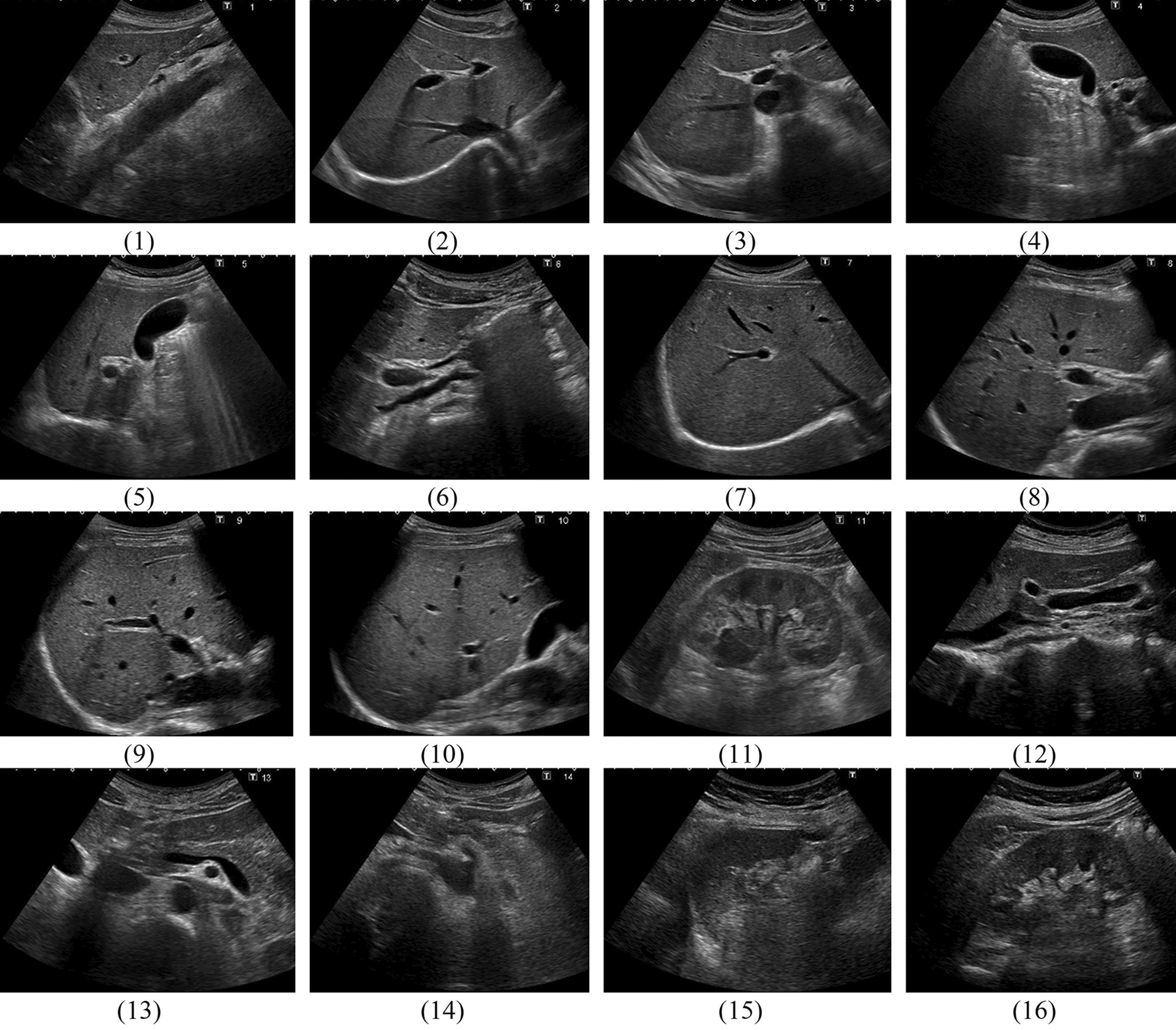
Patients who underwent abdominal US performed by the radiology department during on-call hours (i.e., between 17.00 p.m. and 8.00 a.m. on weekdays, in weekends, or on official holidays) between January 2005 and November 2017 were considered for inclusion in this study. All of these US examinations during on-call hours were performed by radiology residents. The organs that were evaluated with US depended on the clinical request and the judgment of the resident based on clinical and live US findings. Residents either independently performed and reported the US examination, or were assisted and supervised by an on-call radiologist when requested by the resident. Note that a radiology residency takes 5 years in the Netherlands, that there is year-round influx of new residents, and that residents are allowed to perform on-call duties after completing the first year of residency and obtaining a sufficient “entrustable professional activity” score for US in this setting.
Using the Random Calendar Date Generator [12], 87 unique calendar dates were randomly selected between January and November. Out of all abdominal US examinations performed during on-call hours on these 87 unique calendar days in each of the years from 2005 to 2017, 250 cases per year were randomly selected. December of all 13 years was dismissed since a change in patient file software did not allow for the extraction of cases from December 2017. This random selection process yielded 3250 cases that were potentially eligible for inclusion. Exclusion criteria were as follows: US not involving the abdomen, routine protocolized US (including routine Focused Assessment with Sonography for Trauma [FAST], intra- and postoperative liver transplant US, postoperative kidney transplant US, and pre-transplant donor assessment), duplicate records of the same US examination, non-clinical US examinations (e.g., US for education or research purposes), lack of an imaging request or imaging report in the electronic patient file, and US-guided diagnostic or therapeutic interventions.
Data collection
Patient files of all 3250 cases, including US reports, were reviewed by a research fellow (T.E.S.). The following parameters were extracted for each included case: patient age and gender, specialty that requested the US examination, and indication for US (categorized as abdominal aorta aneurysm, acute bowel pathology, acute liver failure, acute oncology, appendicitis, gallbladder and biliary ducts, infection/inflammation, mixed, other, transplant [outside of routine protocols], trauma [non-FAST] and urolithiasis/postrenal obstruction.
Each US examination was classified as either positive (i.e., findings that were related to the reason the US examination was made) or negative (i.e., the absence of findings related to the reason that the US examination was made and no disease deterioration or other new and clinically relevant findings compared to a previous imaging examination if available) [13]. If an US examination could not be clearly classified as positive or negative, it was classified as indeterminate.
For each US examination it was also recorded whether it revealed an incidentaloma or not. Incidentalomas were defined as incidental US findings serendipitously diagnosed in an asymptomatic patient or symptomatic patient undergoing imaging for an unrelated reason [3]. Both incidental findings that may potentially cause morbidity or death when left untreated, and incidental findings for which it is unknown if they require treatment, were considered incidentalomas, in line with previous literature [14]. Clearly benign findings such as simple liver or renal cysts were not considered incidentalomas [14]. Incidentalomas that had already been detected on previous imaging examinations were not counted either.
Data analysis
The proportions of US examinations that were negative and those that yielded at least one incidentaloma were calculated, for all years together and for each year between 2005 and 2017. Temporal changes in aforementioned proportions between 2005 and 2017 were analyzed using the Mann–Kendall test. Logistic regression analyses were performed to determine the association between a negative US examination with the following variables: patient age, patient gender, specialty that requested the US examination, and indication for US. Logistic regression analyses were also performed to determine the association between the detection of an incidentaloma on US (i.e., at least one incidentaloma on a patient level) with the following variables: patient age, patient gender, and organ(s) evaluated with US. Variables that were significant on univariate analysis were subjected to multivariate analysis. Categories with the highest number of observations were used as reference for nominal variables. Categories with less than 10 counts were excluded from logistic regression analyses. Two-sided P-values < 0.05 were considered statistically significant. Statistical analyses were executed using R version 4.1.1. software (https://www.r-project.org) and MedCalc version 19.1.6 software (MedCalc, Mariakerke, Belgium).


留言 (0)