
記住我


Cellular identity is maintained by the constellation of trans- and cis-acting factors that regulate gene expression programmes. Among these, epigenetic mechanisms, including histone modifications and DNA methylation, play a key role in establishing and perpetuating transcription states during development (Atlasi & Stunnenberg, 2017; Grosswendt et al, 2020). For example, heterochromatin domains facilitate stable transcriptional silencing, and are characterised by repressive H3K9me3 and DNA methylation or by H3K27me3 (Allshire & Madhani, 2018). Once established, DNA methylation patterns propagate through cell divisions via the maintenance methylase DNMT1, while histone modifications, such as H3K27me3 and H3K9me3, utilise self-reinforcing feedback loops (Smith & Meissner, 2013; Reinberg & Vales, 2018). These entail “read-write” modules that associate with the replication fork to reinstate modification patterns and mutual cross-talk between epigenetic systems, which together are thought to promote stable “epigenetic” inheritance. Nevertheless, chromatin marks are also subject to active reversal mechanisms and imperfect maintenance during replication, and are consequently rendered in a dynamic equilibrium of opposing influences (Stewart-Morgan et al, 2020). Thus, while chromatin states can convey a degree of heritable memory through reinforcing loops, they also exhibit plasticity in response to extrinsic cues.
These dual properties have implicated epigenetic systems as potential mechanisms that underlie genome–environment interactions (Cavalli & Heard, 2019). Indeed, across phyla and model organisms, environmental changes can induce specific epigenetic alterations—known as epialleles—that drive major phenotypic responses and adaptations (Seong et al, 2011; Simola et al, 2016; Jiang & Berger, 2017; Yang et al, 2017; Ge et al, 2018; Duempelmann et al, 2019; Torres-Garcia et al, 2020). In mammals, emergent phenotypes have also been linked with chromatin changes as a response to environmental contexts, for example, hypoxia (Batie et al, 2019; Chakraborty et al, 2019) or availability of metabolic intermediates (Haws et al, 2020). Chromatin perturbations more generally are additionally associated with human disease susceptibility (Feinberg, 2018; Panzeri & Pospisilik, 2018). Understanding the potential prevalence and heritability of epialleles (or epimutations) in mammals is therefore of great interest.
Early embryogenesis is considered a susceptibility window for induction of epialleles (Cavalli & Heard, 2019; Bertozzi & Ferguson-Smith, 2020). Importantly, if epigenetic perturbations occur during development they have the potential to be inherited throughout adult tissues, possibly influencing disease risk (Walker & Shuk-mei, 2012; Hitchins, 2015). Furthermore, evidence is accruing across model organisms that adverse environments even prior to conception can induce epigenetic perturbations that are intergenerationally inherited, and influence offspring phenotype (Carone et al, 2010; Ost et al, 2014; Rechavi et al, 2014; Huypens et al, 2016; Ciabrelli et al, 2017; Klosin et al, 2017). However, in mammals, pre-implantation development entails reprogramming of parentally inherited epigenomes, including rewiring of chromatin and global DNA demethylation (Hackett & Surani, 2013). While this reprogramming is often believed to be directly linked with—or even prerequisite for—emergence of naïve pluripotency, epigenome reprogramming could alternatively function as a barrier to inheritance of acquired or ectopic chromatin states (epialleles) (Kazachenka et al, 2018). In any case, the potential for heritable epialleles in mammals and the underlying mechanisms that enable or antagonise this during development are relatively uncharacterised.
The advent of epigenome editing tools has provided a means to programme precise epigenetic perturbations at regulatory loci that can model environmentally induced epialleles. Previous reports have shown that targeted H3K9me3 and DNA methylation, or Polycomb, can exhibit stable propagation (Hathaway et al, 2012; Amabile et al, 2016; Bintu et al, 2016; Saunderson et al, 2017; Moussa et al, 2019; O'Geen et al, 2019; Nunez et al, 2021), however, these studies do not extend in vivo, generally involve epigenetically abnormal cancer cell lines, and/or manipulate the underlying genetic context, while other studies found contradictory results (Kungulovski et al, 2015; Braun et al, 2017; Policarpi et al, 2021). Thus, the potential for epigenetic inheritance at endogenous loci in normal developmental contexts is unresolved. By exploiting an optimised and releasable CRISPR-dCas9 epigenetic editing tool to deposit broad heterochromatin domains, we reveal that developmental phases of naïve pluripotency function as a blockade to heritable silencing memory in mammals. Coupling our epigenetic memory assay with genome-wide genetic screens, we pinpoint Dppa2 as a key “surveyor” that restricts inheritance of epialleles in naïve cells at specific loci. During subsequent developmental transitions, however, inheritance of induced epialleles is supported both in vitro and in vivo. The results reveal heritable memory of induced chromatin states is viable in differentiated contexts, which has implications for disease risk, but places naïve pluripotency and Dppa2 as an intergenerational safeguard against epiallele propagation in mammals.
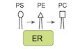
Results A dynamic traceable system to programme de novo epiallelesTo investigate the potential for memory of de novo epigenetic states, we first developed an optimised CRISPR-based epigenetic programming tool. Here, we employed a catalytically dead (d)Cas9 fused with an array of five optimally spaced GCN4 repeats (dCas9GCN4) (Morita et al, 2016). These serve as docking sites to recruit up to five “effectors” to a specific genomic locus via their single-chain antibody (scFv) domain (Fig 1A). This modular system amplifies both the quantitative level and domain size of ON-target epigenome editing, relative to dCas9 effector fusions, while minimising OFF-target effects (Pflueger et al, 2018). To target de novo heterochromatin, we generated KRABGFP-scFv and DNMT3A/3LGFP-scFv effectors, which promote direct deposition of H3K9me3 and DNA methylation respectively (Quenneville et al, 2012).

Figure 1. Programming an epiallele promotes a robust heterochromatin domain and gene silencing
Schematic showing recruitment of the modular epigenetic editing system to an endogenous promoter upon addition of DOX. A knock-in reporter downstream facilitates single-cell analysis. The relative abundance of indicated histone modifications assayed by CUT&RUN-qPCR after KRABGFP-scFv (blue) or control GFPscFv (light brown) targeting, relative to untransfected (grey). Shown are independent quantifications at two genomic positions on the Esg1 promoter, indicated relative to TSS (−300 bp, −700 bp). Data are mean of two or three independent biological replicates. Histograms showing the average DNA methylation across three genomic regions of the Esg1 promoter determined by bisulphite pyrosequencing in two biological replicates. CUT&RUN genome tracks for H3K4me3, H4K20me3 and H3K4me3 in untransfected (grey), control GFPscFv (light brown) or KRABGFP-scFv (blue) targeted ESC after 7 days of DOX treatment. Grey box highlights the region of heterochromatin spreading induced by epigenetic editing. Scatterplots demonstrating specificity and magnitude of programmed modifications at Esg1 by CUT&RUN-seq. Shown are all promoters genome wide. Epifluorescence images of Esg1-tdTomato ESC in −DOX (top) or +DOX conditions (bottom), where targeted heterochromatin is deposited. Single-cell expression of Esg1-tdTomato in control (GFPscFv) or upon heterochromatin induction (KRABGFP-scFv) using a gRNA targeting close to the TSS (+87 bp) or further upstream on the promoter (−475 bp). Each data point indicates a cell, percentage indicates the proportion of fully silenced cells (mean of four biological replicates) and bars represent median. P-values calculated by comparing +DOX with −DOX conditions.Data information: In all panels, asterisks indicate P-values by unpaired t-test over biological replicates; *P < 0.05, **P < 0.01, ***P < 0.001. Error bars ± SD.
We placed all components under a DOX-inducible promoter and destabilised dCAS9GCN4 protein and effectors with d2 domains, which together facilitate precise temporal control over epigenome editing activity. This is important to assess subsequent epigenetic memory without confounding reiterative targeting. To track the temporal ON-OFF dynamics in real-time, all effectors are tagged with superfolder GFP, which also enables cell isolation via flow cytometry (Fig EV1A–C). Finally, we used an “enhanced” gRNA scaffold (AT-flip, extended stem loop) linked with a tagBFP (Chen et al, 2013), which further amplifies ON-target activity and facilitates tracking respectively. In summary, dCas9GCN4 and KRABGFP-scFv expression can be induced by DOX treatment and traced in real-time via GFP, while BFP is constitutively expressed. Reciprocally, the system is destabilised and can be rapidly switched back OFF by removal of DOX.
Click here to expand this figure.
Figure EV1. iCRUSH induces domains of de novo histone modification levels comparable to endogenous heterochromatin loci
A. PiggyBac constructs used to deliver the epigenetic editing tool (iCRUSH) into ESC. From left to right: the enhanced gRNA scaffold is under control of a constitutive U6 promoter, the same construct also drives constitutive expression of a puromycin selection gene and blue fluorescent protein (BFP) separated by the self-cleaving peptide T2A; the dCas9GCN4 fusion gene is under control of the TRE3G DOX-inducible promoter, the same construct also drives constitutive expression of the rtTA trans-activator and a hygromycin resistance gene separated by a T2A; the epigenetic effector constructs (KRABGFP-scFv or Dnmt3a3LGFP-scFv ) are also under control of the TRE3G DOX-inducible promoter and it is fused with a green fluorescent protein (GFP) and an scFv domain specific for GCN4, it also drives constitutive expression of the neomycin resistance gene. These constructs are genomically integrated into ESC by co-transfection with the PiggyBac transposase. B. Density plots, obtained by flow cytometry analysis, show gRNABFP and KRABGFP-scFv expression in the cell population prior to and upon DOX induction, revealing its dynamic activation. C. Fluorescence distribution measured by flow cytometry showing Esg1 reporter silencing after recruitment of KRABGFP-scFv for 4 or 7 days (blue) compared to a reference untargeted sample (red), demonstrating order of magnitude of silencing. Cells were gated for the presence of both KRABscFv-GFP (GFP positive) and gRNA-BFP (BFP positive). D, E. Genome views comparing the magnitude of de novo peaks of histone marks deposited by iCRUSH at the Esg1 promoter (D) with endogenous representative examples (Mbnl and Oct4) (E). Programmed heterochromatin epialleles fully silence gene activity in single cellsTo follow programmed epialleles at endogenous loci, we initially used a naïve pluripotent ESC line wherein the endogenous Esg1 gene carries a knock-in tdTomato (Fig 1A) (Hackett et al, 2018). We introduced dCas9GCN4::KRABGFP-scFv and a single gRNABFP that targets the Esg1 promoter (+87 bp of TSS) via piggyBac, and assessed the extent of de novo programmed epigenetic states after 7 days (7 d) induction with DOX. Quantitative CUT&RUN qPCR demonstrated a highly significant deposition of heterochromatic H3K9me3 (P = 0.014) marks across the Esg1 promoter specifically with KRABGFP-ScFv, relative to either untransfected or control GFPscFv targeting (Fig 1B). This was paralleled by enrichment of another heterochromatic mark, H4K20me3 (P = 0.002), which often co-localises with H3K9me3 (Schotta et al, 2004), and complete loss of endogenous H3K4me3 modification (P = 0.0013). Moreover, bisulphite pyrosequencing revealed a highly significant increase in DNA methylation (P = 0.0005) across the entire Esg1 promoter region (Fig 1C). These results indicate that upon single-gRNA tethering of a flexible array of five KRABGFP-scFv effectors, a de novo domain of heterochromatic modifications is established.
To further investigate the extent and specificity of programmed heterochromatin we performed CUT&RUN-seq. We observed that our epigenetic editing system deposits a broad domain encompassing ~12 kb of H3K9me3 and H4K20me3 around the endogenous Esg1 locus, while previously abundant H3K4me3 is undetectable (Fig 1D). Importantly the de novo peaks of H3K9me3 and H4K20me3 are of a magnitude comparable to the strongest peaks throughout the genome, suggesting they recapitulate robust physiological heterochromatin status (Figs 1E and EV1D). Moreover, targeting was highly specific, since we observed minimal OFF-target changes in H3K9me3, H4K20me3 and H3K4me3 (Figs 1E and EV1D). Taken together, these data reveal that a substantial epigenomic domain (> 10 kb), which bears the key hallmarks of repressive heterochromatin, is specifically programmed at an endogenous genomic locus.
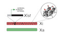
We next asked whether this de novo heterochromatin domain is associated with induction of transcriptional silencing. Esg1 is highly active in pluripotent cells, and the endogenous reporter facilitates dynamic single-cell analysis over time. As expected, addition of DOX led most cells to become GFP positive, indicative of activation of the epigenetic editing system (Fig 1F). Strikingly, this concomitantly led to complete loss of Esg1tdTomato-positive cells, which was additive with time (Figs 1F and EV1C). Quantitative single-cell expression revealed > 99% cells exhibited transcriptional repression, with > 85% reaching a complete OFF state, indistinguishable from ESC that do not carry the tdTomato reporter (Neg), and corresponding to > 500-fold transcriptional silencing (Fig 1G). In contrast, the GFPscFv control exhibited only modest repression, which likely reflected steric hindrance due to TSS binding, since a gRNA targeting upstream (−475 bp) elicited full silencing with KRABGFP-scFv (> 500 fold) but its control GFPscFv exhibited no effect on transcription (Fig 1G). In summary, these data indicate that our system is able to ectopically programme heterochromatin states at Esg1, which is directly linked with powerful transcriptional silencing at the single-cell level, implying high penetrance of deposition across a broad domain. We refer to this enhanced epigenetic tool as inducible CRISPR unleashing of silencing by heterochromatin (iCRUSH) (Fig 1A).
Induced heterochromatin is progressively erased in ESCExtant paradigms suggest that large domains of heterochromatic H3K9me3, H4K20me3 and DNA methylation are heritable, and self-propagate via “read-write” feedback machinery (Hathaway et al, 2012; Reinberg & Vales, 2018). To understand this in a developmental context, we next investigated the potential for propagation of induced heterochromatin epialleles in naïve ESC, which faithfully recapitulate in vivo epiblast when maintained in 2i/L (Hackett & Surani, 2014). Withdrawal of DOX resulted in a rapid switch off of the iCRUSH epigenetic editing system as determined by quantitative cytometry for GFP, and consistent with dCas9GCN4 and KRABGFP-scFv being destabilised, therefore fully releasing the inducing signal (Fig 2A). In parallel, we observed a progressive loss of Esg1tdTomato silencing (Fig 2B). Interestingly, 4 days after DOX washout (D-wo (4 days)), we observed a graded distribution of Esg1 expression among single cells, indicative of probabilistic reactivation dynamics. By 7 days after release (D-wo (7 days)), however, all cells reverted to the ON state, reflecting > 500-fold increase in Esg1 expression (Fig 2B).

Figure 2. Induced heterochromatin epialleles are progressively erased in naïve pluripotent cells
Representative flow cytometry density plots showing gRNABFP (+87 bp of TSS) and KRABGFP-scFv expression after DOX treatment (7 days) and DOX washout (4 days). Esg1-tdTomato expression at single-cell resolution during DOX washout in control (GFPscFv) or induced heterochromatin (KRABGFP-scFv) cells. Horizontal bars indicate median, each data point a single cell. Histograms of mean DNA methylation levels across the Esg1 promoter (6 CpG sites) during DOX washout in two biological replicates. CUT&RUN tracks at +DOX, and 4 and 7 days of DOX washout in control GFPscFv or KRABGFP-scFv for indicated histone marks. Grey boxes highlight the domain of heterochromatin spreading in +DOX. CUT&RUN qPCR quantification of the relative abundance of each mark at Esg1 promoter in two or three independent biological replicates, normalised to a positive control region and untransfected cells. p53-tdTomato expression in single cells during DOX washout in control (GFPscFv) or induced heterochromatin (KRABGFP-scFv) cells. Each data point indicates a cell, and horizontal lines represent the median. Bisulphite pyrosequencing quantification of DNA methylation at the p53 promoter (4 CpG sites) at indicated time point in two biological replicates. CUT&RUN qPCR quantification of the relative abundance of H3K9me3 and H3K4me3 at p53 promoter in biological replicates. Heatmap representing relative expression by qRT-PCR of each indicated gene upon heterochromatin targeting (+DOX) or after DOX washout (D-wo (7 days)), normalised to the untransfected control in three biological replicates. Statistics are measured between KRABGFP-scFv and control (GFPscFv) at DOX-wo time point.Data information: In all panels, asterisks indicate P-values by unpaired t-test; *P < 0.05, **P < 0.01 ***P < 0.001. Error bars ± SD.
To determine if transcriptional re-expression corresponds to loss of programmed epigenetic states, we used bisulphite pyrosequencing and CUT&RUN. Consistent with the reactivation dynamics, we found that DNA methylation is partially maintained at the Esg1 promoter at the early time point (D-wo (4 days)) but is almost completely erased by 7 days washout (Fig 2C). However, we found that the high levels of deposited H3K9me3 and H4K20me3 are largely erased by 4 days after DOX withdrawal (Fig 2D and E). Following 7 days release of the epigenetic editing trigger, the Esg1 chromatin state completely reverts to the initial configuration, including erasure of H3K9me3, H4K20me3 and DNA methylation, and reacquisition of the endogenous H3K4me3 mark (Fig 2D and E). While our system deposits high levels of DNA methylation, we additionally checked whether co-targeting KRABGFP-scFv together with the catalytic domain of Dnmt3a and its cofactor Dnmt3L (3a3LGFP-scFv) would enhance epigenetic inheritance, since such effects have been reported in cancer and primed cell lines (Fig EV2A) (Amabile et al, 2016; Nunez et al, 2021). Although we found a slight further increase in DNA methylation by compound recruitment (Fig EV2B), we observed equivalent or faster erasure of epigenetic memory (Fig EV2C). Taken together, our data argue that induction of a robust heterochromatin domain, and consequently extensive epigenetic silencing, is readily reversible from OFF→ON in naïve pluripotent cells.
Click here to expand this figure.
Figure EV2. Cell cycle inhibition slows but does not block epigenetic erasure in naïve ESC
Schematics of the iCRUSH epigenetic tool used in the experiment. KRABGFP-scFv is either recruited alone or in combination with Dnmt3a/Dnmt3L (3a3LGFP-scFv) or the catalytically mutant Mut3a3LGFP-scFv. Bar plot showing the average of the percentage DNA methylation at 6 CpG sites on the Esg1 promoter assayed by bisulphite pyrosequencing. Violin plots show the single-cell distribution of Esg1-tdTomato expression (log scale) in each condition (−DOX, 7 days +DOX and 7 days DOX washout) upon epigenetic editing with KRABGFP-scFv (blue) +/− 3a3LGFP-scFv (aqua green) or Mut3a3LGFP-scFv (purple). Each data point indicates a cell, and horizontal lines represent the median. Esg -tdTomato reporter expression in KRABGFP-scFv or untransfected control after gRNA transient transfection and 3 days of DOX induction. Bar plots show percentage of CpG methylation or relative abundance of histone modifications normalised to a positive control region and untransfected control after 3 days of DOX (gRNA transiently transfected) on one, two or three biological replicates. Statistics calculated by unpaired t-test over two or three independent biological replicates (*P < 0.05; **P < 0.01). Error is measured as ± SD. Timeline of the experiment. Briefly, cells are treated for 72 h (3 days) with DOX to induce iCRUSH-mediated silencing. tdTomato-negative cells are sorted and plated back in culture in duplicate experiments without DOX. After 24 h of DOX washout, one sample is treated with the cell cycle inhibitor RO3306 for further 24 h. After a total of 48 h from DOX washout, the cell cycle is released and cells further analysed at 12 h interval up to 96 h. In parallel, cells are cultured in the absence of the inhibitor. Time course of the percentage of Esg1-tdTomato-silenced cells in the + or -RO3306 conditions at 12 h intervals of DOX washout. Error is measured as ± SD between two biological replicates. Line plots indicate log growth of cells in + or – RO3306 conditions. Epigenetic inheritance is restricted by naïve ESCTo confirm that failure to propagate robust heterochromatin in ESC is not a phenotype specific to Esg1, we generated a second endogenous reporter ESC line by inserting tdTomato downstream of the p53 gene, separated by a T2A self-cleavable domain (Fig 2F). Targeting KRABGFP-scFv to the p53tdTomato promoter recapitulated the same extensive heterochromatin deposition including DNA methylation, H3K9me3 and loss of H3K4me3, and robust (> 100-fold) single-cell silencing, as achieved at Esg1tdTomato (Fig 2F–H). Upon 7 days DOX withdrawal, we found that p53 expression becomes fully reactivated in ESC (Fig 2F–H). This is paralleled by erasure of targeted DNA methylation and H3K9me3, and reacquisition of endogenous H3K4me3, consistent with heterochromatin failing to confer epigenetic memory in naïve ESC.
To examine this across further genomic locations, we programmed heterochromatin to additional endogenous loci, selected to represent different regulatory features (e.g. imprinting control regions, promoters). We imposed strong epigenetic silencing with iCRUSH, yet most loci (Pten, Cdh1, Greb1, Adamts7, Smoc1 and Jade1) reverted to their original expression status within 7 days DOX withdrawal (Fig 2I). Nevertheless, we did observe that imprinted genes (Peg3, Mest and Plagl1) are exceptions and, uniquely, maintain memory of de novo silencing in naïve ESC (Fig 2I). This suggests that heterochromatin domains at ectopic sites can be epigenetically inherited in a genomic context-dependent manner, with imprinted loci providing the necessary sequence substrate for propagation. However, in general, we find de novo chromatin states at endogenous single-copy loci are not heritable over extended periods in naïve ESC. This supports a dynamic competition of opposing activities that generally disfavours epigenetic inheritance during the phase of naïve pluripotency, potentially as a safeguard to restrict intergenerational transmission of aberrant epialleles.
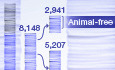
We next asked if this principle in naïve ESC holds for other epigenetic silencing pathways by exploiting a hybrid female ESC line carrying a DOX-inducible Xist allele on the BL6-derived X-chromosome (TX1072) (Schulz et al, 2014) (Fig 3A). Activation of Xist leads to programmed silencing of X-linked genes in cis via recruitment of repressive epigenetic systems, with a principal role for polycomb (Zylicz et al, 2019). In differentiated cells, cis repression propagates independently, resulting in stable silencing memory (X-Chromosome inactivation (XCI)), even after withdrawal of Xist (Loda & Heard, 2019). However, using transcriptomics, we observed that while strong epigenetic silencing is initially imposed in naïve ESC, withdrawal of DOX led to an almost complete reactivation of X-linked genes after 3 days (Fig 3B), extending a previous finding based on two marker genes (Wutz & Jaenisch, 2000). Hierarchical clustering revealed the majority of genes (81%) exhibit fast reactivation dynamics (< 3 days) in ESC (Fig 3C). A second cluster (8% of genes) also reactivated but with slower dynamics (< 7 days), and these overlapped with X-linked loci that reactivate late in vivo, for example, Fmr1b and Pnma5 (Borensztein et al, 2017). A third cluster (4%) was resistant to initial silencing in naïve cells (ESC escapees), while the final cluster (7% of genes) did exhibit memory of silencing following DOX withdrawal (Fig 3C). This “memory” cluster was enriched for tandem gene families such as the Rhox, Mage and Xlr clusters. Overall, however, these data suggest that the vast bulk of X-linked genes cannot propagate programmed epigenetic silencing in ESC, which is in contrast to differentiated cells, and supports the principle that naïve pluripotency specifically antagonises epiallele memory.

Figure 3. Induced X-linked gene silencing is reversible in naïve pluripotent cells
Schematic representing the workflow of the experiment: wild-type TX1072 female mESC cells (mixed Cast/BL6), carrying a DOX-inducible Xist on the BL6 allele, are treated with DOX for 6 days to induce Xist overexpression driving allele-specific epigenetic silencing of X-linked genes in cis (XCI). Subsequent DOX washout enables silencing memory to be investigated. Scatterplots showing allele-specific expression of X-linked genes on the Cast (green) or BL6 (orange) X-chromosome following DOX. The dynamics of reactivation of repressed BL6 genes are shown at indicated time points of DOX washout. Grey dots indicate all autosomal genes, which are unaffected. Heatmap of BL6 X-linked genes in three independent clones at the indicated time points arranged by unsupervised hierarchical clusters: fast reactivation (81%), slow reactivation (8%), escapees (4%) and memory (7%). Line plots indicate the expression trend of each individual gene from the cluster, with black line representing the mean, and error bars 95% CI. CRISPR screen reveals key factors that antagonise epigenetic memory in ESCTo investigate whether the reversal of repressive epialleles in naïve cells is driven by passive dilution during cell divisions, or by active erasure, we transiently transfected iCRUSH to epigenetically silence the Esg1 reporter for 3 days with DOX. This led to ~100-fold silencing, deposition of significant levels of DNA methylation, H3K9me3 and H4K20me3, and loss of H3K4me3 (Fig EV2D and E). We then released the epigenetic editing system by DOX washout and concomitantly treated the cells with or without the cell cycle inhibitor RO3306 (Fig EV2F), which blocks cells at the G2/M phase boundary. We observed Esg1 reactivation is only weakly impaired by cell cycle inhibition (Fig EV2G), spanning at least 60 h (Fig EV2H). Thus, while passive dilution may partially contribute to reversion of epigenetic memory, active mechanisms play a key role in erasing de novo epialleles in naïve ESC.
We therefore sought to identify the putative factors that actively counteract epigenetic memory in pluripotent phases by designing a genome-wide loss-of-function CRISPR screen (Fig 4A). We introduced a single copy of Cas9 nuclease tagged with GFP (Cas9T2A-GFP) into Esg1tdTomato ESC that also carry dCas9GCN4 in the OFF state, and infected these cells with a pooled lentiviral library of exon-targeting gRNA covering 19,674 genes (Doench et al, 2016). We subsequently induced self-inactivation of the Cas9T2A-GFP with a pair of specific gRNAs, which we confirmed by flow sorting cells according to loss of GFP (GFPneg) (Fig 4A). This cell population is now composed of a heterogeneous pool of knockout cells, to which we could apply our epigenetic editing system to identify the factors that antagonise epigenetic inheritance.

Figure 4. A CRISPR screen identifies key factors that restrict epigenetic inheritance in ESC
A. Schematic of screen design and workflow: an Esg1-tdTomato cell line carrying constitutive Cas9T2A-GFP nuclease and DOX-inducible (off)-dCas9GCN4 is transduced with a lentiviral gRNA library. The Cas9T2A-GFP is self-inactivated via introducing specific gRNAs, and GFP-negative cells are isolated. Subsequently, gRNABFP and KRABGFP-ScFv are introduced and cells treated with DOX to induce Esg1 heterochromatin-mediated silencing (TOMneg). TOMneg ESC are flow sorted and re-cultured in absence of DOX to isolate cells that inherit epigenetic silencing, which are subject to NGS to identify the gene knockout they carry. B, C. Significant hits for factors that permit epigenetic memory when knocked-out, showing the -log relative ranking algorithm (RRA) score at 4 or 7 days of DOX washout. False discovery rate (FDR) is indicated. D. Histograms showing the percentage of Esg1-tdTomato-negative cells in WT or knockout ESC lines after programming heterochromatin (+DOX) and during DOX washout. Each data point indicates a biological independent knockout line. Error bars ± SD, asterisks indicate P-values relative to −DOX by unpaired t-test; **P < 0.01. E. Quantitative expression of Esg1-tdTomato among single cells in WT or in independent clonal knockout ESC lines. Each data point indicates a cell, and bars represent the median.To achieve this, we targeted heterochromatin to Esg1tdTomato and isolated cells that subsequently retained silencing memory (TOMneg) following release of dCas9GCN4::KRABGFP-scFv using a gating strategy to distinguish between cells remaining fully silenced (bottom 2.5% (TOMneg-2.5%) (Fig EV3A) and those that retain a degree of repression memory (TOMneg-wide) (Fig EV3B). We then used model-based analysis of genome-wide CRISPR-Cas9 knockout (MAGeCK) to identify the gene knockouts enriched in the TOMneg populations that retained epigenetic memory relative to the complementary TOMpos population over short (3 days) and extended (7 days) timescales (Li et al, 2014). As expected, top hits across both gates were associated with roles in transcriptional or translational regulation, and comprised many candidates with established or predicted epigenetic functions. This included the SWI/SNF histone remodeller Smarcc1 (FDR 0.03), the H3K79 methyltransferase Dot1L (FDR 0.16) and the H3K4 histone methyltransferase Kmt2d (FDR 0.13), although this latter was enriched only at the shorter time point (Figs 4B and C and EV3B). Additionally, we noted cells that propagated silencing memory also exhibited significant enrichment for knockouts of the X-linked zinc-finger protein Zmym3 (FDR 0.01), the NSL complex subunit Kansl2 (FDR 0.05) and Dppa2 (FDR 0.03) (Figs 4B and C, and EV3B and C), which is a pluripotency-specific gene recently linked with regulating de novo DNA methylation and bivalency (Eckersley-Maslin et al, 2020; Gretarsson & Hackett, 2020).
Click here to expand this figure.
Figure EV3. Gating strategy for CRISPR screen and supplementary scatterplots of candidate factors that antagonise epigenetic inheritance
Histogram showing the gate used to sort cells that strictly retained epigenetic silencing memory in the CRISPR screen (tdTomato negative) using a stringent gating threshold (bottom 2.5% TOM- cells (TOM2.5%neg). The results of significant candidate genes that enable this memory when knocked out are detailed in main Fig 4B and C. Middle histogram: A wider gate was used to include all tdTomato-negative cells (TOMnegwide) and was designed according to a tdTomato-positive control sample to capture all cells exhibiting full or partial silencing memory. Scatterplots: show significant hits from the screen displayed by -log relative ranking algorithm (RRA) score from the cells captured using the TOMnegwide gate at short-term time point (3 days DOX washout; left) or longer term (7 aysd DOX washout; right). These hits are linked with enabling epigenetic inheritance when abrogated. Line plot for gRNA count from the CRISPR screen after silencing (+DOX) and during memory phase (DOX washout for 3 or 7 days) for the candidate genes indicated. Each line represents a different gRNA from the pool for the same target gene. A concordant enrichment during DOX washout indicates all gRNAs (and therefore independent knockouts of the target gene) promote epigenetic inheritance. Note Kmt2d is only enriched at the earlier time point.To validate these candidates, we generated independent clonal knockout ESC lines of each. Deletion of these factors did not affect Esg1tdTomato basal activity prior to imposition of epigenetic silencing, and all knockouts also exhibited a comparable extent of programmed silencing as WT after 7 days DOX, implying no changes in initial parameters (Fig 4D). Following DOX washout, Smarcc1−/− and Dot1L−/− reverted to the active state with a similar kinetics to WT, implying false positives. In contrast, Kmt2d−/− cells showed penetrant memory at 4 days of DOX washout, but reverted to an ON state after 7 days, suggesting absence of Kmt2d impacts the rate of memory erasure, potentially by affecting re-deposition of H3K4me3 (Fig 4D and E). Interestingly, while some Zmym3−/− lines exhibited memory, and independent gRNAs were concordant (Fig EV3C), there was high heterogeneity between independent knockout clones, indicating a complex regulatory response that we did not follow further. However, all Dppa2−/− lines fully maintained epigenetic memory after 3d DOX withdrawal, with the majority of cells remaining in the OFF state after 7 days (Figs 4D and E, and EV3C). This suggests that abrogating Dppa2 changes the balance of activates in ESC to generate an environment that is permissive for epigenetic inheritance.
Epigenetic inheritance is permitted by deletion of DPPA2To examine the role of Dppa2 further, we traced the single-cell dynamics of transcriptional memory in multiple-knockout ESC lines (Fig EV4A). While WT cells rapidly lose silencing memory after 7 days DOX washout, the majority of Dppa2−/− cells remain fully silenced at this stage (Fig 5A). Importantly, inheritance of this silenced state in the absence of Dppa2 is subsequently maintained across a consistent fraction of cells for at least 43 days after DOX withdrawal (> 100 cell replications), with the population therefore exhibiting a bimodal distribution (Fig 5B). Importantly, population doubling time was similar between wild-type and knockout cells (Fig EV4B). This implies that abrogation of Dppa2 facilitates heritability of ectopic silencing in a probabilistic manner, potentially by shifting the odds against reversion, and thus promoting steady-state inheritance. Notably, flow sorting TOMneg and TOMpos fractions after 26 days of DOX washout revealed that, while the TOMpos remained positive, the TOMneg reacquired a bimodal distribution, supporting a stochastic memory function (Fig EV4C–E).
Click here to expand this figure.
Figure EV4. Epigenetic inheritance is probabilistic upon Dppa2 knockout
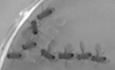
Western blot comparing DPPA2 protein expression in WT and Dppa2 knockout individual ESC clonal lines. β-TUBULIN is used as loading control. Line plot indicates exponential growth of wild-type or Dppa2−/− cells. Timeline of the experiment in D and E. After induction for 7 days, tdTomato-negative (TOMneg) and -positive (TOMpos) fractions of cells are sorted following 26 days of DOX washout in Dppa2−/− a
留言 (0)