
記住我


Male and female gregarious desert locusts were obtained from colonies reared at Philipps-Universität Marburg. Animals were kept at a constant temperature of 28 °C under a 12 h:12 h light/dark cycle. Animals were mounted onto a metal holder and legs and wings were cut off. A window was cut frontally into the head capsule, and fat tissue and air sacs were removed to get access to the brain. The esophagus was cut and the gut was removed via the abdomen, which was sealed afterwards with dental wax. The brain was stabilized from posterior with a small twisted metal wire inserted into the window of the head. Finally, the neural sheath of the brain was partly removed to allow penetration of the recording electrode. The brain was kept submerged in locust saline (Clements and May 1974) during preparation, recording and dissection.
ElectrophysiologyIntracellular recordings were performed with sharp microelectrodes drawn from borosilicate capillaries (Hilgenberg, Malsfeld, Germany), with a Flaming/Brown horizontal puller (P-97, Sutter Instrument, Novato, CA). The tip of the electrodes was loaded with 4% Neurobiotin (Vector Laboratories, Burlingame, CA) in 1 mol l−1 KCl and the shanks were loaded with 1 mol l−1 KCl. Signals were amplified 10 × with a BA-01 × amplifier (npi electronic instruments, Tamm, Germany), and monitored with a custom-built audio monitor (University of Marburg) and an oscilloscope (Hameg, Frankfurt/Main, Germany). Data were digitized with a Power1401-mkII (Cambridge Electronic Design, Cambridge, GB) and stored on a PC using Spike2 (Cambridge Electronic Design, Cambridge, UK) with a sampling rate of 20 kHz. After the recording, Neurobiotin was injected into the cell by application of a positive current of 0.5–2 nA for at least 2 min.
Histology and image acquisitionBrains were dissected and fixed in 4% paraformaldehyde, 0.25% glutaraldehyde, and 2% saturated picric acid diluted in 0.1 mol l−1 phosphate buffered saline (PBS) at 4 °C overnight. Following rinses in PBS (4 × 15 min) brains were incubated in Cy3-conjugated streptavidin (1:1000) in PBS containing 0.3% Triton X-100 (PBT) at 4 °C for 3 days. Following rinses in PBT (2 × 20 min) and PBS (3 × 20 min) brains were dehydrated in an ascending ethanol series (30%, 50%, 70%, 90%, 95%, 100%, 15 min each) and cleared in a 1:1 mixture of 100% ethanol and methyl salicylate (Merck, Darmstadt, Germany) for 15 min and in pure methyl salicylate for 1 h. Finally, they were mounted in Permount (Fisher Scientific, Pittsburgh, PA) between two coverslips. Samples were scanned with a confocal laser scanning microscope (Leica, TCS SP5, Leica Microsystems, Wetzlar, Germany) with a 20 × immersion objective (HC PL APO 20 ×/0.75 Imm Corr CS2, Leica). A diode pumped solid state laser (561 nm) was used to excite Cy3. Scanning frequency was 400 Hz and the voxel size was 0.54 × 0.54 × 2.0 µm3.
StimulationAnimals were stimulated from the zenith by light from a blue light emitting diode (LED; Oslon SSL 80, LDCQ7P, 452 nm, Osram Opto Semiconductors GmbH, Regensburg, Germany). The light was linearly polarized by a dichroic polarizer sheet (HNP’B, Polaroid, Cambridge, MA). To generate degrees of polarization between 0.002 and 0.9, diffusor sheets were placed in different combinations between the LED and the polarizer or between the animal and the polarizer (Fig. 2a). At the highest degree of polarization, all four diffusor sheets were placed between the LED and the polarizer, while at the lowest degree of polarization, the four diffusor sheets were placed between the animal and the polarizer. With the different arrangements, five stimuli could be tested: DoP = 0.99 (maximally polarized light, 1.9 × 10¹³ photons cm−2 s−1), DoP = 0.35 (1.6 × 10¹³ photons cm−2 s−1), DoP = 0.1 and DoP = 0.05 (1.5 × 10¹³ photons cm−2 s−1), and DoP = 0.002 (1.7 × 10¹³ photons cm−2 s−1). The stimuli covered a visual angle of 12.6°. To monitor the angle and the degree of polarization, a HNP’B polarization filter was placed in front of an OPT101 photodiode/transimpedance amplifier (Texas Instruments, Dallas, TX) positioned close to the animal’s head (Fig. 2a). The output of the OPT101 was continuously recorded via the digitizer. The stimulation device rotated 360° clockwise and counterclockwise at 40°/s. In four recordings from TL neurons, we painted the DRAs of the animal during the recording with acrylic black paint using a fine paintbrush. Removing the paint was also done while recording and was either done with forceps or with a paintbrush, depending on whether the paint was already dry or not.
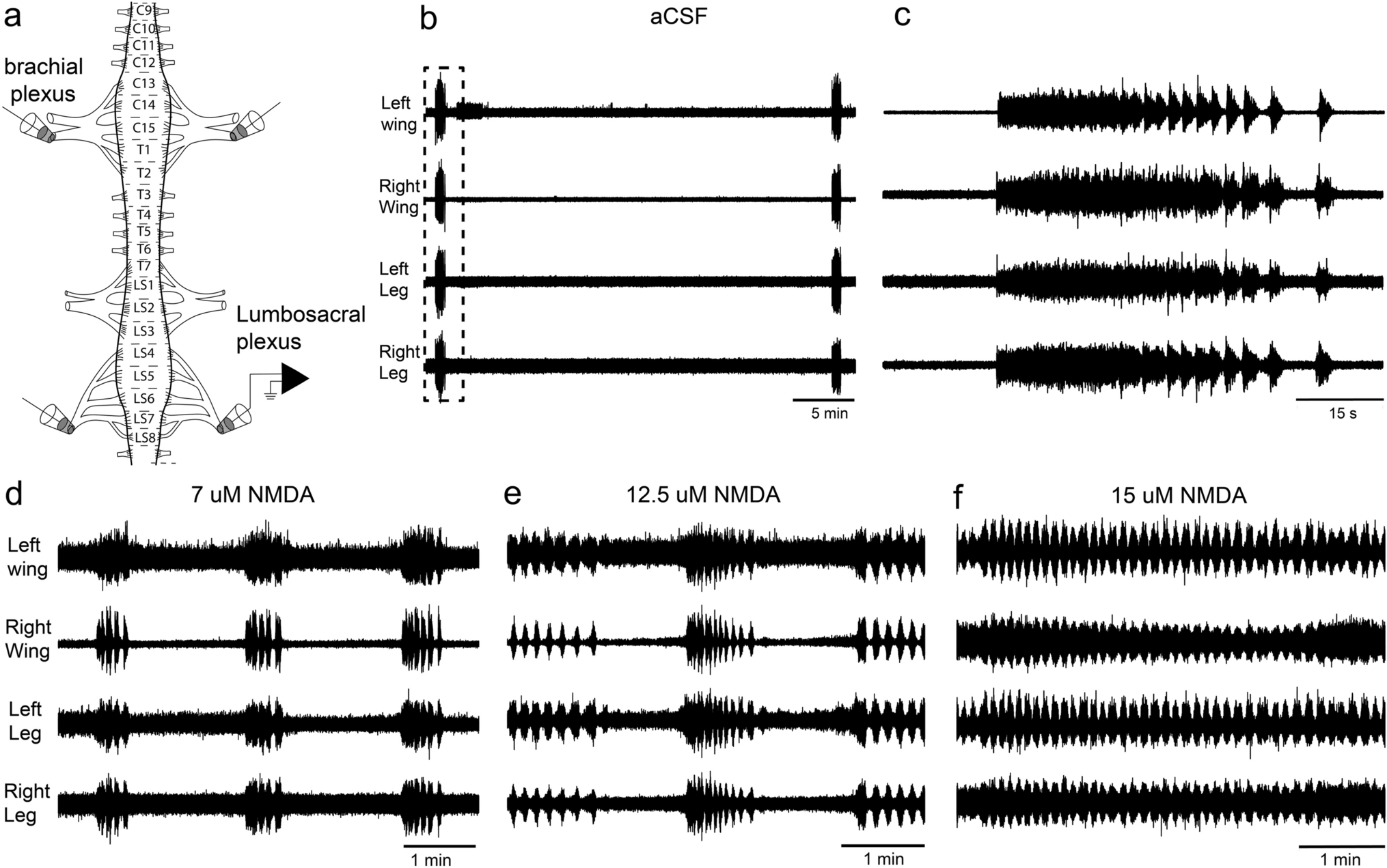
Fig. 2
a Schematic illustration of the stimulus setup. Unpolarized light emitted by a light-emitting diode (LED) was linearly polarized by a polarizer. Diffusors were placed between the LED and the polarizer or between the polarizer and the animal to achieve different degrees of polarization (DoPs). With four diffusors between the LED and the polarizer (small image inset) maximally polarized light (DoP = 0.99) was generated. The degree and angle of polarization were measured via a photodiode/transimpedance amplifier placed behind a polarization filter. b Spike train showing the response of a CL1a neuron to two full rotations of the polarizer in clockwise and counterclockwise direction (0°–360°, 360°–0°). The blue bar indicates the time window during which polarized blue light was presented. The mean spiking frequency is indicated as moving average with a window size of 1 s above the spike train
Data evaluationRecording data were only analyzed when the recorded neuron was successfully labeled and the stained cell type unequivocally identified. Physiological data were preprocessed using Spike2 and exported as mat-files for further analysis in MATLAB (Version 2020a, The MathWorks, Natick, MA, USA) using custom scripts. Circular histograms were created with the CircHist package (Zittrell 2019). Confocal image stacks were processed in Amira 5.6 (ThermoFisher Scientific, Waltham, MA; RRID:SCR_007353). Images showing raw data were exported from Spike2 and processed with Affinity photo and Affinity Designer (Serif, Nottingham, UK; RRID:SCR_016951). Diagrams were generated with Microsoft Excel or MATLAB and were exported to Affinity photo to create figure panels.
Background activityOwing to fluctuations of background activity (BA) in some neurons we calculated the BA for comparison with firing activity during stimulation within a time window of 5 s preceding the respective stimulus. Spikes were binned in 1 s bins and these spike counts were used to calculate the median and the 2.5th and 97.5th percentile of BA.
Stimulus responsivenessWe used linear-circular correlation analysis (CircStat; Berens 2009) to determine whether the modulation of spike rate was correlated to changes in AoP. Time points of action potentials during each 360° rotation were assigned to the respective orientation of the polarizer and these angles were doubled to allow using circular statistics on these axial data (Zar 1999). The resulting angles were averaged and the result was halved, yielding the preferred AoP (Фmax) in circular space. At least one clockwise and one counterclockwise rotation of the polarizer were included to measure the responsiveness to a stimulus with a particular DoP. To determine a correlation between firing rate and AoP, spike angles were binned in 10° bins and counts were tested for correlation with bin angles. A resulting P value < 0.05 indicated significant modulation by AoP. To specify properties of significant responses, we calculated the mean resultant vector length r and the response amplitude A. The vector length r describes the directedness of the response and ranges from 0 to unity, with unity indicating that all vectors are of the same direction (Batschelet 1981). It was calculated with the CircStat toolbox (Berens 2009). A describes the absolute amplitude of spike frequency modulation during stimulation, with higher A values indicating stronger modulation. A was calculated as follows according to Labhart (1996) and Pfeiffer et al. (2011):
$$A= \sum_^\left| _-\overline\right|,$$
where ni is the number of spikes in bin i and \(\overline\) is the number of spikes during the 360° rotation divided by the number of bins. Firing rates at Фmax and Фmin were estimated by fitting a bimodal von Mises distribution model to the binned data (Fitak and Johnsen 2017) and taking the model’s firing rate at the respective angles.
Regression analysisTo test whether modulation amplitude A, length of the mean vector r, and mean spiking activity were dependent on the DoP, respective data were pooled and tested for linear regression. Only cells that were stimulated with at least three different DoPs were included for this analysis. If the resulting residuals were not normally distributed (based on the Lilliefors test), the data were logarithmically transformed and the regression was done again. If the residuals of this regression were not normally distributed, the two linear models were compared regarding their R2 values and the one with the higher value was chosen.
Threshold for reliable coding of the AoPTo estimate the DoP threshold for reliable coding of the AoP we compared the mean resultant vector length r obtained during the stimulus with the upper 95% confidence limit of r obtained without stimulation (Pfeiffer et al. 2011). We defined the threshold as the lowest DoP at which the r values of all responses exceeded the upper 95% confidence limit of the estimated average r value of the no-stimulus controls.
留言 (0)